New Botryosphaeriaceae fruit rot of mango in Taiwan: identification and pathogenicity
←
→
Page content transcription
If your browser does not render page correctly, please read the page content below
Botanical Studies (2012) 53: 467-478.                                                                      physiology

New Botryosphaeriaceae fruit rot of mango in Taiwan:
identification and pathogenicity
Hui-Fang NI1,2, Hong-Ren YANG1, Ruey-Shyang CHEN3, Ruey-Fen LIOU2,*, and Ting-Hsuan
 HUNG2,*
1
  Department of Plant Protection, Chiayi Agricultural Experiment Station, Taiwan Agricultural Research Institute, Chiayi,
  Taiwan
2
  Department of Plant Pathology and Microbiology, National Taiwan University, Taipei, Taiwan
3
  Department of Biochemical Science and Technology, National Chiayi University, Chiayi, Taiwan

       (Received February 21, 2012; Accepted May 29, 2012)

       Abstract. Mango is an important fruit crop in Taiwan. Recently, severe fruit rot disease was found fre-
       quently on harvested mango fruits. To monitor the incidence of disease and to characterize the causal agent,
       we performed a field survey in the major mango-producing areas of southern Taiwan, including Guntain, Fan-
       shan, and Yujing, during 2009-2011. The results showed a disease incidence ranging from 18.7% to 58.1%,
       with those of Guntain significantly greater than the incidence found in Yujing and Fanshan. Based on morpho-
       logical characteristics and nucleotide sequences of the internal transcribed spacer (ITS), β-tubulin gene (TUB)
       and elongation factor 1-alpha (EF1-α) gene, we identified four Botryosphaeriaceae species, including Fusi-
       coccum aesculi, Neofusicoccum mangiferae, N. parvum, and Lasiodiplodia theobromae. Pathogenicity tests
       indicated that all of these fungal species were pathogenic to harvested mango fruits, and L. theobromae was
       the most aggressive pathogen. Moreover, when attached, immature mango fruits were inoculated with conidia
       of Botryosphaeriaceae species, disease symptoms characteristic of fruit rot appeared on the fruits after harvest
       and ripening. These findings indicated that L. theobromae, F. aesculi, N. mangiferae, and N. parvum were all
       causal agents of the new fruit rot of mango. Furthermore, their conidia may serve as important sources of in-
       ocula causing fruit rot disease in mango orchards.

       Keywords: Botryosphaeriaceae; Fruit rot; Mango.

INTRODUCTION                                                     Phomopsis mangiferae (Ploetz, 1994; Ko et al., 2009)
                                                                 are usually considered to be the most severe postharvest
   Mango (Mangifera indica) is an economically im-               disease of mango worldwide. However, we have recently
portant fruit crop in Taiwan. According to Agricultural          found that many harvested mango fruits displayed brown
Statistics Yearbook 2010 (http://www.coa.gov.tw/view.            soft lesions on the body surface of ripe mango fruit, rather
php?catid=23771), the total area of mango cultivation            than at the pedicel end, which indicated that fruit rot could
in Taiwan was 16,796 ha, which led to the production of          be another serious postharvest disease in Taiwan.
135,293 metric tons of mango fruits, with a yearly value             Botryosphaeriaceae species are known to occur world-
over 157 million US dollars. The main area for mango cul-        wide, causing dieback, cankers, shoot blights, leaf spot,
tivation is located in the southern part of Taiwan, including    gummosis, and fruit rots in a wide range of plant hosts
Tainan, Kaohsiung, and Pingtung.                                 which play important roles in agriculture and forestry
   Postharvest diseases, which cause serious problems            (Phillips, 2002; van Niekerk et al., 2004; Slippers et al.,
during storage and transportation of mango fruits, are the       2005; Damn et al., 2007; de Macedo and Barreto, 2008;
major factors that limit the thriving mango industry. Both       Marincowitz et al., 2008; Javier-Alva et al., 2009; Yu et
anthracnose disease caused by Colletotrichum gloeospo-           al., 2009; Wang et al., 2011).
rioides (Ploetz, 1994; Yang and Leu, 1988; Arauz, 2000)              In Taiwan, the first plant pathogenic species of Botry-
and stem-end rot caused by Lasiodiplodia theobromae              osphaeriaceae that caused mango stem-end rot was re-
(Liao, 1975; Johnson and Cooke, 1991; Ploetz, 1994) or           ported by Liao (1975). He isolated the pathogen Diplodia
                                                                 natalensis (syns. Lasiodiplodia theobromae and Botryodi-
*Corresponding authors: E-mail: rfliou@ntu.edu.tw; Fax:         plodia theobromae (teleomorph: Botryosphaeria rhodina))
  886-2-23620271; Tel: 886-2-33665208 (Ruey-Fen Liou);           and confirmed its pathogenicity. Recent studies from
 E-mail: thhung@ntu.edu.tw; Fax: 886-2-23636490; Tel:            several laboratories demonstrated that a complex of Botry-
 886-2-33664600 (Ting-Hsuan Hung).                               osphaeriaceae pathogens (Slippers et al., 2005; de Oliveira
468 Botanical Studies, Vol. 53, 2012
Costa et al., 2010), including L. theobromae, N. mangifer- Fruits with typical symptoms were then selected for fun-
ae, Neofusicoccum parvum and Fusicoccum aesculi, are gal isolation. The epidermis of fruit was first disinfested
associated with stem-end rot of mango. Take for example with 70% (v/v) ethanol and air-dried. Subsequently, small
Australia, where F. aesculi (= B. dothidea), N. mangiferae, pieces (2-3 mm2) of necrotic tissue were dissected from
N. parvum, L. theobromae, and Fusicoccum sp. cause the margins of lesions on fruit and placed on an acidified
stem-end rot of mango (Slippers et al., 2005). In Brazil, L. (750 μL of a 50% (v/v) solution of lactic acid per 300 mL
theobromae, F. aesculi, and N. parvum have been reported of potato dextrose agar medium) (APDA) (Merck KGaA,
as pathogens of mango stem-end rot and dieback disease Darmstadt, Germany). The plates were incubated at room
(de Oliveira Costa et al., 2010). temperature for 1-2 weeks. Putative Botryosphaeriaceae
Differentiation of Botryosphaeriaceae species were species isolates, recognized by their rapidly growing colo-
in the past chiefly based on the morphology of their ana- nies with gray mycelium (Lazzizera et al., 2008), were
morphs (Jacobs and Rehner, 1998; Denman et al., 2000). subcultured on potato-dextrose agar (PDA) plates. Isolates
However, morphological characteristics of these fungi were stored on PDA slants at 8°C. The frequency of oc-
may vary within the species, and in some cases, they currence of the fungi in the collected fruits, which showed
may look very similar between species, making identi- characteristic symptoms of fruit rot, was calculated ac-
fication of the fungus even more difficult. For efficient cording to the following formula: Frequency of occurrence
identification, DNA-based techniques have been applied (%) = (Number of fruits colonized by a specific pathogen/
to the taxonomy of Botryosphaeriaceae (Denman et al., Total number of fruits with fruit rot symptoms) x 100%.
2000; Slippers et al., 2004; Alves et al., 2005; Taylor
et al., 2005; Crous and Groenewald, 2005; Crous et al., Morphological characterization
2006; De Wet et al., 2008). Combination of the molecular For studies on colony morphology, isolates were grown
techniques with morphological characteristics has been on PDA and incubated at 25°C in darkness. The morphol-
used to successfully identify F. aesculi, N. parvum, and N. ogy of mycelium and conidia (dimensions, shape, color,
ribis, all of which were previously classified as F. aesculi presence of septa and longitudinal striations) were record-
(= B. dothidea) (sensu von Arx and Müller, 1954) (Jacobs ed. To induce sporulation, putative Botryosphaeriaceae
and Rehner, 1998; Smith and Stanosz, 2001; Slippers et isolates were grown on 2% water agar plates containing
al., 2004; Crous et al., 2006). sterilized pine (Pinus morrisonicola) needles (Smith et al.,
Currently, fungal pathogens known to cause mango 1996), and incubated at 25°C with a 12-h light (near UV)/
fruit rot include only Alternaria alternata, Phytophthora dark cycle. For further purification, conidia released from
nicotianae, Pestalotiopsis mangiferae, and Phyllosticta pycnidia on the pine needles were spread on water agar.
anacardiacearum, according to “Common Names of Plant After 12-24 h, single germinating conidia were picked and
Diseases” posted on the website of the American Phyto- transferred to PDA. Morphology of the conidia formed on
pathological Society (http://www.apsnet.org/publications/ pine needles was also examined under a stereoscopic mi-
commonnames/Pages/Mango.aspx). Information regarding croscope (Nikon SMZ 1500, Tokyo, Japan). To determine
the role of Botryosphaeriaceae species on mango fruit rot the average length and width of conidia, at least 50 conidia
has been very limited. The aims of this study were to: a) from each isolate were analyzed using a light microscope
identify Botryosphaeriaceae isolates collected from fruit (Nikon, ECLIPSE 80i, Tokyo, Japan); their images were
rot of mango fruits, b) investigate the incidence of fruit rot photographed with a pixel camera system (Pixera Penguin
and frequency of Botryosphaeriaceae species in three ma- 600CL, Los Gatos, CA, USA) and the length and width
jor mango producing areas of Taiwan, including Fanshan of conidia were measured by using a Simple PCI software
(Kaohsiung), Yujing, and Guntain (both in Tainan) during Rev. 3.6 (Compix Inc., Cranberry Township, PA, USA).
2009-2011; c) test the pathogenicity and compare the viru- The measurements of conidia were subjected to statistical
lence of Botryosphaeriaceae isolates obtained from mango analyses and presented as average ± standard deviations.
fruits with fruit rot.
Molecular characterization
MATERIALS AND METHODS The genomic DNA of fungal mycelia was isolated
using the method described by Wang et al. (1993). For
Field survey, disease symptoms, and fungal comparative phylogenetic study, partial sequences of three
isolation housekeeping genes were amplified by PCR, including
Field surveys were conducted at 77 orchards located in ribosomal internal transcribed spacer (ITS), β-tubulin
the Pintung (Fanshan) and Tainan (Yujing and Guntain) (TUB), and elongation factor 1-α (EF1-α). The PCR
areas of Taiwan during 2009-2011, and 15-20 mango fruits mixture contained 1X PCR buffer (10 mM Tris-HCl, pH
were randomly collected from each orchard. The incidenc- 8.0, 50 mM KCl, 1.5 mM MgCl2, 0.1% (w/v) gelatin, 1%
es of fruit rot disease were calculated 7 days after harvest Triton X-100), 100 μM of each dNTP, 0.2 μM of each
according to the following formula: Disease incidence (%) primer, 0.4 U of Prozyme DNA polymerase (Protech Tech-
= (Number of fruits which showed only fruit rot but not nology Enterprise, Taipei, Taiwan). Primers used for the
anthracnose symptoms/ Total number of fruits) × 100%. amplification of each gene were: ITS1 (5’-TCC GTA GGTNI et al. — New Botryosphaeriaceae fruit rot of mango 469 GAA CCT GCG G-3’) and ITS4 (5’-TCC TCC GCT TAT developed 7 days after inoculation. Statistical analyses of TGA TAT GC-3’) (White et al., 1990; Wang et al., 2010) the data were performed by using SAS (version 8, SAS for ITS, Bt2a (5’-GGT AAC CAA ATC GGT GCT GCT Institute) with the Fisher’s protected test, and an F value TTC-3’) and Bt2b (5’-ACC CTC AGT GTA GTG ACC with P
470 Botanical Studies, Vol. 53, 2012
Table 1. The incidence of fruit rot disease and occurrence frequency of Botryosphaeriaceae species on mango fruits exhibiting
symptoms of fruit rot during 2009-2011.
Frequency of occurrence (%)b
a
Region Year Incidence (%) Lasiodiplodia Neofusicoccum Neofusicoccum Fusicoccum
theobromae parvum mangiferae aesculi
Fanshan 2009 20.1 1.8 16.1 28.6 12.5
2010 18.7 6.4 25.5 4.3 34.0
2011 22.5 0.0 37.8 20.0 37.8
Yujing 2009 35.0 9.7 3.2 3.2 25.8
2010 25.6 2.8 30.6 0.0 47.2
2011 23.8 10.5 26.3 7.9 13.2
Guntain 2009 46.0 7.5 0.0 2.5 17.5
2010 53.8 3.3 5.0 0.0 8.3
2011 58.1 13.1 3.6 2.2 12.4
a
Incidence of mango fruit rot in each area was calculated by the following formula: Disease incidence (%) = (Number of fruits
which showed only symptoms of fruit rot but not anthracnose/Total number of fruits) × 100%.
b
Frequency of occurrence (%) = (Number of fruits colonized by a pathogen/Total number of fruits with fruit rot symptoms) × 100%.
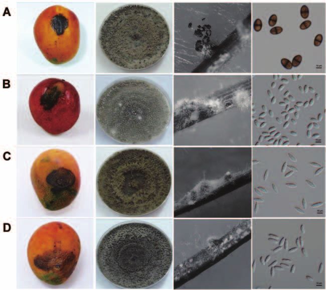
Figure 1. Symptoms of mango fruit rot and morphology of fungal colony, pycnidia, and conidia. A: Lasiodiplodia theobromae; B:
Neofusicoccum mangiferae; C: Fusicoccum aesculi; D: N. parvum. Bar = 10 μm.NI et al. — New Botryosphaeriaceae fruit rot of mango 471
Table 2. Representative isolates of Botryosphaeriaceae species collected from mango fruits with fruit rot in southern Taiwan.
GenBank Accession No.
Isolate Identity Locality
ITS EF1-α TUB
B961 Lasiodiplodia theobromae Guntain GQ502453 GQ979999 GU056845
B965 L. theobromae Guntain GQ502454 GQ980000 GU056854
B838 L. theobromae Fangshan GQ502456 GQ980001 GU056852
B852 L. theobromae Guntain GQ502457 GQ980002 GU056851
B918 L. theobromae Guntain GQ502458 GQ980003 GU056850
B902 L. theobromae Guntain GQ502459 GQ980004 GU056849
B878 L. theobromae Guntain GQ502460 GU056848
B607 L. theobromae Guntain GQ502461 GU056846
B845 Neofusicoccum parvum Fangshan GQ861434 GQ985316 GU062771
B946 N. parvum Yujing GQ861432 GQ985313 GU062768
B1314 N. parvum Fangshan GU073291 GU121436 GU111537
B794 N. parvum Fangshan GQ861433 GQ985312 GU062767
B1260 N. parvum Fangshan GU073287 GU121432 GU111533
B1272 N. parvum Yujing GU073288 GU121433 GU111534
B1296 N. parvum Yujing GU073289 GU121434 GU111535
B1307 N. parvum Fangshan GU073290 GU121435 GU111536
B809 N. mangiferae Fangshan GQ848323 GQ998898 GU071122
B793 N. mangiferae Fangshan GQ848320 GQ998900 GU071120
B808 N. mangiferae Fangshan GQ848322 GQ998899 GU071121
B979 N. mangiferae Yujing GQ848315 GQ998897 GU071123
B763 N. mangiferae Fangshan GQ421486 GQ998896 GU071119
B964 Fusicoccum aesculi Guntain GQ861429 GU002157 GU071124
B811 F. aesculi Fangshan GU453689 GU002164 GU071125
B844 F. aesculi Fangshan GU453690 GU002163 GU071126
B922 F. aesculi Yujing GQ421485 GU002162 GU071127
B833 F. aesculi Fangshan GQ861430 GU002161 GU071129
B801 F. aesculi Fangshan GQ861431 GU002160 GU071130
B932 F. aesculi Yujing GQ861428 GU002159 GU071131
B1113 F. aesculi Yujing GU453691 GU002158 GU071132
Isolates of N. mangiferae initially produced white, ap- days. Pycnidia were formed on WA supplemented with
pressed mycelium. Four days after incubation on PDA, sterilized pine needles after 1-2 weeks. Conidia were hya-
the middle of colony turned to pale olivaceous gray and line, thin-walled, aseptate, and fusiform (Figure 1C), with
produced pycnidia 10-14 days after incubation. When vi- an average length and width of 18.72-22.10 × 5.72-6.63
sualized from the bottom of Petri dish, the colonies were μm (L/W= 3.05-3.52) (Table S2).
olivaceous to black. This fungus grew slower than the Cultures of N. parvum were initially white with aerial
other three Botryosphaeria species found in this study. mycelium. The middle of colony then became pale oliva-
Pycnidia were formed on WA supplemented with sterilized ceous gray after 3-4 days. When visualized from the bot-
pine needles after 3-7 days. The conidia were hyaline and tom of Petri dish, the colony was deep olivaceous gray to
ovoid (Figure 1B), with an average length and width of black after 4-7 days of incubation. Pycnidia were formed
11.98-12.93 × 6.25-6.98 μm (L/W= 1.85-1.95) (Table S2). on WA supplemented with sterilized pine needles for 2-3
Colonies of F. aesculi were initially white with aerial weeks. Conidia were hyaline, aseptate, and fusiform (Fig-
mycelium. They became pale olivaceous gray from the ure 1D). The average length and width of 416 conidia were
center of colony after 3-4 days, and turned black after 7 15.85-19.25 × 4.49-6.61 μm (L/W= 2.70-3.68) (Table S2).472 Botanical Studies, Vol. 53, 2012
Phylogenetic analysis nucleotide sequence of ITS, TUB, and EF1-α showed a
To confirm the morphometric identifications and to respective identity range of 93-98%, 91-98%, and 75-96%
infer the evolutionary relatedness among these Botry- among different species.
osphaeriaceae species, partial nucleotide sequences of In addition, three unrooted phylogenetic trees were
ITS, TUB, and EF1-α from L. theobromae, N. mangiferae, constructed based on multiple sequence alignment of ITS,
N. parvum, and F. aesculi were amplified by PCR. The TUB, and EF1-α. The overall topology of these trees was
respective length of amplified products of ITS, TUB, and similar, with each tree composed of four major clades
EF1-α for L. theobromae were approximately 540, 460, (Figure 2). Isolates from the same species formed a single,
and 320 bp, respectively, while those for N. mangiferae, monophyletic group with a bootstrap support ranging
N. parvum, and F. aesculi were 580, 460, and 300 bp, from 90% to 100%. Moreover, the clade representing N.
respectively. Sequences of these amplified products were mangiferae is close to that of N. parvum in all three trees.
compared with those deposited in GenBank (Table 2), and
performed multiple sequence alignments to detect differ- Frequency of Botryosphaeriaceae species in
ences among these species. Sequences of the ITS, TUB, southern Taiwan
and EF1-α genes from isolates of the same species showed To know the frequency of occurrence of each fungal
an identity ranging from 98% to 100%. In contrast, pathogen in different mango producing areas, the per-
Figure 2. Unrooted phylogenetic trees of Botryosphaeriaceae
species based on the nucleotide sequences of internal transcribed
spacer (ITS) (A), β-tubulin (TUB) (B), and elongation factor
1-α (EF1-α) (C). Sequence alignments were conducted by using
Clustal X, and phylogenetic trees were then constructed by the
neighbor-joining method. Bootstrap values from 1000 replicates
were given above the nodes. Lt: Lasiodiplodia theobromae;
Fa: Fusicoccum aesculi; Np: Neofusicoccum parvum; Nm:
Neofusicoccum mangiferae.NI et al. — New Botryosphaeriaceae fruit rot of mango 473
centage of fungal isolates collected from the diseased Table 4. Mean length of lesions caused by the isolates of
mango fruits were calculated. As shown in Table 1, at Botryosphaeriaceae species following inoculation on wounded
least three Botryosphaeriaceae species were found in Fan- or unwounded mango fruits (cultivars ‘Irwin’) for 7 days.
shan, Yujing, and Guntain, respectively, but their relative Mean lesion Mean lesion
prevalence was different. In 2009, the major pathogen Isolate length (mm)a length (mm)
in Fanshan was N. mangiferae, while that for Yujing and (wounded) (unwounded)
Guntain was F. aesculi. In 2010, F. aesculi and N. parvum Neofusicoccum mangiferae (B763) 43.4 bc 18.9 b
were predominant in Fanshan and Yujing. They were also
found in Guntain, but only with a low frequency. In 2011, Neofusicoccum mangiferae (B793) 42.3 bc 9.7 b
both F. aesculi and N. parvum were found frequently in Fusicoccum aesculi (B811) 41.4 c 11.3 b
Fanshan, while only N. parvum was predominant in Yu- Fusicoccum aesculi (B932) 45.5 bc 8.0 b
jing. In Guntain, both L. theobromae and F. aesculi oc-
curred more frequently than N. mangiferae and N. parvum. Lasiodiplodia theobromae (B826) 101.9 a 56.7 a
It was obvious that, even in the same region, incidence of Lasiodiplodia theobromae (B878) 114.4 a 73.3 a
mango fruit rot caused by some pathogens differed greatly Neofusicoccum parvum (B837) 66.5 b 55.3 a
between years. For example, the occurrence frequency of
N. mangiferae in Fanshan varied from 28.6% in 2009 to Neofusicoccum parvum (B1001) 60.8 bc 26.0 b
4.3% in 2010, and then to 20.0% in 2011. Moreover, the Neofusicoccum parvum (B010) 60.4 bc 29.3 b
frequency of N. mangiferae in Yujing and Guntain during LSD (P = 0.05) 25.0 25.4
2009-2011 was less than 8% and 3% of the total rotted a
mangos, respectively. Means followed by the same letter are not significantly
different.
Pathogenicity tests
To evaluate the pathogenicity of the Botryosphaeri-
aceae isolates, inoculation experiments on attached, the previous section. These results indicated that symp-
immature mango fruits was performed with conidial toms development on inoculated fruit was indeed caused
suspensions of L. theobromae, N. mangiferae, N. parvum by the specific Botryosphaeriaceae isolate originally used
or F. aesculi. Four to 6 weeks after inoculation, the in- as the inoculum.
oculated fruits were harvested and ripened by a treatment Furthermore, inoculation experiments were also per-
using calcium carbide. Fruit rot symptoms appeared on formed with harvested mango fruits. When inoculated on
the fruit body as fruits ripened gradually, no matter which the harvested fruits, all Botryosphaeriaceae isolates re-
pathogen was used as the inoculum. When examined 2 sulted in the formation of black-brown lesions of irregular
weeks after harvest, most of the inoculated fruits showed shape on the surface of both wounded and unwounded
symptoms characteristic of fruit rot (Table 3). In contrast, fruits within 7 days post inoculation. In contrast, no le-
the mock-inoculated fruits appeared healthy and intact. To sion developed on wounded or unwounded fruits of the
make sure that lesions on the diseased fruits were caused control experiment. Recovery of fungal isolates from the
by Botryosphaeriaceae species, the pathogens were reiso- lesion edge of the diseased fruits confirmed that the fruit
lated from the lesion edge and examined as described in rot symptoms was indeed caused by a Botryosphaeriaceae
isolate which was used as inoculum. Measurement of le-
sion lengths on the inoculated mango fruits followed by
Table 3. Incidence of fruit rot on mature mango fruits after statistical analyses indicated that the size of lesions caused
inoculation of attached mango fruits with L. theobromae, N. by F. aesculi, N. mangiferae, and N. parvum showed no
mangiferae, F. aesculi, or N. parvum.a significant difference on both wounded and unwounded
fruits (Table 4). In contrast, the lesion size caused by L.
No. diseased/ No. harvested fruitsb
Pathogen theobromae was significantly larger than those of the other
Exp. 1 Exp. 2 three pathogens. Nonetheless, when the inoculation experi-
L. theobromae 18/20 17/20 ment was performed on unwounded fruits, isolates of the
N. mangiferae 20/20 12/15 same Botryosphaeriaceae species differed in virulence as
seen in the case of N. parvum. The size of lesions caused
F. aesculi 15/17 15/19
by isolate B837 on unwounded fruits (55.3 mm) was sig-
N. parvum 17/20 18/20 nificantly larger than those caused by the other two N. par-
Control 0/20 0/15 vum isolates, B1001 and B010, but showed no significant
a difference from those of L. theobromae. It is also worth
Conidia of the fungal pathogen were inoculated on the imma-
ture fruits which were still attached on the mango trees.
mentioning that, when the inoculation was performed with
b
Four to 6 weeks post inoculation, the fruits were harvested wounded fruits, the lesions caused by all four pathogens
and ripened by the treatment with calcium carbide. Incidence developed faster and ended up with the formation of le-
of fruit rot was recorded 2 weeks after harvest of the mango sions with a bigger size compared to those observed on
fruits. unwounded fruits (Table 4).474 Botanical Studies, Vol. 53, 2012
DISCUSSION survey. N. parvum is known to be associated with stem-
end rot of mango in Australia (Slippers et al., 2005) and
Mango fruit rot is characterized by the appearance of Brazil (de Oliveira-Costa et al., 2010), and dieback of
brown to dark spots on the epidermis of the fruit body. mango in Peru (Javier-Alva et al., 2009). In addition, it has
Subsequently, the affected areas will rapidly split open and been reported as the pathogen of stem canker and dieback
become soft and watery. Therefore, once infected, mango of Asian pear trees in Taiwan (Shen et al., 2010). On the
fruits completely lose their commercial values. As revealed other hand, F. aesculi is known to cause canker and fruit
by its high incidence, fruit rot has become an important rot in numerous woody plants, such as fruit rot of olives
post-harvest disease of mango in Taiwan. However, little is (Phillips et al., 2005), canker in grapevines (Úrbez-Torres
known about pathogens causing this disease. Based essen- et al., 2006), shoot and panicle blight in eucalyptus (Yu et
tially on characteristics of the anamorph, such as morphol- al., 2009), stem-end rot of mango in Brazil (de Oliveira
ogy of fungal colonies and conidia, we have identified four Costa et al., 2010) and Australia (Slippers et al., 2005). In
pathogens which belong to Botryosphaeriaceae, including Taiwan, F. aesculi caused the ring rot of Pyrus and stem
L. theobromae, N. mangiferae, N. parvum, and F. aesculi. canker of Salix (Tsai et al., 1991). In this study, we found
Nonetheless, F. aesculi and N. parvum remained difficult that, in 2010 and 2011, N. parvum and F. aesculi were the
to identify, because of their similarity in colony morphol- major Botryosphaeriaceae species that caused mango fruit
ogy as well as size and shape of conidia. Indeed, these two rot in Fanshan and Yujing. In 2009, the dominant pathogen
species have in former years been treated as part of the B. in Fanshan was N. mangiferae, while that in Yujing was F.
dothidea complex (Smith and Stanosz, 2001; Crous et al., aesculi. N. mangiferae has been found as the pathogen of
2006). In this study, phylogenetic analysis of the sequenc- stem-end rot of mango (Slippers et al., 2005), and the ca-
es of ITS, TUB, and EF1-α clearly separated N. parvum sual agent of avocado fruit rot in Taiwan (Ni et al., 2009).
from F. aesculi, indicating that molecular characteristics The reason determining which fungus is the dominant
were useful for the differentiation of Botryosphaeriaceae pathogen for causing mango fruit rot can be complicated,
species. In support of our results, L. theobromae, F. aes- because factors such as survival of fungus in the field,
culi, N. mangiferae, and N. parvum are known to form dis- inoculum source, inoculum density, climate, and cultiva-
tinct clades by phylogenetic analysis (Slippers et al., 2005; tion practices all could affect the dynamics of pathogens
Damm et al., 2007; de Oliverira Costa et al., 2010). as well as the frequency of disease incidence in the field
In culture, isolates of L. theobromae grew much faster (Niekerk et al., 2011; Sakalidis et al., 2011).
than the other three fungal species, able to fully colonize a Furthermore, it is also interesting to note that the to-
90-mm Petri dish within 48 h. Furthermore, they produced tal frequency of Botryosphaeriaceae species in Guntain
conidia with pigment and longitudinal striations, which was lower than that in Fanshan and Yujing, despite of the
were quite different from those of the other species. As an high incidence of fruit rot in this area. Indeed, Phomopsis
important pathogen of woody hosts, L. theobromae has spp. were isolated at a high rate in Guntain, suggesting
been reported to cause cankers, dieback, fruit rot, and root the roles of additional fungal pathogens other than Botry-
rots on over 500 different hosts, including perennial fruits, osphaeriaceae species as causal agents of mango fruit rot
nut trees, vegetable crops, and ornamental plants (Punithal- (unpublished data).
ingam, 1980; Alves et al., 2008; Úrbez-Torres et al., 2008; Although the causal agents of both mango fruit rot and
Abdollahzadeh et al., 2010). In Taiwan, L. theobromae is stem-end rot are L. theobromae, F. aesculi, N. mangiferae,
also known to cause stem canker and fruit rot of guava and N. parvum, fruit rot is a disease distinct from stem-
as well as stem-end rot of mango and papaya (Wang and end rot. Johnson and Cooke (1991) suggested that these
Hsieh, 2006; Wang et al., 2007). In the present study, we pathogens may occur as endophytes in mango stem tissue
verified that L. theobromae is one of the fungal pathogens and colonize the stem end of mango fruits during matura-
that causes mango fruit rot. Moreover, as revealed by the tion, thereby causing stem-end rot. In this study, however,
inoculation experiment, L. theobromae was more virulent lesions caused by Botryosphaeriaceae species were usually
than the other three Botryosphaeriaceae pathogens, which found on the fruit body, suggestive of an infection pathway
was also noticed by de Oliverira-Costa et al. (2010). It is different from that of stem-end rot. Inoculum may come
also interesting to notice that, despite L. theobromae being from conidial ooze generated from dead twigs of mango
highly virulent, incidence of fruit rot caused by this patho- trees (unpublished data), or fungal spores in soil and leaf
gen was relatively low in Fanshan, Yujing, and Guntain, litter around mango orchards (Johnson, 2008). In sup-
compared to those caused by the other three pathogens. It port of this idea, as shown in the inoculation experiments
is likely that the inoculum density of L. theobromae was performed with attached, immature mango fruits, conidia
low in these areas. Further investigation will be conducted of these Botryosphaeriaceae species were able to infect
to investigate the relationship between the inoculum den- the fruits successfully. Therefore, to reduce the incidence
sity and disease incidence. of fruit rot, it is important to remove the dead twigs and
Neofusicoccum parvum and F. aesculi, both are first branches for routine maintenance of orchard hygiene, and
reported as pathogens of mango in Taiwan, were the domi- also to wrap developing fruits inside paper bags for pro-
nant species associated with mango fruit rot during this tection against conidia residing in soil and diseased plantNI et al. — New Botryosphaeriaceae fruit rot of mango 475
tissues on the ground. When the inoculation experiments Plant Pathol. 127: 509-519.
were performed with wounded mango fruits, the symp- de Macedo, D.M. and R.W. Barreto. 2008. First record of Botry-
toms developed faster than that on unwounded fruits. It is osphaeria ribis associated with leaf spots on Magonlia aff.
likely that fruit sap released by the wounds may serve as Candollei in Brazil. Brazil. J. Microbiol. 39: 321-324.
nutrients of the mycelium, and thus lead to rapid growth of Deman, S., P.W. Crous, J.E. Taylor, J.C. Kang, I. Pascoe, and
the fungus onto the plant tissue (Amponsah et al., 2011). M.J. Wingfield. 2000. An overview of the taxonomic his-
It is thus also important to avoid the making of any wound tory of Botryosphaeria, and re-evaluation of its anamorphs
on the mango fruits to reduce the incidence of fruit rot. based on morphology and ITS rDNA phylogeny. Stud. My-
The present work is the first comprehensive study col. 45: 129-140.
of Botryosphaeriaceae species that are associated with Damm, U., P.W. Crous, and P.H. Fourie. 2007. Botryosphaeri-
mango fruit rot. Correct identification of these pathogens aceae as potential pathogens of Prunus in South Africa, with
is helpful for more effective management of fruit rot dis- descriptions of Diplodia africana and Lasiodiplodia plu-
ease of mango. Further screening of effective fungicides rivora sp. nov. Mycologia 99: 664-680.
and understanding of the epidemiology of these fungal
pathogens will help to reduce financial loss to the mango De Wet, J., B. Slippers, O. Preisig, B.D. Wingfield, and M.J.
industry in Taiwan. Wingfield. 2008. Phylogeny of the Botryosphaeriaceae re-
veals pattern of host association. Mol. Phylogen. Evol. 46:
Acknowledgement. This study was supported by the 116-126.
Council of Agriculture of Taiwan. We thank S. L. Hsu and Glass, N.L. and G.C. Donaldson. 1995. Development of primer
S. Y. Lai for their excellent technical supports. sets designed for use with the PCR to amplify conserved
genes from filamentous Ascomycetes. Appl. Environ. Mi-
crobiol. 61: 1323-1330.
LITERATURE CITED Hsu, S.T., T.T. Chang, C.A. Chang, J.L. Tsai, and T.T. Tsai (eds.).
Abdollahzadeh, J., A. Javadi, E. Mohammadi Goltapeh, R. Zare, 2002. List of plant diseases in Taiwan, 4th ed. The Phyto-
and A.J.L. Phillips. 2010. Phylogeny and morphology of pathological Society of the Republic of China, Taiwan, 386
four new species of Lasiodiplodia from Iran. Persoonia 25: pp.
1-10. Jacobs, K.A. and S.A. Rehner. 1998. Comparison of cultural and
Alves, A., A.J.L. Phillips, I. Henriques, and A. Correia. 2005. morphological characters and ITS sequences in anamorphs
Evaluation of amplified ribosomal DNA restriction analysis of Botryosphaeria and related taxa. Mycologia 90: 601-610.
as a method for the identification of Botryosphaeria species. Javier-Alva, J., D. Gramaje, L.A. Alvarez, and J. Armengol.
FEMS Microbiol. Lett. 245: 221-229. 2009. First report of Neofusicoccum parvum associated with
Alves, A, P.W. Crous, A. Correia, and A.J.L. Phillips. 2008. dieback of mango trees in Peru. Plant Dis. 93: 426.
Morphological and molecular data reveal cryptic speciation Johnson, G.I. 2008. Status of mango postharvest disease man-
in Lasiodiplodia theobromae. Fungal Div. 28: 1-13. agement R&D: Options and solutions for the Australian
Amponsah, N.T., E.E. Jones, H.J. Ridgway, and M.V. Jaspers. mango industry. Horticulture Australia Final report for proj-
2011. Identification, potential inoculum sources and patho- ect MG08017, pp. 1-130.
genicity of Botryosphaeriaceous species associated with Johnson, G.I. and A.W. Cooke. 1991. Stem end rot mango in
grapevine dieback disease in New Zealand. Eur. J. Plant Australia: cause and control. ISHS Acta. Hort. 291: 288-
Pathol. 131: 467-482. 295.
Arauz, L.F. 2000. Mango anthracnose: economic impact and Ko, Y., C.W. Liu, C.Y. Chen, S. Maruthasalam, and C.H. Lin.
current options for integrated management. Plant Dis. 84: 2009. First report of stem-end rot of mango caused by Pho-
600-611. mopsis mangiferae in Taiwan. Plant Dis. 93: 764.
Carbone, I. and L.M. Kohn. 1999. A method for designing prim- Lazzizera, C., S. Frisullo, A. Alves, and A.J.L. Phillips. 2008.
er sets for speciation studies in filamentous ascomycetes. Morphology, phylogeny and pathogenicity of Botryospha-
Mycologia 91: 553-556. eria and Neofusicoccum species associated with drupe rot
Crous, P.W. and J.Z. Groenewald. 2005. Hosts, species and gen- of olives in southern Italy. Plant Pathol. 57: 948-956.
otypes: opinions versus data. Austral. Plant Pathol. 34: 463- Liao, J.X. 1975. Mango disease in Taiwan-stem-end rot. Scien-
470. tific Agriculture 23: 415-416.
Crous, P.W., B. Slippers, M.J. Wingfield, J. Rheeder, W.F.O. Marincowitz, S., J.Z. Groenewald, M.J. Wingfield, and P.W.
Marasas, A.J.L. Philips, A. Alves, T. Burgess, P. Barber, and Crous. 2008. Species of Botryosphaeriaceae occurring on
J. Z. Groenewald. 2006. Phylogenetic lineages in the Botry- Proteaceae. Persoonia 21: 111-118.
osphaeriaceae. Stud. Mycol. 55: 235-253. Ni, H.F., R.F. Liou, T.H. Hung, R.S. Chen, and H.R. Yang. 2009.
de Oliveira Costa, V.S., S.J. Michereff, R.B. Martins, C.A.T. First report of a fruit rot disease of avocado caused by Neo-
Gava, E.S.G. Mizubuti, and M.P.S. Câmara. 2010. Species fusicoccum mangiferae. Plant Dis. 93: 760.
of Botryosphaeriaceae associated on mango in Brazil. Eur. J. Niekerk, J.M., W. van Bester, F. Halleen, P.W. Crous, and P.H.476 Botanical Studies, Vol. 53, 2012
Fourie. 2011. The distribution and symptomatology of Thompson, J.D., T.J. Gibson, F. Plewniak, F. Jeanmougin, and
grapevine trunk disease pathogens are influenced by cli- D.G. Higgins. 1997. The CLUSTAL_X windows interface:
mate. Phytopath. Medit. 50 (sup.): 98-111. Flexible strategies for multiple sequence alignment aided by
Phillips, A.J.L. 2002. Botryosphaeria species associated with quality analysis tools. Nucl. Acids Res. 25: 4876-4882.
diseases of grapevines in Portugal. Phytopathol. Mediterr. Úrbez-Torres, J.R., G.M. Leavitt, J.C. Guerrero, J. Guevara,
41: 3-18. and W.D. Gubler. 2008. Identification and pathogenicity of
Phillips, A. J. L., I. C. Rumbos, A. Alves, and A. Correia. 2005. Lasiodiplodia theobromae and Diplodia seriata, the causal
Morphology and phylogeny of Botryosphaeria dothidea agents of bot canker disease of grapevines in Mexico. Plant
causing fruit rot of olives. Mycopathologia 159: 433-439. Dis. 92: 519-529.
Ploetz, R.C., G.A. Zentmyer, W.T. Nishijima, K.G. Rohrbach, Úrbez-Torres, J.R., G.M. Leavitt, T.M. Voegel, and W.D. Gubler.
and H.D. Ohr. 1994. Compendium of Tropical Fruit Dis- 2006. Identification and distribution of Botryosphaeria spp.
eases. American Phytopathological Society Press. St. Paul, associated with grapevine cankers in California. Plant Dis.
Minnesota, 118 pp. 90: 1490-1503.
Punithalingam, E. 1980. Plant diseases attributed to Botryodi- van Niekerk, J. M., P.W. Crous, J.Z. Groenewald, P.H. Fourie,
plodia theobromae. In Biblioteca Mycologica. J. Cramer, and F. Halleen. 2004. DNA phylogeny, morphology and
Berlin. pathogenicity of Botryosphaeria species on grapevines.
Mycologia 96: 781-798.
Sakalidis, M.L., J.D. Ray, V. Lanoiselet, G.E.S. Hardy, and T.I.
Burgess. 2011. Pathogenic Botryosphaeriaceae associated Wang, C.L. and H.Y. Hsieh. 2006. Occurrence and Pathogenicity
with Mangifera indica in the Kimberley region of western of Stem Canker of Guava in Taiwan Caused by Botryospha-
Australia. Eur. J. Plant Pathol. 130: 379-391. eria rhodina. Plant Pathol. Bull. 15: 219-230.
Shen, Y.M., C.H. Chao, and H.L. Liu. 2010. First report of Neo- Wang, F., L. Zhao, and G. Li. 2011. Identification and character-
fusicoccum parvum associated with stem canker and die- ization of Botryosphaeria spp. causing gummosis of peach
back of Asian pear trees in Taiwan. Plant Dis. 94: 1062. trees in Hubei Province, central China. Plant Dis. 95: 1378-
1384.
Slippers, B. and M.J. Wingfield. 2007. Botryosphaeriaceae as
endophytes and latent pathogens of woody plants: diversity, Wang, H., M. Qi, and A.J. Culter. 1993. A simple method of pre-
ecology and impact. Fungal Biol. Rev. 21: 90-106. paring plant samples for PCR. Nucl. Acids Res. 21: 4153-
4154.
Slippers, B., G.I. Johnson, P.W. Crous, T.A. Coutinho, B.D.
Wingfield, and M.J. Wingfield. 2005. Phylogenetic and Wang, H.L., P.H. Chen, H.F. Ni, and R.S. Chen. 2007. Physi-
morphological re-evaluation of the Botryosphaeria species ological characterization and screen of control chemicals
causing diseases of Mangifera indica in Australia. Mycolo- for Lasiodiplodia theobromae of papaya. Plant Pathol. Bull.
gia 97: 99-110. 16: 71-77.
Slippers, B., P.W. Crous, S. Denman, T.A. Coutinho, B.D. Wing- Wang, P.H., Y.S. Chen, M.J. Lin, Y.J. Tsou, and W.H. Ko. 2010.
field, and M.J. Wingfield. 2004. Combined multiple gene Severe decline of wax apple trees caused by Fusarium so-
genealogies and phenotypic characters differentiate several lani in northern Taiwan. Bot. Stud. 51: 75-80.
species previously identified as Botryosphaeria dothidea. White, T.J., T. Bruns, S. Lee, and J.W. Taylor. 1990. Amplifica-
Mycologia 96: 83-101. tion and direct sequencing of fungal ribosomal RNA genes
Smith, D.R. and G.R. Stanosz. 2001. Molecular and morphologi- for phylogenetic. In M.A. Innis, D.H. Gelfand, J.J. Sninsky
cal differentiation of Botryosphaeria dothidea (anamorph and T.J. White (eds.), PCR Protocols: A Guide to Methods
Fusicoccum aesculi) from some other fungi with Fusicoc- and Applications. Academics Press. San Diego, pp. 315-
cum anamorphs. Mycologia 93: 505-515. 322.
Smith, H., M.J. Wingfield, P.W. Crous, and T.A. Coutinho. 1996. Yang, H.C. and L.S. Leu. 1988. The morphological and physi-
Sphaeropsis sapinea and Botryosphaeria dothidea endo- ological characteristics of the causal agent of mango anthra-
phytic in Pinus spp. and Eucalyptus spp. in South Africa. S. cnose, Colletotrichum gloeosporioides Penzig. Plant Prot.
Afr. J. Bot. 62: 86-88. Bull. 30: 323-336.
Taylor, A., G.E. St J. Hardy, P. Wood, and T. Burgess. 2005. Yu, L., X.L. Chen, L.L. Gao, H.R. Chen, and Q. Huang. 2009.
Identification and pathogenicity of Botryosphaeria species First report of Botryosphaeria dothidea causing canker and
associated with grapevine decline in Western Australia. shoot blight of Eucalyptus in China. Plant Dis. 93: 764.
Austral. Plant Pathol. 34: 187-195.NI et al. — New Botryosphaeriaceae fruit rot of mango 477
Botryosphaeriaceae 在臺灣引起之檬果果腐病:病原種類
鑑定及其病原性
1,2 1 3 2 2
1
2
3
2009-2011 ( )
18.7% 58.1%
iTS, β-tubulin (TUB) EF1-α
Lasiodiplodia theobromae, Neofusicoccum mangiferae, N. parvum
Fusicoccum aesculi
L. theobromae
L. theobromae, F. aesculi, N. mangiferae N. parvum
關鍵詞:
Table S1. Sequences of Botryosphaeriaceae species from GenBank used in the phylogenetic analysis.
GenBank Accession No.
Species
ITS EF1-α TUB
Lasiodiplodia theobromae AY640255 AY236901 AY236930
DQ458891 AY640258 EU673110
GQ502452 GQ980005 GU056847
GQ502455 GU056853
Neofusicoccum parvum AY259098 AY573221 AY615169
AY615182 DQ487158 EU673095
GQ861435 GQ985315 GU062770
N. mangiferae AY615186 DQ093221 AY615172
DQ316081 AY615173
Fusicoccum aesculi AY615191 EF585562 AY615178
(Botryosphaeria dothidea) EF638769 EF638727 EU673106478 Botanical Studies, Vol. 53, 2012
Table S2. Dimensions of conidia of selected isolates of Botryosphaeriaceae species collected from mango fruits with fruit rot.
Conidium length Conidium width
Isolate Identity L/W
(μm ± SD) (μm ± SD)
B961 Lasiodiplodia theobromae 24.35±1.48(50)a 12.47±1.40 1.98±0.32
B965 L. theobromae 23.40±1.62(50) 12.75±0.95 1.84±0.18
B838 L. theobromae 24.86±1.38(50) 14.42±0.78 1.73±0.12
B852 L. theobromae 24.99±1.90(50) 15.59±0.87 1.61±0.15
B918 L. theobromae 23.49±1.22(50) 12.90±0.66 1.82±0.13
B902 L. theobromae 24.73±1.98(50) 13.35±0.99 1.86±0.18
B878 L. theobromae 24.77±1.19(50) 13.07±0.77 1.87±0.16
B607 L. theobromae 27.18±1.86(50) 15.08±1.75 1.82±0.21
B845 Neofusicoccum parvum 15.85±1.16(58) 4.49±0.11 3.52±0.30
B946 N. parvum 16.54±1.63(50) 5.57±0.40 2.97±0.28
B1314 N. parvum 17.71±1.68(56) 6.61±0.80 2.70±2.24
B794 N. parvum 18.87±2.11(50) 5.25±0.65 3.65±0.65
B1260 N. parvum 19.25±1.53(50) 5.97±0.54 3.26±0.37
B1272 N. parvum 19.24±2.23(50) 6.13±1.52 3.26±0.40
B1296 N. parvum 18.50±1.65(52) 5.07±0.57 3.68±0.52
B1307 N. parvum 19.08±1.34(50) 5.68±0.48 3.39±0.47
B809 N. mangiferae 12.01±0.94(50) 6.35±0.55 1.86±0.20
B793 N. mangiferae 11.98±0.80(50) 6.25±0.47 1.92±0.15
B808 N. mangiferae 12.26±0.77(60) 6.37±0.34 1.93±0.13
B979 N. mangiferae 12.50±1.00(50) 6.42±0.48 1.95±0.15
B763 N. mangiferae 12.93±0.93(50) 6.98±0.40 1.85±0.13
B964 Fusicoccum aesculi 21.56±2.18(50) 6.40±2.22 3.50±0.53
B811 F. aesculi 19.31±1.59(50) 6.10±0.36 3.17±0.26
B844 F. aesculi 19.86±1.20(50) 6.38±0.36 3.12±0.24
B922 F. aesculi 22.10±1.92(50) 6.28±0.30 3.52±0.30
B833 F. aesculi 18.72±2.70(50) 6.14±0.36 3.05±0.43
B801 F. aesculi 19.67±1.04(50) 5.72±0.38 3.46±0.31
B932 F. aesculi 21.34±1.47(50) 6.63±0.70 3.26±0.41
B1113 F. aesculi 20.51±1.31(54) 5.92±0.32 3.48±0.31
a
Numerals inside the parenthesis indicate the number of conidia used for the measurement of length and width.You can also read