Phosphodiesterase 4D promotes angiotensin II-induced hypertension in mice via smooth muscle cell contraction - Nature
←
→
Page content transcription
If your browser does not render page correctly, please read the page content below

ARTICLE
                   https://doi.org/10.1038/s42003-022-03029-0              OPEN

                  Phosphodiesterase 4D promotes angiotensin
                  II-induced hypertension in mice via smooth
                  muscle cell contraction
                  Tianfei Fan1,2, Yangfeng Hou1,2, Weipeng Ge1, Tianhui Fan1, Xiaohang Feng1, Wenjun Guo1, Xiaomin Song1,
                  Ran Gao 1 ✉ & Jing Wang 1 ✉

                  Hypertension is a common chronic disease, which leads to cardio-cerebrovascular diseases,
                  and its prevalence is increasing. The cyclic adenosine monophosphate (cAMP)-protein kinase
1234567890():,;

                  A (PKA) pathway participates in multiple cardiovascular diseases. Phosphodiesterase (PDE)
                  4 has been shown to regulate PKA activity via cAMP specific hydrolysis. However, whether
                  PDE4-cAMP-PKA pathway influences hypertension remains unknown. Herein, we reveal that
                  PDE4D (one of PDE4 isoforms) expression is upregulated in the aortas of experimental
                  hypertension induced by angiotensin II (Ang II). Furthermore, knockout of Pde4d in mouse
                  smooth muscle cells (SMCs) attenuates Ang II-induced hypertension, arterial wall media
                  thickening, vascular fibrosis and vasocontraction. Additionally, we find that PDE4D deficiency
                  activates PKA-AMP-activated protein kinase (AMPK) signaling pathway to inhibit myosin
                  phosphatase targeting subunit 1 (MYPT1)-myosin light chain (MLC) phosphorylation,
                  relieving Ang II-induced SMC contraction in vitro and in vivo. Our results also indicate that
                  rolipram, a PDE4 inhibitor, may be a potential drug for hypertension therapy.

                  1 State
                        Key Laboratory of Medical Molecular Biology, Institute of Basic Medical Sciences, Chinese Academy of Medical Sciences, Department of
                  Pathophysiology, School of Basic Medicine, Peking Union Medical College, 100005 Beijing, China. 2These authors contributed equally: Tianfei Fan,
                  Yangfeng Hou. ✉email: ggrr1991@outlook.com; wangjing@ibms.pumc.edu.cn

                  COMMUNICATIONS BIOLOGY | (2022)5:81 | https://doi.org/10.1038/s42003-022-03029-0 | www.nature.com/commsbio                                         1
ARTICLE                                                                COMMUNICATIONS BIOLOGY | https://doi.org/10.1038/s42003-022-03029-0

H
         ypertension is defined as an arterial systolic and diastolic          pulmonary arterial hypertension15–18. In addition, the association
         blood pressure (BP) >140/90 mmHg by European Society                 between cAMP and these diseases was mainly established through its
         of Cardiology/European Society of Hypertension1. While               effector protein kinase A (PKA)17. However, the roles of PKA and its
generally asymptomatic, hypertension is a severe risk factor for              regulator PDE in hypertension remain unknown. Furthermore, PKA
cardiovascular diseases, strokes, and kidney diseases2. Hyperten-             has been shown to activate AMPK19. AMPK inhibitor aggravated
sion occurs through multiple pathogeneses, including sympathetic              SMCs contraction and hypertension by activating MYPT1-MLC
activation3, the renin-angiotensin-aldosterone system disorder4,              signaling pathway20. As known, one of the pathological processes of
inflammation5, and endothelial cell (EC) and smooth muscle cell                hypertension is vasoconstriction, and MYPT1-MLC is a classical cell
(SMC) dysfunction6,7. Presently, most hypertension medicines                  contraction signaling pathway21. Therefore, it is hypothesized that
have adverse effects—headaches, oedema, and hyperkalaemia—                    PDE4 may affect SMCs contraction by PKA-AMPK-MYPT1-MLC
which limit their application and lead to reduced patient                     pathway and thus affect hypertension.
compliance8. Besides, there are some hypertension patients who                   In this study, we found that PDE4D expression was upregu-
are insensitive to existing antihypertensive drugs, ultimately lead to        lated in aortic tissues of hypertensive mice. Furthermore, PDE4D,
resistant hypertension9. It is therefore imperative to develop                expressed in SMCs instead of ECs, contributed to hypertension
potential hypertension treatments.                                            development. PDE4D deficiency in SMCs and PDE4 inhibitor
   Phosphodiesterase (PDE), consisting of 11 subfamilies (PDE1–               rolipram reduced Ang II-induced hypertension, and the protec-
PDE11), is the hydrolase of cyclic adenosine monophosphate                    tive effect of rolipram on hypertension was mainly through
(cAMP) and cyclic guanosine monophosphate. Then, PDE4, con-                   PDE4D in SMCs. In addition, we demonstrated that PDE4D
sisting of four isoforms (PDE4A-D), are cAMP specific hydrolases10.            promoted SMCs contraction and vasocontraction via PKA-
PDE4 participates in a variety pathophysiological processes11, pro-           AMPK-MYPT1-MLC signaling pathway.
moting SMCs’ phenotypic switch and neointima formation in
atherosclerosis12, as well as aggravating pulmonary arterial hyper-           Results
tension through the regulation of vascular tone and inflammatory               Phosphodiesterase 4D (PDE4D) expression is upregulated in
factors13. In addition, PDE4 inhibitors is an effective treatment             angiotensin (Ang) II-induced hypertensive mice. We first
strategy for a variety of diseases, including asthma, chronic                 established a hypertensive model in wild-type (WT, C57BL/6J) male
obstructive pulmonary disease, and psoriasis14. Exploring the role of         mice (Supplementary Fig. 1a, b). To initially investigate PDE4
PDE4 isoenzymes in hypertension is vital to the development of new            expression after hypertension, we evaluated mRNA levels of each
treatment strategies.                                                         PDE4 isoform (Pde4a-d) in control and hypertensive mice aortas.
   As a second messenger, cAMP is related to cardiovascular diseases:         The results revealed a increase in Pde4d mRNA level of hyperten-
cardiac fibrosis, abdominal aortic aneurysm, atherosclerosis, and              sive mice (Fig. 1a). The western blot and immunohistochemical

Fig. 1 PDE4D expression is upregulated in hypertensive mice. a Real-time polymerase chain reaction (RT-PCR) was used to measure mRNA expression
of PDE4A, PDE4B, PDE4C, and PDE4D in the aorta tissues of control mice and hypertensive mice (fold change vs. one of controls). b Representative
western blot showing PDE4D expression in aorta tissues. c Quantification of PDE4D expression normalized to glyceraldehyde-3-phosphate dehydrogenase
(GAPDH) protein. d Representative immunohistochemical staining of PDE4D. Arrows indicate positive areas. e Quantification of the percentage of PDE4D-
positive area. n = 4 in control group, n = 8 in hypertensive group. Data are expressed as mean ± standard error of mean (SEM). Two-tailed Student’s t test
was performed to compare differences between two groups. *p < 0.05, ***p < 0.001. L lumen.

2                           COMMUNICATIONS BIOLOGY | (2022)5:81 | https://doi.org/10.1038/s42003-022-03029-0 | www.nature.com/commsbio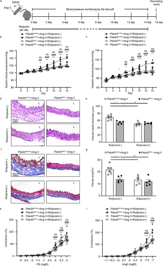
COMMUNICATIONS BIOLOGY | https://doi.org/10.1038/s42003-022-03029-0                                                           ARTICLE

staining tests showed that PDE4D expression was increased in                  PDE4D in smooth muscle cells (SMCs) contributes to Ang II-
hypertensive mice (Fig. 1b–e). However, there was no change in                induced mice hypertension. Among vascular intrinsic cell types,
other PDE4 isoforms (Fig. 1a and Supplementary Fig. 2a, b).                   EC and SMC are known to play crucial roles in hypertension
Together, these findings indicate that PDE4D expression is elevated            development6,7. To explore whether PDE4D, via SMCs or ECs,
in Ang II-induced hypertensive mice aortas.                                   plays a role in hypertension, we generated Pde4d SMC-specific

COMMUNICATIONS BIOLOGY | (2022)5:81 | https://doi.org/10.1038/s42003-022-03029-0 | www.nature.com/commsbio                                 3

ARTICLE                                                                    COMMUNICATIONS BIOLOGY | https://doi.org/10.1038/s42003-022-03029-0

Fig. 2 PDE4D in SMCs contributes to Ang II-induced mouse hypertension. a Scheme of hypertensive mice inducement. b SBP and c DBP were measured
in Pde4dflox/flox and Pde4dEC−/− mice, with or without Ang II treatment: n = 5 in Pde4dflox/flox + saline group, n = 8 in Pde4dflox/flox + Ang II group, n = 5
in Pde4dEC−/− + saline group, and n = 8 in Pde4dEC−/− + Ang II group. d SBP and e DBP were measured in Pde4dflox/flox and Pde4dSMC−/− mice, with
or without Ang II treatment: n = 5 in Pde4dflox/flox + saline group, n = 10 in Pde4dflox/flox + Ang II group, n = 5 in Pde4dSMC−/− + saline group, and n = 8 in
Pde4dSMC−/− + Ang II group. Data are expressed as mean ± SEM. Two-way ANOVA with Bonferroni’s post hoc test was performed to compare the difference
between the multiple groups. ***p < 0.001 for Pde4dflox/flox + Ang II group vs. Pde4dflox/flox + saline group, and ###p < 0.001 for Pde4dSMC−/− + Ang II group vs.
Pde4dflox/flox + Ang II group. f Representative H&E staining under the indicated experimental conditions. g Measurement of arterial wall media thickness including
all mice in f. h Representative masson-trichrome staining under the indicated experimental conditions. i Quantification of the positively stained area to the aortic
wall area including all mice in h. Data are expressed as mean ± SEM. Two-way ANOVA with Bonferroni’s post hoc test was performed to compare the difference
between the multiple groups. **p < 0.01, ***p < 0.001. L lumen.

Fig. 3 Pde4d deficiency in SMCs affects vasocontraction. Concentration-response curves for a PE and b Ang II induced vasocontraction of mesenteric
resistance artery from Pde4dflox/flox and Pde4dSMC−/– mice with or without Ang II treatment (n = 5 in Pde4dflox/flox + saline group, n = 10 in Pde4dflox/flox + Ang II
group, n = 5 in Pde4dSMC−/− + saline group, and n = 8 in Pde4dSMC−/− + Ang II group). Data are expressed as mean ± SEM. Two-way ANOVA with
Bonferroni’s post hoc test was performed to compare the difference between the multiple groups. **p < 0.01 and ***p < 0.001 for Pde4dflox/flox + Ang II group vs.
Pde4dflox/flox + saline group. #p < 0.05, ##p < 0.01, and ###p < 0.001 for Pde4dSMC−/− + Ang II group vs. Pde4dflox/flox + Ang II group.

knockout mice (Tagln-Cre mice; Pde4dSMC−/−) and Pde4d EC-                          vasocontraction was markedly suppressed in Pde4dSMC−/− mice
specific knockout mice (Tek-Cre mice; Pde4dEC−/−) mice via                          compared with Pde4dflox/flox mice (Pde4dflox/flox + Ang II group
Cre-LoxP recombinase system, and confirmed the mice genotypes                       vs. Pde4dSMC−/− + Ang II group; 10−5 M PE: 175.53% ± 5.52%
by agarose gel electrophoresis genotyping and western blot                         vs. 130.42% ± 10.79%, 25.7% reduced by Pde4dSMC−/− + Ang II
(Supplementary Fig. 3a–e).                                                         group; 10−7 M Ang II: 214.38% ± 25.3% vs. 132.08% ± 12.6%,
   We then induced hypertension in two knockout mice                               38.39% reduced by Pde4dSMC−/− + Ang II group; Fig. 3a, b).
groups. Using the tail-cuff method, we measured BP on the first                     These findings further support that SMC Pde4d deficiency
day and every other day during Ang II infusion. After 2 weeks, we                  relieves vasocontraction.
harvested the aorta tissues (Fig. 2a). Notably, EC Pde4d deficiency
(Pde4dflox/flox + Ang II group vs. Pde4dEC−/− + Ang II group)
did not affect the occurrence or development of the Ang                            PDE4D promotes SMCs contraction via the PKA-AMPK-
II-induced systolic blood pressure (SBP; 139.3 ± 2.47 mmHg                         MYPT1-MLC signaling pathway in vitro. To determine
vs. 139.45 ± 2.56 mmHg) or diastolic blood pressure (DBP;                          PDE4D’s role in regulating SMC contraction, we evaluated its
116.85 ± 2.47 mmHg vs. 117.1 ± 3.73 mmHg; Fig. 2b, c). Howe-                       impact on rat aorta smooth muscle cells (RASMCs) in vitro. First,
ver, SMC Pde4d deficiency (Pde4dflox/flox + Ang II group vs.                          we verified that PDE4D protein expression but not PDE4A-C was
Pde4dSMC−/− + Ang II group) inhibited the Ang II-induced increase                  elevated by 5.37-fold in RASMCs after Ang II stimulation
of both SBP (149.98 ± 1.78 mmHg vs. 127.43 ± 2.97 mmHg,                            (100 nM, 24 h; Fig. 4a, b and Supplementary Fig. 4a, b). Con-
15.04% reduced by Pde4dSMC−/− + Ang II group) and DBP                              sistently, we found that it was Pde4d upregulation instead of other
(122.48 ± 1.35 mmHg vs. 99.4 ± 2.55 mmHg, 18.84% reduced by                        PDE4 isoforms in RASMCs treated with Ang II in mRNA level
Pde4dSMC−/− + Ang II group; Fig. 2d, e). Hematoxylin and eosin                     (Supplementary Fig. 4c). Immunofluorescence staining also
(H&E) staining revealed that Ang II-induced vessel wall media                      showed that PDE4D was increased by Ang II in vitro (Supple-
thickening, which was reduced in Pde4dSMC−/− Ang II infused mice                   mentary Fig. 4d). Besides, PDE4 activity was increased by 2.34-
(Fig. 2f, g). In addition, masson-trichrome staining demonstrated that             fold with Ang II stimulation (Supplementary Fig. 4e). These
SMC Pde4d deficiency reversed Ang II-induced vascular fibrosis                       results indicated that PDE4D was upregulated by Ang II in vitro.
(Fig. 2h, i). These results indicate that PDE4D in SMCs, but not in                Then, we introduced PDE4D small-interfering RNA (siRNA) to
ECs, contribute to Ang II-induced mice hypertension.                               validate whether Ang II induces SMCs contraction via PDE4D.
                                                                                   After PDE4D siRNA administration, only PDE4D expression,
                                                                                   instead of other PDE4 isoforms, was reduced in RASMCs’ mRNA
SMC Pde4d deficiency reduces vasocontraction. To further                            and protein levels (Supplementary Fig. 5a–c). PDE4D was
explore how PDE4D in SMCs influences hypertension, we                               reduced by PDE4D siRNA through immunofluorescence staining
examined direct vascular function in the knockout mice. Speci-                     (Supplementary Fig. 5d). Indeed, PDE4 activity was also sup-
fically, the ex vivo vascular function of mesenteric arterioles from                pressed when PDE4D was knockdown (Supplementary Fig. 5e).
Pde4dflox/flox and Pde4dSMC−/− mice with or without Ang II                           We next performed a collagen gel cell contraction assay to explore
treatment. The vasoconstriction of mesenteric arterioles, known                    RASMCs contraction, which revealed that while Ang II promoted
as precapillary resistance vessels, causes vascular remodeling and                 RASMC contraction, the addition of PDE4D siRNA inhibited it
hypertension22. Using phenylephrine (PE) and Ang II to                             (si-control + Ang II vs. si-PDE4D + Ang II: 72.68% ± 2.11% vs.
induce mesenteric arterioles contraction, we found that the                        58.65% ± 1.76%) (Fig. 4c, d).

4                            COMMUNICATIONS BIOLOGY | (2022)5:81 | https://doi.org/10.1038/s42003-022-03029-0 | www.nature.com/commsbio
COMMUNICATIONS BIOLOGY | https://doi.org/10.1038/s42003-022-03029-0                                                             ARTICLE

Fig. 4 PDE4D promotes SMC contraction through the PKA-AMPK-MYPT1-MLC signaling pathway in vitro. a Representative western blot showing
PDE4D expression in control and Ang II-stimulated RASMCs. b Quantification of PDE4D expression normalized to GAPDH protein. c Representative
images showing RASMCs contraction. d Quantification of RASMCs contraction. e Protein kinase A (PKA) kinase activity in RASMCs. f Representative
western blot exhibiting phosphor-AMPK (pAMPK), AMPK, phospho-MYPT1 (pMYPT1), MYPT1, phospho-MLC (pMLC), and MLC expression in RASMCs.
g Quantification of pAMPK expression normalized to AMPK protein. h Quantification of pMYPT1 expression normalized to MYPT1 protein. i Quantification
of pMLC expression normalized to MLC protein. n = 3 per group. Data are expressed as mean ± SEM. Two-tailed Student’s t test was performed to
compare differences between two groups and two-way ANOVA with Bonferroni’s post hoc test was performed to compare the difference between the
multiple groups. *p < 0.05, **p < 0.01, and ***p < 0.001.

   PDE4 family specifically hydrolyzes cAMP to inhibit PKA                        Additionally, AMPK activation inhibits phosphorylation of
activity23–25. PKA phosphorylates AMPKα at Thr-172 through                    myosin phosphatase targeting subunit 1 (MYPT1) and myosin
LKB1 signaling, ultimately leading to AMPK activation19,26–30. As             light chain (MLC), consequently attenuating SMCs contraction20.
such, we investigated whether PDE4D inhibited PKA and AMPK                    Therefore, we hypothesized that PDE4D may further increase
activity in RASMCs. Consistently, we found that Ang II stimulation            MYPT1 and MLC phosphorylation by suppressing AMPK
reduced PKA activity in RASMCs, and that PDE4D siRNA reversed                 activation, promoting Ang II-induced SMCs contraction. Indeed,
this effect (Fig. 4e). Moreover, we observed that PDE4D siRNA                 Ang II increased MYPT1 and MLC phosphorylation, whereas
increased Ang II-reduced AMPK phosphorylation (Fig. 4f, g).                   PDE4D siRNA suppressed MYPT1 and MLC phosphorylation
Meanwhile, we used PKA inhibitor (PKI, 10 μM, 60 min) to further              (Fig. 4f, h, i). Moreover, the effect of PDE4D siRNA on reducing
confirm whether PDE4D regulated pAMPK relying on PKA. We                       MYPT1 and MLC phosphorylation was largely reversed by PKI
found that the protective effect of PDE4D siRNA on pAMPK was                  (Supplementary Fig. 6a, c, d). We also used AMPK inhibitor
blocked by PKI (Supplementary Fig. 6a, b), suggesting that PDE4D              (Compound C, 20 μM, 2 h) for further validation. The inhibitory
negatively regulated AMPK phosphorylation via PKA.                            effect of PDE4D siRNA on pMYPT1 and pMLC was blocked by

COMMUNICATIONS BIOLOGY | (2022)5:81 | https://doi.org/10.1038/s42003-022-03029-0 | www.nature.com/commsbio                                     5
ARTICLE                                                               COMMUNICATIONS BIOLOGY | https://doi.org/10.1038/s42003-022-03029-0

Fig. 5 PDE4D promotes vasocontraction through the PKA-AMPK-MYPT1-MLC signaling pathway in vivo. a PKA kinase activity in aorta tissues.
b Representative western blot showing pAMPK, AMPK, pMYPT1, MYPT1, pMLC, and MLC expression in aorta tissues from Pde4dflox/flox and Pde4dSMC−/−
mice with or without Ang II treatment. c Quantification of pAMPK expression normalized to AMPK protein. d Quantification of pMYPT1 expression
normalized to MYPT1 protein. e Quantification of pMLC expression normalized to MLC protein. n = 3 per group. Data are expressed as mean ± SEM. Two-
way ANOVA with Bonferroni’s post hoc test was performed to compare the difference between the multiple groups. *p < 0.05, **p < 0.01, and ***p < 0.001.

Compound C (Supplementary Fig. 6e–g). These results suggest                  age with rolipram (3 mg kg−1 day−1) or vehicle daily via
that PDE4D promotes SMCs contraction via inhibition of PKA                   gavage for 14 days (Fig. 6a). Notably, rolipram reduced Ang
activity and AMPK phosphorylation, and conversely promotes                   II-induced increase of BP [Rolipram (−) + Ang II group vs.
MYPT1 and MLC phosphorylation.                                               Rolipram (+) + Ang II group; SBP: 140.67 ± 1.69 mmHg vs.
                                                                             113.74 ± 0.80 mmHg, 19.14% reduced by Rolipram (+) + Ang II
PDE4D promotes vasocontraction through the PKA-AMPK-                         group; DBP: 110.76 ± 1.79 mmHg vs. 90.76 ± 0.87 mmHg, 18.06%
MYPT1-MLC signaling pathway in Ang II-induced mice                           reduced by Rolipram (+) + Ang II group; Fig. 6b, c]. Besides,
hypertension. To further validate the mechanism identified                    Ang II-induced vessel wall media thickening was inhibited by
in vitro above, we detected PKA activity and AMPK, MYPT1, and                rolipram accroding to H&E staining (Fig. 6d, e). In addition,
MLC phosphorylation in mice aorta tissues. Consistently, Ang II              masson-trichrome staining demonstrated that rolipram reversed
infusion reduced PKA activity in mice aortas, and this reduction             Ang II-induced vascular fibrosis (Fig. 6f, g). To further confirm
was reversed in Pde4dSMC−/− Ang II mice (Fig. 5a). Western blot              the effect of rolipram on vasocontraction, we detected the ex vivo
also exhibited that Ang II infusion reduced AMPK phosphoryla-                vascular function by using PE and Ang II to induce mesenteric
tion in the aorta, an effect which was recovered in Pde4dSMC−/−              arterioles contraction. We found that rolipram also suppressed
mice (Fig. 5b, c). Ang II infusion also increased MYPT1 and MLC              vasocontraction observably [Rolipram (−) + Ang II group vs.
phosphorylation, and again Pde4dSMC−/− mice exhibited reduced                Rolipram (+) + Ang II group; 10−5 M PE: 167.04% ± 11.79% vs.
Ang II-induced MYPT1 and MLC phosphorylation (Fig. 5b, d, e).                126.79% ± 5.31%, 24.1% reduced by Rolipram (+) + Ang II
These results suggest that PDE4D promotes vasocontraction, and               group; 10−7 M Ang II: 227.06% ± 14.38% vs. 172.43% ± 10.56%,
thus contributes to Ang II-induced hypertension in mice, through             23.62% reduced by Rolipram (+) + Ang II group; Fig. 6h, i].
the PKA-AMPK-MYPT1-MLC signaling pathway.                                    These results indicate a pharmacological impact of rolipram in
                                                                             preventing hypertension in mice.
PDE4 inhibitor rolipram attenuates Ang II-induced hyper-
tension. As known, there are several PDE4 inhibitors used in                 Rolipram attenuates Ang II-induced hypertension through the
clinical therapy, for example, rolipram as the first generation of            inhibition of SMC PDE4D. Rolipram has pan inhibitory effects on
PDE4 inhibitor for neuroinflammation31. However, there is no                  PDE4A-D, and the inhibition of PDE4 subfamily by rolipram was
specific inhibitor for PDE4D in pre-clinical experiment. Then, we             not tissue or cell specific. To further explore whether the therapeutic
tried to determine the pharmacological effect of PDE4 inhibitor              effect of rolipram mainly through SMC PDE4D, we established the
rolipram on hypertension. We treated WT male mice at 8 weeks                 hypertensive model in Pde4dflox/flox and Pde4dSMC−/− mice, and

6                          COMMUNICATIONS BIOLOGY | (2022)5:81 | https://doi.org/10.1038/s42003-022-03029-0 | www.nature.com/commsbio
COMMUNICATIONS BIOLOGY | https://doi.org/10.1038/s42003-022-03029-0 ARTICLE gavaged mice with rolipram (3 mg kg−1 day−1) or vehicle Pde4dSMC−/− + Ang II + Rolipram (+) group (SBP: (Fig. 7a). Consistently with Figs. 2 and 6, SMC Pde4d deficiency 119.0 ± 1.09 mmHg vs. 117.9 ± 0.81 mmHg; DBP: or rolipram inhibited Ang II-induced increase of both SBP 94.60 ± 0.93 mmHg vs. 93.20 ± 1.14 mmHg; Fig. 7b, c). H&E and DBP (Fig. 7b, c). However, there was no difference staining revealed that SMC Pde4d deficiency, rolipram or between Pde4dSMC−/− + Ang II + Rolipram (−) group and Pde4dSMC−/− mice with rolipram inhibited Ang II-induced vessel COMMUNICATIONS BIOLOGY | (2022)5:81 | https://doi.org/10.1038/s42003-022-03029-0 | www.nature.com/commsbio 7
ARTICLE                                                                    COMMUNICATIONS BIOLOGY | https://doi.org/10.1038/s42003-022-03029-0

Fig. 6 Effect of rolipram on Ang II-induced hypertension in mice. a Scheme of hypertensive mice treated with vehicle or rolipram. Rolipram (3 mg kg−1 day−1)
was orally administered daily for 14 days. b SBP and c DBP were measured in wild-type (WT) mice with or without Ang II/rolipram treatment: n = 5 in Rolipram
(−) + saline group, n = 9 in Rolipram (−) + Ang II group, n = 6 in Rolipram (+) + saline group, and n = 10 in Rolipram (+) + Ang II group. Data are expressed as
mean ± SEM. Two-way ANOVA with Bonferroni’s post hoc test was performed to compare the difference between the multiple groups. *p < 0.05, ***p < 0.001 for
Rolipram (−) + Ang II group vs. Rolipram (−) + saline group, and #p < 0.05, ###p < 0.001 for Rolipram (+) + Ang II group vs. Rolipram (−) + Ang II group.
d Representative H&E staining under the indicated experimental conditions. e Measurement of arterial wall media thickness including all mice in d. f Representative
masson-trichrome staining under the indicated experimental conditions. g Quantification of the positively stained area to the aortic wall area including all mice in
f. Data are expressed as mean ± SEM. Two-way ANOVA with Bonferroni’s post hoc test was performed to compare the difference between the multiple groups.
*p < 0.05, **p < 0.01, ***p < 0.001 in e, g. Concentration-response curves for h PE and i Ang II induced vasocontraction of mesenteric resistance artery in WT mice
with or without Ang II/rolipram treatment including all mice. Data are expressed as mean ± SEM. Two-way ANOVA with Bonferroni’s post hoc test was performed
to compare the difference between the multiple groups. *p < 0.05, **p < 0.01, ***p < 0.001 for Rolipram (−) + Ang II group vs. Rolipram (−) + saline group group.
#p < 0.05, ##p < 0.01, ###p < 0.001 for Rolipram (+) + Ang II group vs. Rolipram (−) + Ang II group. L lumen.

wall media thickening, but there was no change among the three                     hypertension via inflammation regulation. Although, further
groups indicated above (Fig. 7d, e). Besides, masson-trichrome                     study would be needed to validate this supposition.
staining demonstrated that Ang II-induced vascular fibrosis was                        While collagen deposition and inflammation may be potential
reversed by SMC Pde4d deficiency, rolipram or Pde4dSMC−/− mice                      additional mechanisms, we demonstrated a link between PDE4D
with Rolipram with no changes among the three groups (Fig. 7f, g).                 and the PKA-AMPK signaling pathway. cAMP is known to be
Furthermore, mesenteric arterioles contraction caused by PE                        involved in signal transduction through PKA regulation46. Recently,
and Ang II was markedly suppressed by SMC Pde4d                                    researchers have found that PKA phosphorylates AMPKα at Thr-
deficiency or rolipram, meanwhile, there was no change                              172 through the widely expressed tumor suppressor liver kinase B1,
between Pde4dSMC−/− + Ang II + Rolipram (−) group and                              ultimately activating AMPK19,30. AMPK activity has been linked to
Pde4dSMC−/− + Ang II + Rolipram (+) group (Pde4dSMC−/− + Ang                       numerous cardiovascular diseases, including hypertension, athero-
II + Rolipram (−) group vs. Pde4dSMC−/− + Ang II + Rolipram (+)                    sclerosis, and heart failure20,47–49. Crucially, AMPK activation low-
group; 10−5 M PE: 141.5% ± 6.57% vs. 135.2% ± 9.89%; 10−7 M Ang                    ers BP and suppresses SMC contractility by inhibiting the MYPT1-
II: 140.5% ± 2.97% vs. 130.4% ± 4.85%; Fig. 7h, i). These results                  MLC signaling pathway20. Consistent with previous reports, our
confirmed that rolipram exerts its therapeutic effect on vascular                   results exhibited that PDE4D upregulated MYPT1 and MLC
remodeling, vasoconstriction and hypertension mainly by inhibiting                 phosphorylation by inhibiting the PKA-AMPK signaling pathway,
PDE4D in SMCs.                                                                     inducing SMCs contraction and thereby, hypertension.
                                                                                      In conclusion, our study provided that PDE4D in SMCs
Discussion                                                                         aggravated Ang II-induced hypertension. We identified the
In this study, we observed upregulated PDE4D expression in                         mechanism by which PDE4D affected SMCs contraction via
hypertensive mice aortas, which showed that PDE4D contributes                      in vitro and in vivo experimental models and verified those results
to hypertension. Furthermore, via EC- and SMC-specific Pde4d                        through several molecular biology approaches. In addition, roli-
knockout hypertensive mice, these models revealed a causal                         pram alleviated hypertension mainly through PDE4D in SMCs.
association between SMC Pde4d and vasocontraction in hyper-                        This study elucidated PDE4D as a potential target for the treatment
tension. To further elucidate this association, we investigated a                  of hypertension and, potentially, other cardiovascular diseases.
potential mechanism for PDE4D involvement in SMC contrac-
tion and hypertension development, and identified the PKA-                          Materials and methods
AMPK-MYPT1-MLC signaling pathway to be a likely candidate.                         Animal models. All animal protocols were reviewed and approved by the Ethics
Importantly, we demonstrated that rolipram, a pan PDE4 inhi-                       Committee of Peking Union Medical College. Pde4d-floxed (flanked by LoxP) mice
bitor, relieved Ang II-induced hypertension mainly by inhibiting                   (Pde4dflox/flox), Tagln-Cre mice, and Tek-Cre mice were generated and obtained by
                                                                                   Shanghai Model Organisms Center, Inc. (Shanghai, China). To generate SMC-
PDE4D in SMCs, which suggested that PDE4D might represent a                        specific knockout mice (Pde4dSMC−/−) or EC-specific knockout mice (Pde4dEC−/−),
potential therapeutic hypertension target (Fig. 8).                                Pde4dflox/flox was crossed with Tagln-Cre mice or Tek-Cre mice. Genotyping was
   Hypertension is well known to be a complex syndrome                             performed by polymerase chain reaction (PCR) using primers (Supplementary
involving multiple organs, tissues, and cells32,33. Among the cell                 Table 1). Pde4dflox/flox, Pde4dSMC−/−, and Pde4dEC−/− littermates were used in this
types associated with hypertension, PDE4D is also expressed in                     research. WT mice (C57BL/6, N11) were obtained from The Jackson Laboratory
                                                                                   (Bar Harbor, ME). All mice were housed in temperature-controlled rooms under a
fibroblasts34,35. Adventitial fibroblasts, another major component of                12-h light-dark cycle with water and food ad libitum.
blood vessels, are the primary cause of collagen deposition and aortic                 To induce hypertension in Pde4dflox/flox, Pde4dSMC−/−, Pde4dEC–/–, and WT mice,
stiffening in hypertension36. In this study, we observed vascular                  8-week old male mice were infused with angiotensin II (Ang II, 490 ng kg−1 min–1;
collagen deposition in Ang II infusion mice. Accordingly, it should                Sigma, Cat#: A9525-50MG) or saline and subcutaneously implanted with osmotic
                                                                                   pumps (Alzet MODEL 2002; DURECT, Cupertino, CA) for 14 days50. Mice groups:
not be discounted that PDE4D could further contribute to the                       WT mice were divided into two groups randomly, WT mice infused with saline (n = 4)
development of hypertension by interfering with collagen produc-                   or Ang II (n = 8); Pde4dEC−/− mice were divided into four groups randomly, Pde4dflox/
tion in fibroblasts, a possibility warranting future investigation.                 flox mice infused with saline (n = 5) or Ang II (n = 8), and Pde4dEC−/− mice infused

   Hypertension is commonly associated with inflammation37,38,                      with saline (n = 5) or Ang II (n = 8); Pde4dSMC−/− mice were divided into four groups
and the cell types involved in inflammation (T lymphocytes39, B                     randomly, Pde4dflox/flox mice infused with saline (n = 5) or Ang II (n = 10), and
                                                                                   Pde4dSMC−/− mice infused with saline (n = 5) or Ang II (n = 8).
lymphocytes40,41, dendritic cells, monocytes, and macrophages42)                       To test the effect of rolipram on hypertension, WT, Pde4dflox/flox and Pde4dSMC−/−
are all known to promote hypertension. PDE4D has been shown                        mice at 8 weeks old were infused with Ang II (490 ng kg−1 min−1) and subcutaneously
to interact with cytokines, regulate the function of inflammatory                   implanted with osmotic pumps for 14 days. In total, 0.375 mg ml−1 rolipram (PDE4
cells, and aggravate the inflammatory response43–45. While                          inhibitor, 8 ml kg−1 day−1, dissolved in ethyl alcohol; Sigma-Aldrich, Cat#: R6520) was
                                                                                   administered orally, via gavage, daily for 14 days. WT mice were divided into four
PDE4D’s role in the inflammation response is outside the scope                      groups randomly: Rolipram (−) mice infused with saline (n = 5) or Ang II (n = 9), and
of this study, our findings, along with the body of literature                      Rolipram (+) mice infused with saline (n = 6) or Ang II (n = 10). Pde4dflox/flox and
evidence, suggest that PDE4D could also contribute to                              Pde4dSMC−/− mice were divided into four groups randomly: Pde4dflox/flox + Ang II

8                            COMMUNICATIONS BIOLOGY | (2022)5:81 | https://doi.org/10.1038/s42003-022-03029-0 | www.nature.com/commsbio
COMMUNICATIONS BIOLOGY | https://doi.org/10.1038/s42003-022-03029-0 ARTICLE
mice with Rolipram (−) (n = 6) or Rolipram (+) (n = 5), and Pde4dSMC−/− + Ang II then threaded through the occlusion cuff and the sensor cuff, which was then
mice with Rolipram (−) (n = 5) or Rolipram (+) (n = 5). attached to the controller. For each measurement, five values of SBP and DBP were
recorded for each mouse and their mean values were used as the final result.
BP measurement by tail-cuff plethysmography. SBP and DBP in mice were
measured using the CODA non-invasive BP system (Kent Scientific Co., Tor- Measurement of mesenteric arteriole tension. Mice were euthanized under
rington, CT, USA) according to the manufacturer’s instructions51. Each mouse was pentobarbital sodium (50 mg kg−1, intraperitoneal). The mesenteric vascular bed
gently placed in a sizeable holder and allowed to acclimate for 5 min. The tail was was quickly removed, and immersed in Krebs bicarbonate buffer (119 mM NaCl,
COMMUNICATIONS BIOLOGY | (2022)5:81 | https://doi.org/10.1038/s42003-022-03029-0 | www.nature.com/commsbio 9ARTICLE COMMUNICATIONS BIOLOGY | https://doi.org/10.1038/s42003-022-03029-0
Fig. 7 Rolipram attenuates Ang II-induced hypertension through the inhibition of SMCs PDE4D. a Scheme of Pde4dflox/flox and Pde4dSMC−/− mice
infused with Ang II and/or treated with rolipram. Rolipram (3 mg kg−1 day−1) was orally administered daily for 14 days. b SBP and c DBP were measured in
Pde4dflox/flox and Pde4dSMC−/− mice infused with Ang II and/or rolipram treatment: n = 6 in Pde4dflox/flox + Ang II + Rolipram (−) group, n = 5 in Pde4dflox/
flox + Ang II + Rolipram (+) group, n = 5 in Pde4dSMC−/− + Ang II + Rolipram (−) group, and n = 5 in Pde4dSMC−/− + Ang II + Rolipram (+) group. Data
are expressed as mean ± SEM. Two-way ANOVA with Bonferroni’s post hoc test was performed to compare the difference between the multiple groups.
*p < 0.05, **p < 0.01, ***p < 0.001 for Pde4dflox/flox + Ang II + Rolipram (−) group vs. Pde4dflox/flox + Ang II + Rolipram (+) group, ##p < 0.01, ###p < 0.001
for Pde4dflox/flox + Ang II + Rolipram (−) group vs. Pde4dSMC−/− + Ang II + Rolipram (−) group, and ns no significant difference for Pde4dSMC−/− + Ang
II + Rolipram (−) group vs. Pde4dSMC−/− + Ang II + Rolipram (+) group. d, e Representative H&E staining and measurement of arterial wall media
thickness including all mice. f, g Representative masson-trichrome staining and quantification of the positively stained area to the aortic wall area including
all mice. Data are expressed as mean ± SEM. Two-way ANOVA with Bonferroni’s post hoc test was performed to compare the difference between the
multiple groups. **p < 0.01, ***p < 0.001, and ns no significant difference in e, g. Concentration-response curves for h PE and i Ang II induced
vasocontraction of mesenteric resistance artery. Data are expressed as mean ± SEM. Two-way ANOVA with Bonferroni’s post hoc test was performed to
compare the difference between the multiple groups. ***p < 0.001 for Pde4dflox/flox + Ang II + Rolipram (−) group vs. Pde4dflox/flox + Ang II + Rolipram (+)
group, ###p < 0.001 for Pde4dflox/flox + Ang II + Rolipram (−) group vs. Pde4dSMC−/− + Ang II + Rolipram (−) group, and ns no significant difference for
Pde4dSMC−/− + Ang II + Rolipram (−) group vs. Pde4dSMC−/− + Ang II + Rolipram (+) group. L lumen.
Fig. 8 PDE4D promotes Ang II-induced hypertension in mice. PDE4D exacerbates Ang II-induced vasocontraction by affecting SMC contraction,
consequently contributing to hypertension. The mechanism by which PDE4D aggravates SMC contraction likely involves the PKA-AMPK-MYPT1-MLC
signaling pathway. PDE4 inhibitor, rolipram, showed a therapeutic effect on Ang II-induced hypertension in mice primarily through PDE4D in SMCs. The
image was created with BioRender.com, and acquired the permission for use and proper accreditation.
25 mM NaHCO3, 4.7 mM KCl, 1 mM MgCl2, 1.2 mM KH2PO4, 2.5 mM CaCl2, and Cell culture and small-interfering RNA (siRNA) transfection. RASMCs were
11 mM D‐glucose) and gassed with a mixture of 95% O2 and 5% CO252. Second- purchased from American Type Culture Collection (Manassas, VA, USA) and
order branches of the mesenteric artery (~2 mm long segments), known as the cultured in SMC medium (ScienCell, Cat#: 1101), containing 5% fetal bovine
mesenteric arterioles, were suspended with two tungsten wires in the organ serum (FBS), 100 U/ml penicillin, and 100 g/ml streptomycin. RASMCs within
chamber containing 5 ml of Krebs buffer solution. The vasocontraction was passage 5 to 12 were used for all experiments. RASMCs were stimulated with
examined by PE or Ang II. Changes in isometric force were recorded with a Multi 100 nM Ang II (Sigma, Cat#: A9525-50MG) for 24 h before harvest. To knockdown
Myograph System (610 M, Danish Myo Technology A/S, Aarhus N, Denmark) PDE4D, PDE4D siRNA (Ribobio, siB180730051733) and control siRNA (Ribobio,
according to the manufacturer’s instruction. The relative contraction was quanti- siN0000001-1-5) were purchased from Ribobio (Guangzhou RiboBio Co., Ltd.,
fied as the ratio of post stimulation tension to baseline tension. Guangzhou, China). RASMCs were transfected with 200 nM PDE4D siRNA in 5 μl
of Oligofectamine (Invitrogen, Carlsbad, CA, USA, Cat#: 12252011) for 48 h. The
siRNA transfection efficiency was determined by real-time polymerase chain
reaction (RT-PCR), western blot and immunofluorescence assay (Supplementary
Histological and immunohistochemical analysis. Mouse aorta segments were cut Fig. 5a–e).
at the thoracic aorta, embedded vertically with OCT compound (SAKURA,
Cat#:4538), and then stored at −80 °C. Ten to 15 serial frozen sections containing
the entire vascular lumen were sectioned using a freezing microtome (Leica Immunofluorescent analysis. RASMCs treated with Ang II (100 nM, 24 h) or
CM1860), and then fixed with 4% paraformaldehyde. Frozen sections (6 μm) were PDE4D siRNA (200 nM, 48 h) were fixed with 4% paraformaldehyde. The cells
stained by immunohistochemical staining of PDE4D (1:100, Abcam, Cat#: were incubated overnight at 4 °C with primary antibodies against PDE4D (1:100,
ab14613) by the 3-amino-9-ethylcarbazole staining method, H&E (Solarbio, Cat#: Abcam, Cat#: ab14613). The antibodies were detected using fluorescein-labeled
G1120), and masson-trichrome staining (Servicebio, Cat#: G1006). Images were secondary antibodies (1:1000, Invitrogen, Cat#: A11037). The nuclei were stained
photographed using a Leica optical microscope (Leica Microsystems, Germany) with 4′,6-diamidino-2-phenylindole (Abcam, Cat#: ab228549). Images were pho-
and the integrated optical density (IOD) values of positive staining analyzed using tographed using a Leica optical microscope (Leica Microsystems, Germany).
Image‐Pro Plus software (Media Cybernetics, USA). For statistical analysis, 5
images per mouse of each group were randomly selected. For PDE4D immuno-
histochemical staining, the content of PDE4D was quantified as the ratio of Real-time polymerase chain reaction (RT-PCR). Total RNA was extracted from
positively stained area to the total cross-sectional area of the aortic wall. For mouse aortic tissues or RASMCs using TRIzol reagent (Invitrogen, Carlsbad, CA,
masson-trichrome staining, the degree of vascular fibrosis was quantified as the Cat#: 15596018) according to the manufacturer’s protocol. Equal quantities of
ratio of the positively stained area to the total cross-sectional area of the aortic wall. RNA (1000 ng) were reverse transcribed into cDNA (TianGen, KR116-02), and
For H&E staining, arterial wall media thickness was measured using Nikon NIS- quantitative RT-PCR was performed in a single-color RT-PCR detection system
Elements image analysis software (Nikon Instruments Inc., Japan). (Bio-Rad, Hercules, CA, USA). The mRNA levels of Pde4a, Pde4b, Pde4c, and
Pde4d were normalized to the level of the housekeeping gene glyceraldehyde-3-
10 COMMUNICATIONS BIOLOGY | (2022)5:81 | https://doi.org/10.1038/s42003-022-03029-0 | www.nature.com/commsbioCOMMUNICATIONS BIOLOGY | https://doi.org/10.1038/s42003-022-03029-0 ARTICLE
phosphate dehydrogenase (Gapdh). Pde4a, Pde4b, Pde4c, and Pde4d mRNA 2. Fuchs, F. D. & Whelton, P. K. High blood pressure and cardiovascular disease.
expression fold changes compared to one of controls, were calculated using the Hypertension 75, 285–292 (2020).
2−ΔΔCt method. The RT-PCR primers are shown in Supplementary Table 1. 3. Grassi, G. & Ram, V. S. Evidence for a critical role of the sympathetic nervous
system in hypertension. J. Am. Soc. Hypertens. 10, 457–466 (2016).
Western blot analysis. Protein was extracted from the aortic tissues or RASMCs 4. Te Riet, L., van Esch, J. H., Roks, A. J., van den Meiracker, A. H. & Danser, A.
in a lysis buffer. Equal quantities of protein extract (30 μg per lane) were separated H. Hypertension: renin-angiotensin-aldosterone system alterations. Circ. Res.
by 8, 10, or 12% SDS–PAGE and transferred to a polyvinylidene fluoride mem- 116, 960–975 (2015).
brane (Merck, Cat#: IPVH00010). The target protein was probed with numerous 5. Guzik, T. J. & Touyz, R. M. Oxidative stress, inflammation, and vascular aging
antibodies: PDE4A (1:1000, Thermo Fisher, Cat#:PA5-115730), PDE4B (1:1000, in hypertension. Hypertension 70, 660–667 (2017).
Cell Signaling Technology, Cat#: 72096S), PDE4C (1:1000, Thermo Fisher, Cat#: 6. Konukoglu, D. & Uzun, H. Endothelial dysfunction and hypertension. Adv.
PA5-106624), PDE4D (1:1000, Abcam, Cat#: ab171750), AMP-activated protein Exp. Med. Biol. 956, 511–540 (2017).
kinase (AMPK; 1:1000, Cell Signaling Technology, Cat#: 2532S), Phospho-AMPK 7. Brown, I. A. M. et al. Vascular smooth muscle remodeling in conductive and
(1:1000, Cell Signaling Technology, Cat#: 2535S), myosin phosphatase targeting resistance arteries in hypertension. Arterioscler. Thromb. Vasc. Biol. 38,
subunit 1 (MYPT1; 1:1000, Cell Signaling Technology, Cat#: 2634S), Phospho- 1969–1985 (2018).
MYPT1 (1:500, Cell Signaling Technology, Cat#: 5163S), MLC (1:1000, Cell Sig- 8. Mahfoud, F. et al. Treatment strategies for resistant arterial hypertension.
naling Technology, Cat#: 8505S), and Phospho-MLC (1:1000, Cell Signaling Dtsch. Arztebl. Int. 108, 725–731 (2011).
Technology, Cat#: 3675S), respectively. Immunoblotting of the housekeeping 9. Carey, R. M., Sakhuja, S., Calhoun, D. A., Whelton, P. K. & Muntner, P.
protein GAPDH (1:5000, Proteintech, Cat#: 60004-1-Ig) was performed to ensure Prevalence of apparent treatment-resistant hypertension in the United States.
equal protein loading. Immunoreactive bands were visualized with SuperSignal™ Hypertension 73, 424–431 (2019).
West Pico PLUS Chemiluminescent Substrate (Pierce, Cat#: 34577). The protein 10. Beavo, J. A. Cyclic nucleotide phosphodiesterases: functional implications of
expression was measured by analyzing the relative protein band intensity with multiple isoforms. Physiol. Rev. 75, 725–748 (1995).
Image-Pro Plus 6.0 software. 11. Bhat, A. et al. Phosphodiesterase-4 enzyme as a therapeutic target in
neurological disorders. Pharmacol. Res. 160, 105078 (2020).
Protein kinase A (PKA) kinase activity assay. PKA kinase activity in aortic 12. Lehrke, M. et al. PDE4 inhibition reduces neointima formation and inhibits
tissues or RASMCs was detected via a PKA kinase activity assay kit (Abcam, Cat#: VCAM-1 expression and histone methylation in an Epac-dependent manner.
ab139435) according to the manufacturer’s instructions. Absorbance was measured J. Mol. Cell. Cardiol. 81, 23–33 (2015).
at OD = 450 nm via a multi-mode microplate reader (BioTek Synergy™ HTX, 13. De Franceschi, L. et al. Protective effects of phosphodiesterase-4 (PDE-4)
BioTek Instrument, Inc., Winooski, USA). The relative activity of PKA was inhibition in the early phase of pulmonary arterial hypertension in transgenic
quantified as the ratio of the active PKA content to the sample total protein sickle cell mice. FASEB J. 22, 1849–1860 (2008).
content. 14. Press, N. J. & Banner, K. H. PDE4 inhibitors—a review of the current field.
Prog. Med. Chem. 47, 37–74 (2009).
PDE4 activity assay. PDE4 activity in RASMCs was detected via a PDE4 activity 15. Delaunay, M., Osman, H., Kaiser, S. & Diviani, D. The role of cyclic AMP
assay kit (Abcam, Cat#: ab139460) according to the manufacturer’s instructions. signaling in cardiac fibrosis. Cells 9 https://doi.org/10.3390/cells9010069
PDE4 activity was inhibited with rolipram (20 uM) during the test. Total PDE (2019).
activity assay and PDE activity assay after inhibition of PDE4 were performed for 16. Qin, X. et al. Smooth muscle-specific Gsα deletion exaggerates angiotensin II-
each sample, and PDE4 specific activity was calculated by subtracting inhibitory induced abdominal aortic aneurysm formation in mice in vivo. J. Mol. Cell.
activity from total activity. Absorbance was measured at OD = 620 nm via a multi- Cardiol. 132, 49–59 (2019).
mode microplate reader (BioTek Synergy™ HTX, BioTek Instrument, Inc., 17. Fantidis, P. The role of intracellular 3'5’-cyclic adenosine monophosphate
Winooski, USA). The relative activity of PDE4 was quantified as the ratio of the (cAMP) in atherosclerosis. Curr. Vasc. Pharmacol. 8, 464–472 (2010).
active PDE4 content to the sample total protein content. 18. Hara, Y. et al. Inhibition of MRP4 prevents and reverses pulmonary
hypertension in mice. J. Clin. Invest. 121, 2888–2897 (2011).
19. Kari, S., Vasko, V. V., Priya, S. & Kirschner, L. S. PKA activates AMPK
Cell contraction assay. RASMCs contraction was detected using a Cell Con-
through LKB1 signaling in follicular thyroid cancer. Front. Endocrinol. 10, 769
traction Assay Kit (Cell Biolabs, Inc., San Diego, CA, USA, Cat#: CBA-201)
(2019).
according to the manufacturer’s instructions53. RASMCs were treated with siRNA
20. Wang, S., Liang, B., Viollet, B. & Zou, M. H. Inhibition of the AMP-activated
for 48 h with or without Ang II for 24 h, and then cultured in collagen gel for 48 h
protein kinase-α2 accentuates agonist-induced vascular smooth muscle
to develop mechanical load. The surface image of the collagen gel was captured via
digital camera, and analyzed using Image‐Pro Plus software (Media Cybernetics, contraction and high blood pressure in mice. Hypertension 57, 1010–1017
USA). The percentage of contraction was the ratio of gel contracted surface area to (2011).
the dish bottom. 21. Yin, L. M. et al. Transgelin-2 as a therapeutic target for asthmatic pulmonary
resistance. Sci. Transl. Med. 10 https://doi.org/10.1126/scitranslmed.aam8604
(2018).
Statistics and reproducibility. Statistical analysis was performed using GraphPad 22. Hilgers, R. H. & Das, K. C. Role of in vivo vascular redox in resistance arteries.
Prism 8 (GraphPad Software Inc., La Jolla, CA). Data are expressed as means ± Hypertension 65, 130–139 (2015).
standard error of mean. Two-tailed Student’s t test was performed to compare 23. Li, H., Zuo, J. & Tang, W. Phosphodiesterase-4 inhibitors for the treatment of
differences between two groups from at least three independent experiments. One- inflammatory diseases. Front. Pharmacol. 9, 1048 (2018).
way ANOVA or two-way ANOVA with Bonferroni’s post hoc test was performed 24. Li, H. et al. DC591017, a phosphodiesterase-4 (PDE4) inhibitor with robust
to compare differences between multiple groups, using at least three independent
anti-inflammation through regulating PKA-CREB signaling. Biochem.
experiments. p value < 0.05 was considered statistically significant.
Pharmacol. 177, 113958 (2020).
25. Houslay, M. D. & Adams, D. R. PDE4 cAMP phosphodiesterases: modular
Reporting summary. Further information on research design is available in the Nature enzymes that orchestrate signalling cross-talk, desensitization and
Research Reporting Summary linked to this article. compartmentalization. Biochem. J. 370, 1–18 (2003).
26. Li, G. et al. Mechanisms underlying the anti-proliferative actions of
adiponectin in human breast cancer cells, MCF7-dependency on the cAMP/
Data availability protein kinase-A pathway. Nutr. Cancer 63, 80–88 (2011).
Raw data of genotyping and western blot are provided in Supplementary Fig. 7. Source
27. Hutchinson, D. S., Chernogubova, E., Dallner, O. S., Cannon, B. & Bengtsson,
data underlying the graphs are provided in Supplementary Data 1. Other relevant data
T. Beta-adrenoceptors, but not alpha-adrenoceptors, stimulate AMP-activated
are available from the corresponding author upon request.
protein kinase in brown adipocytes independently of uncoupling protein-1.
Diabetologia 48, 2386–2395 (2005).
Received: 19 March 2021; Accepted: 23 December 2021; 28. Medina, E. A. et al. PKA/AMPK signaling in relation to adiponectin’s
antiproliferative effect on multiple myeloma cells. Leukemia 28, 2080–2089
(2014).
29. Shaw, R. J. et al. The tumor suppressor LKB1 kinase directly activates AMP-
activated kinase and regulates apoptosis in response to energy stress. Proc.
Natl Acad. Sci. USA 101, 3329–3335 (2004).
References 30. Huang, Y. et al. Resveratrol prevents sarcopenic obesity by reversing
1. Williams, B. et al. 2018 ESC/ESH guidelines for the management of arterial mitochondrial dysfunction and oxidative stress via the PKA/LKB1/AMPK
hypertension. Eur. Heart J. 39, 3021–3104 (2018). pathway. Aging 11, 2217–2240 (2019).
COMMUNICATIONS BIOLOGY | (2022)5:81 | https://doi.org/10.1038/s42003-022-03029-0 | www.nature.com/commsbio 11ARTICLE COMMUNICATIONS BIOLOGY | https://doi.org/10.1038/s42003-022-03029-0
31. Kumar, A. & Singh, N. Inhibitor of Phosphodiestearse-4 improves memory 52. Wang, X. et al. Nuciferine relaxes rat mesenteric arteries through
deficits, oxidative stress, neuroinflammation and neuropathological alterations endothelium-dependent and -independent mechanisms. Br. J. Pharmacol.
in mouse models of dementia of Alzheimer’s Type. Biomed. Pharmacother. 88, 172, 5609–5618 (2015).
698–707 (2017). 53. Malhotra, R. et al. HDAC9 is implicated in atherosclerotic aortic calcification and
32. Brown, M. J. & Haydock, S. Pathoaetiology, epidemiology and diagnosis of affects vascular smooth muscle cell phenotype. Nat. Genet. 51, 1580–1587 (2019).
hypertension. Drugs 59(Suppl 2), 1–12 (2000).
33. Abrams, W. B. Pathophysiology of hypertension in older patients. Am. J. Med.
85, 7–13 (1988). Acknowledgements
34. Martin-Chouly, C. A. et al. Modulation of matrix metalloproteinase This work was financially supported by the Chinese Academy of Medical Sciences
production from human lung fibroblasts by type 4 phosphodiesterase Innovation Fund for Medical Sciences (2021-I2M-1-016), the National Natural Science
inhibitors. Life Sci. 75, 823–840 (2004). Foundation of China (82100514) and National Key Research and Development Program
35. Dunkern, T. R., Feurstein, D., Rossi, G. A., Sabatini, F. & Hatzelmann, A. of China Grants (2019YFA0801804 and 2019YFA0801703).
Inhibition of TGF-beta induced lung fibroblast to myofibroblast conversion by
phosphodiesterase inhibiting drugs and activators of soluble guanylyl cyclase. Author contributions
Eur. J. Pharmacol. 572, 12–22 (2007). TF.F. and Y.H. designed experiments and analyzed data. TF.F., Y.H., WP.G., TH.F., X.F.,
36. Kuwabara, J. T. & Tallquist, M. D. Tracking adventitial fibroblast contribution WJ.G., and X.S. performed experiments and analyzed data. TF.F. and Y.H. wrote the
to disease: a review of current methods to identify resident fibroblasts. manuscript. R.G. and J.W. conceived research question and oversaw the entirety of
Arterioscler. Thromb. Vasc. Biol. 37, 1598–1607 (2017). research.
37. Dinh, Q. N., Drummond, G. R., Sobey, C. G. & Chrissobolis, S. Roles of
inflammation, oxidative stress, and vascular dysfunction in hypertension.
BioMed. Res. Int. 2014, 406960 (2014). Competing interests
38. Savoia, C. & Schiffrin, E. L. Inflammation in hypertension. Curr. Opin. The authors declare no competing interests.
Nephrol. Hypertens. 15, 152–158 (2006).
39. Ren, J. & Crowley, S. D. Role of T-cell activation in salt-sensitive hypertension.
Am. J. Physiol. Heart Circ. Physiol. 316, H1345–h1353 (2019).
Additional information
Supplementary information The online version contains supplementary material
40. Dingwell, L. S. et al. B-cell deficiency lowers blood pressure in mice.
available at https://doi.org/10.1038/s42003-022-03029-0.
Hypertension 73, 561–570 (2019).
41. Chan, C. T. et al. Obligatory role for B cells in the development of angiotensin
Correspondence and requests for materials should be addressed to Ran Gao or Jing
II-dependent hypertension. Hypertension 66, 1023–1033 (2015).
Wang.
42. Drummond, G. R., Vinh, A., Guzik, T. J. & Sobey, C. G. Immune mechanisms
of hypertension. Nat. Rev. Immunol. 19, 517–532 (2019).
Peer review information Communications Biology thanks George Baillie and the other,
43. Yun, S. et al. Interaction between integrin α5 and PDE4D regulates endothelial
anonymous, reviewers for their contribution to the peer review of this work. Primary
inflammatory signalling. Nat. Cell Biol. 18, 1043–1053 (2016).
Handling Editors: Tami Martino and Eve Rogers.
44. Ariga, M. et al. Nonredundant function of phosphodiesterases 4D and 4B in
neutrophil recruitment to the site of inflammation. J. Immunol. 173,
Reprints and permission information is available at http://www.nature.com/reprints
7531–7538 (2004).
45. Jin, S. L., Ding, S. L. & Lin, S. C. Phosphodiesterase 4 and its inhibitors in Publisher’s note Springer Nature remains neutral with regard to jurisdictional claims in
inflammatory diseases. Chang Gung Med. J. 35, 197–210 (2012). published maps and institutional affiliations.
46. Ould Amer, Y. & Hebert-Chatelain, E. Mitochondrial cAMP-PKA signaling:
What do we really know? Biochimica et biophysica acta. Bioenergetics 1859,
868–877 (2018).
47. Jiang, S. et al. AMPK: potential therapeutic target for ischemic stroke. Open Access This article is licensed under a Creative Commons
Theranostics 8, 4535–4551 (2018). Attribution 4.0 International License, which permits use, sharing,
48. Daskalopoulos, E. P., Dufeys, C., Beauloye, C., Bertrand, L. & adaptation, distribution and reproduction in any medium or format, as long as you give
Horman, S. AMPK in cardiovascular diseases. Experientia Suppl. 107, 179–201 appropriate credit to the original author(s) and the source, provide a link to the Creative
(2016). Commons license, and indicate if changes were made. The images or other third party
49. Dong, Y. et al. Reduction of AMP-activated protein kinase alpha2 increases material in this article are included in the article’s Creative Commons license, unless
endoplasmic reticulum stress and atherosclerosis in vivo. Circulation 121, indicated otherwise in a credit line to the material. If material is not included in the
792–803 (2010). article’s Creative Commons license and your intended use is not permitted by statutory
50. Lob, H. E., Schultz, D., Marvar, P. J., Davisson, R. L. & Harrison, D. G. Role of regulation or exceeds the permitted use, you will need to obtain permission directly from
the NADPH oxidases in the subfornical organ in angiotensin II-induced the copyright holder. To view a copy of this license, visit http://creativecommons.org/
hypertension. Hypertension 61, 382–387 (2013). licenses/by/4.0/.
51. Daugherty, A., Rateri, D., Hong, L. & Balakrishnan, A. Measuring blood
pressure in mice using volume pressure recording, a tail-cuff method. J. Vis.
Exp. https://doi.org/10.3791/1291 (2009). © The Author(s) 2022
12 COMMUNICATIONS BIOLOGY | (2022)5:81 | https://doi.org/10.1038/s42003-022-03029-0 | www.nature.com/commsbioYou can also read