Postural and respiratory function of the abdominal muscles: A pilot study to measure abdominal wall activity using belt sensors
←
→
Page content transcription
If your browser does not render page correctly, please read the page content below
Isokinetics and Exercise Science 29 (2021) 175–184                                                                                 175
DOI 10.3233/IES-203212
IOS Press

Postural and respiratory function of the
abdominal muscles: A pilot study to measure
abdominal wall activity using belt sensors
Jakub Novaka,∗ , Andrew Buschb , Pavel Kolara and Alena Kobesovaa
a
  Department of Rehabilitation and Sports Medicine, Second Faculty of Medicine, Charles University and University
Hospital Motol, Prague, Czech Republic
b
  Department of Health and Human Kinetics, Ohio Wesleyan University, Delaware, OH, USA

Received 15 August 2020
Accepted 1 October 2020

Abstract.
BACKGROUND: The abdominal muscles play an important respiratory and stabilization role, and in coordination with other
muscles regulate intra-abdominal pressure (IAP) to stabilize the spine.
OBJECTIVE: To examine a new, non-invasive method to measure activation of the abdominal wall and compare changes in
muscle activation during respiration while breathing under a load, and during instructed breathing.
METHODS: Thirty-five healthy individuals completed this observational crossover study. Two capacitive force sensors registered
the abdominal wall force during resting breathing stereotype, instructed breathing stereotype and under a load.
RESULTS: Mean abdominal wall force increased significantly on both sensors when holding the load compared to resting
breathing (Upper Sensor: P < 0.0005, d = −0.46, Lower Sensor: P < 0.0005, d = −0.56). The pressure on both sensors also
significantly increased during instructed breathing compared to resting breathing (US: P < 0.0005, d = −0.76, LS: P < 0.0005,
d = −0.78).
CONCLUSIONS: The use of capacitive force-sensors represent a new, non-invasive method to measure abdominal wall activity.
Clinically, belts with capacitive force sensors can be used as a feedback tool to train abdominal wall activation.

Keywords: Spinal stabilization, respiration stereotype, intra-abdominal pressure (IAP), capacitive force sensors

1. Introduction                                                        which forms an important spinal stabilization mecha-
                                                                       nism [7–9]. The pelvis and lumbar spine are reflexively
   Over the past two decades, numerous authors have                    stabilized before the limb movements start [10–12].
investigated lumbar spine stabilization [1,2], motor con-              This feed forward stabilization mechanism is secured
trol of the trunk muscles [3,4], and the regulation of                 by the trunk muscles. The diaphragm works in conjunc-
intra-abdominal pressure [5,6]. Balanced coordination                  tion with the pelvic floor and deep intrinsic muscles of
between the diaphragm, pelvic floor muscles, and ab-                   the spine and the transversus abdominis to create stiff-
dominal wall musculature is critical for IAP regulation                ness and minimize other intrinsic and extrinsic stressors
                                                                       to the spine during motion [13]. Biomechanical studies
                                                                       confirm that pressurization of the abdomen increases
   ∗ Corresponding author: Jakub Novak, Department of Rehabili-
                                                                       lumbar spinal stability, but the degree of spinal stability
tation and Sports Medicine, Second Faculty of Medicine, Charles
University and University Hospital Motol, V Uvalu 84, Prague 5, 150
                                                                       is not substantially influenced by selective activation
06, Czech Republic. Tel.: + 420 22443 9264; Fax: +420 22443 9220;      of certain abdominal muscles [7]. Forced activity of
E-mail: kuba-novak@seznam.cz.                                          transversus or obliques may cause reductions in the ac-

ISSN 0959-3020 c 2021 – The authors. Published by IOS Press. This is an Open Access article distributed under the terms of the Creative
Commons Attribution-NonCommercial License (CC BY-NC 4.0).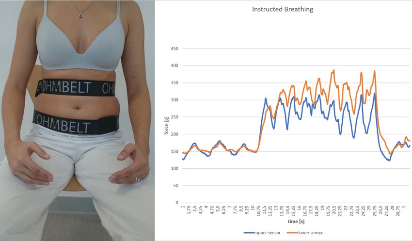
176                           J. Novak et al. / Postural and respiratory function of the abdominal muscles

                                                                                                      Table 1
tivation of other abdominal muscles and even produce                                    Descriptive statistics of the sample
decreased lumbar stability [7].
                                                                                    Age (years)     Height (cm)     Weight (kg)   BMI
   Appropriate functional tone and conditioning the ab-
                                                                          Mean      21.26           170.51          63.17         24.07
dominal wall to work as one harmonic unit with the                        SD        1.62            6.49            7.94          3.02
pelvic floor musculature is a critical stabilization com-                 Min       19              160             47            17.27
ponent that optimizes the “push back” of the viscera                      Max       25              185             80            27.62
up into the diaphragm, helping expansion of the lower
rib cage for respiration [13]. According to Wallden a                     This paper presents a new, non-invasive method to
deconditioned or inhibited abdominal wall causes vis-                   measure force of the abdominal wall using capacitive
ceroptosis, reducing the pressure the viscera exerts into               force sensors, which may help medical practicioners
the diaphragm to open the lower rib cage for inhala-                    provide quick, visual information to patients regarding
tion, and driving the diaphragm back upward during                      their postural-respiratory function.
full exhalation [13]. The postural-respiratory function                   Therefore, the purpose of this research was to com-
of the trunk muscles is inseparable. With heavy load                    pare how activation of abdominal muscles changes dur-
lifting, trunk muscle activation in the abdominal and                   ing respiration while breathing under a load and when
thoracic cavities act like a rigged-walled cylinder, pro-               being instructed to modify breathing stereotype in a
viding increased spinal stability [14]. The regulation                  healthy population.
of IAP within the abdominal wall in coordination with
diaphragm and pelvic floor muscles significantly con-
tributes to spinal stabilization and protects the spine dur-            2. Methods
ing loading [15,16]. Perhaps not only the amount of the
abdominal wall activation but also the type of contrac-                 2.1. Participants
tion, i.e. eccentric vs. concentric plays a role in spinal
stabilization. With postural activites, the diaphragm                      This study included 35 healthy individuals, 8 males
descends caudally to pressurize intra-abdominal con-                    and 27 females, aged 19–25 years. Table 1 shows de-
tents [17]. The pelvic floor must support the viscera                   scriptive statistics of the sample. The participants have
from bellow. Since the viscera are non-compressible,                    not experienced acute or chronic musculoskeletal pain,
one can assume that the abdominal wall must react to                    reported no pain during the measurements, never suf-
diaphragmatic descend by eccentric contraction. Insuf-                  fered from any serious trunk pathologies, never under-
ficient distension or exccesive initial concentric con-                 gone any trunk operations and have never received any
traction of the abdominals preventing diaphragmatic                     therapy or training focusing on intra-abdominal pres-
descend during postural activites may compromise the                    sure activation or abdominal wall expansion. Individ-
whole stabilization mechanism. Poor coordination of                     uals with body mass index (BMI) over 30 were ex-
postural muscles and insufficient stabilizing function of               cluded from the study. Written informed consent was
deep back muscles, diaphragm, abdominal and pelvic                      obtained from each participant, and this study was ap-
floor muscles may result in spinal disorders associated                 proved by an Institutional Ethics Committee (Ethics
with back pain, such as deformation spondyloarthro-                     Committee of the University Hospital Motol and 2nd
sis, intervertebral disc protrusion or spondylolisthe-                  Faculty of Medicine, Charles University in Prague. No.
sis [18,19].                                                            1263.1.15/19; approval date: November 6, 2019). The
   Various measurement procedures have been used to                     study conforms with The Code of Ethics of the World
investigate the postural-respiratory function of the ab-                Medical Association.
dominal muscles and related IAP changes. Esohpageal,
gastric [20], intravesical [21], anal [22,23], and vagi-                2.2. Instruments
nal [24] probes can measure IAP, yet these methods
are often time consuming and uncomfortable for pa-                         For the noninvasive examination of abdominal mus-
tients. Electromyography [23] and ultrasound [25] as-                   cles function, a unique device called Ohm Belt (Nilus
sessments have also been used to analyze activity of the                Medical LLC, 2019 c OHMBELT, Redwood City, CA,
abdominal muscles, but these methods can be burden-                     USA). A research version of the device was designed
some, technically demanding to perform while the for-                   by the manufacturer for the trial purposes, which dif-
mer poses serious difficulty regarding its reproducibility              fers from the commercial version in that two sensors
and hence interpretation.                                               recorded data simultaneously with a software app to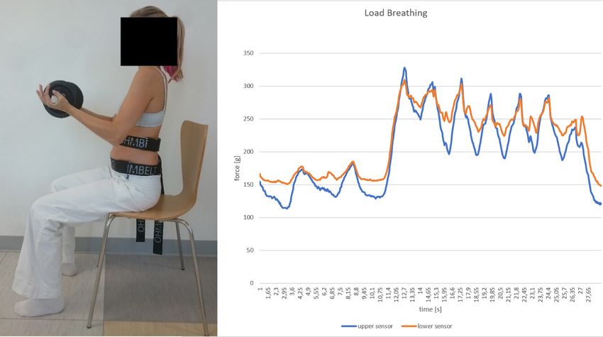
J. Novak et al. / Postural and respiratory function of the abdominal muscles                                177

Fig. 1. Close-up picture of the capacitive force sensor and a scheme of abdominal cavity with expansion of the abdominal wall and attached sensor.

display and record both sensor force data. It consists                       signal that is transmitted wirelessly via bluetooth to the
of two capacitive force sensors of 15 mm diameter,                           computer where the software graphically displays the
0.35 mm thickness, full scale range 0.45 kg, minimal                         results. The program records any time sequences with
detectable force 0.9 g (Fig. 1), attached to the abdomi-                     the numerical values being automatically exported into
nal wall by adjustable straps. The device utilizes force-                    MS Excel. Immediate data analysis, graphical imaging
sensing capacitor type of sensor, which consists of a                        and data saving is available.
material whose capacitance changes when a force is ap-
plied. Such sensors are also known as “force-sensitive                       2.3. Assessments
capacitors” and reported to be used in other medical
research projects [26–28]. This sensor is an example                            The assessments of all participants were performed
                                                                             under the same conditions (daytime, assessment room,
of parallel plate capacitator (see Fig. 1). For small de-
                                                                             temperature), and by the same examiner. Each partic-
flections, there is a linear relationship between applied
                                                                             ipant was in an upright seated position, with hips and
force and change in capacitance. Force sensor, facing
                                                                             knees flexed at 90◦ , and both feet supported on the
the subject’s skin, is pressed against the abdominal wall
                                                                             floor (Fig. 2). First, a pilot study was performed on 20
by the adjustable firm strap (see Fig. 1). Abdominal
                                                                             healthy individuals to measure abdominal wall activity
wall expansion and retraction is recorded by the sensor
                                                                             in various postural and breathing situations, using dif-
as a force (The “abdominal wall force”). The sensors
                                                                             ferent ways of fixation and placement of the sensors to
register the force exerted by the abdominal wall during                      achieve maximal measurement accuracy and sensitivity.
respiration and various postural tasks. The abdominal                        By repeated measurements, it was determined that fix-
wall force is measured in grams over a period of time,                       ation with firm but flexible belts under the pressure of
where 1 g = 0.01 N. The gram scale was selected to                           of 120 grams ± 10 g for the upper sensor (US) and 140
provide users with feedback in integer rather than deci-                     grams ± 10 g for the lower sensor (LS) allows for suffi-
mal values. Abdominal wall force was measured by the                         ciently accurate measurements while not limiting trunk
sensor and recorded in grams over a period of time. The                      and abdominal wall movements. Before each measure-
force displayed on the graph scale in grams is, there-                       ment, boths sensors were calibrated to a baseline of zero
fore, technically in the gram-force unit (gf). The dual-                     and positioned on each participant using palpation by
channel pressure sensor consists of two sensors which                        a skilled manual therapist. The US was placed on the
monitor simultaneously the instantaneous muscle force                        superior trigonum lumbale, bellow the floating ribs, and
at two different locations. Both the amount of the force                     the LS was placed above the groin at the intersection
and its dynamics over time can be analyzed. The sensors                      of the mammilar and bispinal connecting line. Sensors
are also equipped with accelerometers to capture any                         were randomly placed on the left or right side of the
kyphotic trunk synkinesis, i.e. substitutive trunk move-                     body. The sensors were fixed during tidal expirium.
ment replacing abdominal muscle activation. A built-in                          The participants were instructed to maintain the up-
tensometric transducer converts the force to the digital                     right sitting position throughout the course of the whole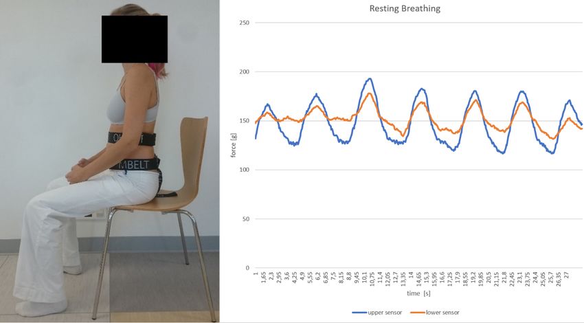
178                              J. Novak et al. / Postural and respiratory function of the abdominal muscles

                                Fig. 2. Resting breathing assessment. Force sensors are placed under the belts.

Fig. 3. Load breathing assessment. At the 10th second the weight was put into participant’s hands (note the steep increase of both lines in the
graph).

assessment, avoiding increased spinal kyphosis or lor-                     each scenario. Abdominal activity fluctuated when each
dosis. In all individuals, the abdominal wall activity                     participant began to hold the load, and upon releasing
was recorded for a total period of 15–20 seconds for                       the load, so only 10-second intervals of stable activation
J. Novak et al. / Postural and respiratory function of the abdominal muscles 179
Fig. 4. Instructed breathing assessment. First, the individual breathes normally, then at the 10th second he/she was instructed to direct the breath
towards both sensors (steep increase of both lines in the graph).
were used for the mean statistical analysis calculated small (< 0.2), small (0.2–0.5), medium (0.5–0.8), or
separately for each sensor. First, the natural stereotype large (> 0.8). Power analysis, using G*Power 3.1, in-
of resting breathing (RB) was monitored (scenario 1, dicated an 80% chance of detecting a medium effect
Fig. 2). Then, the participants were breathing naturally size of 0.5 in 34 subjects with statistical significance
when sitting upright and holding a load corresponding determined a priori at p < 0.05 (two-tailed). Data anal-
to 20% of their body weight with elbows flexed at 90◦ . yses were conducted using the Statistical Package for
This was scenario 2, i.e. load breathing (LB), (Fig. 3). the Social Sciences (SPSS version 26.0 for Mac; IMB
And finally, instructed breathing (IB) was monitored Corp, Armonk, NY).
(Fig. 4). The participants were instructed to voluntarily
and maximally expand their abdominal wall, directing
inspiration towards the sensors and maintaining maxi- 3. Results
mum pressure contact with the sensors during expira-
tion. Thirty-five participants completed the study. Table 2
presents the amount of force of the abdominal wall
2.4. Statistical analysis (g) during resting breathing, loaded breathing and in-
structed breathing (n = 35) for both sensors. The paired
Descriptive statistics were calculated for each mea- sample t-test indicated that the mean abdominal wall
sure. Paired-samples t tests were used to compare ab- force increased significantly on both sensors when hold-
dominal wall pressure in both sensors during resting ing the load compared to the resting breathing (P for
breathing with the interventions, and also to compare both sensors < 0.0005). The force on both sensors also
inter-sensor differences between measurements during significantly increased during instructed breathing com-
each condition. Cohen’s d effect sizes were calculated pared to the resting breathing (P for both sensors <
for the differences between breathing conditions as the 0.0005). Figure 5 depicts the results.
difference between groups divided by the pooled stan- The inter-sensor difference was also compared for
dard deviation. Effect sizes were interpreted as very all three measured scenarios (resting, under load, and180 J. Novak et al. / Postural and respiratory function of the abdominal muscles
Table 2
External force changes of the abdominal wall (g) during loaded breathing and instructed breathing (n = 35)
Breathing Resting 10 second Mean 95% CI of
Measure Effect size P Value
intervention mean (SD) mean (SD) difference (SD) difference
Upper sensor Loaded 123.75 (21.95)∗∗ 165.07 (39.44)∗∗ −41.32 (38.00) (−54.38, −28.27) −0.46 < 0.0005∗
Instructed 228.31 (64.96) −104.56 (60.64) (−125.39, −83.73) −0.76 < 0.0005∗
Lower sensor Loaded 141.98 (18.19)∗∗ 191.74 (40.51)∗∗ −49.76 (39.79) (63.43, −36.09) −0.56 < 0.0005∗
Instructed 249.02 (66.09) −107.04 (63.79) (−128.95, −85.13) −0.78 < 0.0005∗
∗ Statisticaldifference observed between conditions (P < 0.05). ∗∗ Statistical difference observed between sensors (P < 0.05). Note: Values are
(g). Effect size = calculated Cohen’s d.
Fig. 5. Statistical comparison of forces on both sensors at resting, loaded, and instructed breathing.
instructed breathing). There was a significantly greater cult to voluntarily activate in an eccentric manner. Also,
force on the lower sensor in the resting condition (p < a similar placement of abdominal wall pressure measur-
0.0005), and in the loaded condition (p = 0.001), but ing device was previously used in studies published by
not in the instructed breathing condition (p = 0.068). other authors [35,36]. Synergy between the diaphragm,
Figure 5 depicts the inter-sensor difference. pelvic floor, abdominal wall and spinal extensors is
necessary to stabilize the spine. During stabilization,
the concentric activity of the diaphragm is followed by
4. Discussion the eccentric activity of the entire abdominal wall. This
synergy increases intra-abdominal pressure (IAP) that
This study presents a new method to analyze activity stabilizes the lumbar spine anteriorly, balancing with
of the abdominal wall using capacitive force sensors. the spinal extensors that secure stabilization posteri-
Originally, the sensors were produced by a medical orly [37]. Based on existing literature [6,38,39], we as-
technology developer to offer feedback to LBP clients sume the pressure monitored by the sensors is not only
training optimal breathing stereotype. Research sug- activity exerted by the abdominal muscles but mainly
gests patients with low back pain (LBP) often demon- the IAP resulting from the complex coordination of all
strate sub-optimal respiratory parameters [29,30] and core stabilizers.
confirms a positive effect of breathing exercises on This study confirms that pressure exerted by the ab-
LBP [31,32]. Clinically, insufficient activation of the dominal wall increases with increased postural load,
latero-dorsal sections of the abdominal wall (i.e. in su- because the activity measured on both sensors during
perior trigonum lumbale and over the lower abdominal LB was significantly higher comparing to RB. This fur-
wall just above the groin) are common findings in LBP ther supports electromyography (EMG) studies report-
patients [33,34]. Therefore, the sensors were placed in ing increased abdominal muscle activity during postu-
these locations because they are usually the most diffi- rally challenging situations [40] and when lifting theJ. Novak et al. / Postural and respiratory function of the abdominal muscles 181 load [41], as well as studies confirming IAP elevation DSSS, arguing that the DSSS works as one functional during postural tasks and load lifting [9,37]. Within- unit and dysfunction of just one DSSS muscle is related limb movements, and coactivation of the diaphragm to total dysfunction of the whole muscle system [44]. with abdominal muscles cause an increase in IAP [37]. Malatova also used the dynamometer to analyze the This is a very important stabilizing mechanism be- effect of a six-week intervention program focusing on cause IAP elevation increases stiffness of the lumbar optimal body posture and DSSS training [46]. Com- spine [9,42]. Applying simple and non-invasive capaci- paring to Malatova’s rather big muscular dynamometer, tive force sensors, we obtained similar results reported these sensors are smaller in size and transmit signals to by other complicated and sophistic methods such as a PC via Bluetooth, allowing practicioners to analyze multichannel EMG analysis or IAP gastric, esophageal more dynamic situations including all types of exer- or intra-annal pressure measurements [23,25]. Further- cises. Also, unlike Malatova’s measuring device, the more, we statistically confirmed that healthy, young sensors used in this study are commercially available individuals are able to voluntarily activate the coor- (Nilus Medical LLC, 2019 c OHMBELT). dination between the diaphragm, abdominal muscles In a study on 45 asymptomatic women, Malatova and pelvic floor muscles, because the force on both identified more excessive activation of the upper sec- sensors significantly increased during IB comparing to tions of the abdominal wall compared to lower sections, RB. This again, supports other studies showing how questioning if this is a natural or pathological stereo- breath control and abdominal muscle control influences type [44]. Our study demonstrated significantly greater IAP [23,43]. We purposefully performed the IB test at pressure on the lower sensor in the RB condition (p < the end of the assessment to exclude any learning pro- 0.0005), and in the LB condition (p = 0.001), for the cess. First resting and loaded breathing spontaneous un- IB condition the activity on the lower sensor was also corrected stereotypes were analyzed and only then were greater but not statistically significant (p = 0.068). This instructions given on how to breath. The individuals topic deserves more research attention. It is necessary were cued to direct the breath towards both sensors. The to determine how much difference can be considered goal was to measure the amount of voluntary pressure physiological and what is already abnormal. Postural re- exerted on sensors during maximum inhalation directed sponses of the abdominal muscles differ between body to the sensors. The second goal was to confirm it is ac- positions, the recruitment and contribution of abdom- tually possible to teach participants how to voluntarily inal muscle regions to stabilize the trunk varies, and activate the abdominal wall sections under the sensors. regional differentiation in abdominal muscle activity This may be of potential benefit in the treatment and may be a natural stabilization mechanism [47]. training of LBP individuals who frequently demonstrate It has been shown that selective activation of the in- an inability to activate such areas of the abdominal wall dividual abdominal muscles does not significantly in- both during breathing and postural tasks [33,34]. Fur- crease spinal stability [7]. Spinal stabilization train- ther studies are needed to confirm. ing should therefore focus on global coordination of Interestingly, this study showed that postural activa- all muscles involved in stabilization function and IAP tion when holding a load corresponding to 20% body regulation rather then addressing individual abdomi- weight (LB scenario) requires, on average, about two- nal muscles. Abdominal wall pressure changes were thirds of the maximum muscle coactivation generated previously monitored by other authors [35,36] report- by the conscious maximum force of the abdominal wall ing inverse significant correlations between ability to against the sensors (IB scenario). Similarly, Essendrop activate the abdominal wall and pain severity in LBP et al. reported IAP increased from 0 to 40% when hold- population [36]. Esophageal, gastric, anal or vaginal ing a load of 15% body weight [42]. However, it re- sensory measuring IAP [9,48] as well as EMG [3,48] mains questionable what amount of activation would or ultrasound [49] examination procedures help to an- occur when holding a heavier gload, which is common alyze activity of the abdominal wall muscles. These during activities of daily living or sport. techniques inform us about muscle activity but do not A similar measurement of the abdominal wall ex- distinguish eccentric from concentric types of contrac- pansion was done by Malatova et al. using an elec- tion and as a result do not provide information about tromechanical dynamometer [44–46]. The authors de- an individual’s ability to expand their abdominal wall. scribe how muscular dynamometers can be used to eval- Abdominal wall expansion can be assessed clinically uate activation of the whole deep stabilizing spine sys- by palpation [33,34], but clinical abdominal assessment tem (DSSS) because abdominal muscles comprise the showed poor sensitivity and accuracy for IAP pressure
182                           J. Novak et al. / Postural and respiratory function of the abdominal muscles

changes [50]. Trunk and abdominal circumference mea-                    Author contributions
surements can be highly influenced by the adipose tis-
sues, therefore may also be unreliable [51]. The pro-                   CONCEPTION: Jakub Novak, Pavel Kolar and Alena
posed method using capacitive force sensors may be-                     Kobesova.
come a new, simple and non-invasive method to mea-                      PERFORMANCE OF WORK: Jakub Novak.
sure abdominal wall force. Future studies are needed to                 INTERPRETATION OR ANALYSIS OF DATA: Jakub
determine its sensitivity, accuracy and correlation with                Novak and Andrew Busch.
direct IAP measurements.                                                PREPARATION OF THE MANUSCRIPT: Jakub No-
   This study has some limitations. Based on available                  vak, Andrew Busch and Alena Kobesova.
research [6,38,39] we assume the detected amount of                     SUPERVISION: Alena Kobesova.
abdominal wall force is related to IAP changes, but IAP
was not directly measured in this study. Future research
correlating IAP with abdominal wall force would be                      Ethical considerations
useful in strengthening the findings of this study. This
method of IAP detection may lose sensitivity in very                       Written informed consent was obtained from each
obese individuals. The exact placement of the sensors                   participant, and this study was approved by the Ethics
would be less consistent due to difficult palpation of                  Committee of the University Hospital Motol and 2nd
the anatomical landmarks, and the pressure measure-                     Faculty of Medicine, Charles University in Prague. No.
ment accuracy limited by a sick adipose tissue. Obese
                                                                        1263.1.15/19; approval date: November 6, 2019. The
clients with BMI > 30 were excluded. Additionally,
                                                                        study conforms with The Code of Ethics of the World
the sensor’s fixation may alter the measurements. Too
                                                                        Medical Association.
loose fixation of the belts reduces the sensitivity of the
sensors, whereas too tight of fixation limits the ability
to expand the abdominal wall, compromising the mea-
                                                                        Conflict of interest
sured function. Placement of each sensor was unilat-
erally randomized, however in future studies we plan
to use four sensors placed on both sides of the trunk to                   There are no conflicts of interest to disclose.
distinguish bilateral differences. Finally, this pilot study
was performed on asymptomatic individuals, which
does not mean all measured subjects performed with op-                  Funding
timal muscular co-activation. In future studies, a larger
number of individuals should be involved, comparing                        This study was supported by The Charles University
asymptomatic subjects with those suffering from LBP                     Grant Agency (GAUK No. 340220), and by Institu-
or other types of musculoskeletal problems.                             tional research program Progres Q41.

5. Conclusions                                                          References

   This study presenst a new non-invasive method to                      [1] Cresswell AG, Oddsson L, Thorstensson A. The influence
measure abdominal wall force using belts with capaci-                        of sudden perturbations on trunk muscle activity and intra-
                                                                             abdominal pressure while standing. Exp Brain Res. 1994;
tive force sensors. The activity of the abdominal wall                       98(2): 336–41.
muscles significantly increases when lifting a load and                  [2] du Rose A. Have studies that measure lumbar kinematics and
with a purposeful activation during instructed breathing                     muscle activity concurrently during sagittal bending improved
stereotype comparing to resting breathing. Future stud-                      understanding of spinal stability and sub-system interactions?
                                                                             A systematic review. Healthcare (Basel). 2018 Sep 8; 6(3).
ies need to confirm if abdominal wall activation mea-
                                                                         [3] Hodges PW, Moseley GL, Gabrielsson A, Gandevia SC.
sured by the sensors correlates with direct IAP mea-                         Experimental muscle pain changes feedforward postural re-
surements.                                                                   sponses of the trunk muscles. Exp Brain Res. 2003 Jul; 151(2):
                                                                             262–71.
                                                                         [4] Hirata RP, Salomoni SE, Christensen SW, Graven-Nielsen T.
                                                                             Reorganised motor control strategies of trunk muscles due to
Acknowledgments                                                              acute low back pain. Hum Mov Sci. 2015 Jun; 41: 282–94.
                                                                         [5] Hodges PW, Cresswell AG, Daggfeldt K, Thorstensson A. In
  We would like to thank Dr. Miroslav Navratil for                           vivo measurement of the effect of intra-abdominal pressure on
providing us with the sensors.                                               the human spine. J Biomech. 2001 Mar; 34(3): 347–53.
J. Novak et al. / Postural and respiratory function of the abdominal muscles 183
[6] Cholewicki J, Juluru K, McGill SM. Intra-abdominal pres- [24] Guaderrama NM, Nager CW, Liu J, Pretorius DH, Mittal RK.
sure mechanism for stabilizing the lumbar spine. Journal of The vaginal pressure profile. Neurourol Urodyn. 2005; 24(3):
Biomechanics. 1999 Jan 1; 32(1): 13–7. 243–7.
[7] Stokes IAF, Gardner-Morse MG, Henry SM. Abdominal mus- [25] Cresswell AG, Grundstrm̈ H, Thorstensson A. Observations
cle activation increases lumbar spinal stability: analysis of con- on intra-abdominal pressure and patterns of abdominal intra-
tributions of different muscle groups. Clin Biomech (Bristol, muscular activity in man. Acta Physiol Scand. 1992 Apr;
Avon). 2011 Oct; 26(8): 797–803. 144(4): 409–18.
[8] Neumann P, Gill V. Pelvic floor and abdominal muscle inter- [26] Egorov V, Sarvazyan AP. Mechanical imaging of the breast.
action: EMG activity and intra-abdominal pressure. Int Urog- IEEE Trans Med Imaging. 2008 Sep; 27(9): 1275–87.
ynecol J Pelvic Floor Dysfunct. 2002; 13(2): 125–32. [27] Salpavaara TJ, Verho JA, Lekkala JO, Halttunen JE. Embedded
[9] Hodges PW, Eriksson AEM, Shirley D, Gandevia SC. Intra- capacitive sensor system for hip surgery rehabilitation: Online
abdominal pressure increases stiffness of the lumbar spine. J measurements and long-term stability. In: 2008 30th Annual
Biomech. 2005 Sep; 38(9): 1873–80. International Conference of the IEEE Engineering in Medicine
[10] Aruin AS, Latash ML. Directional specificity of postural mus- and Biology Society. 2008. pp. 935–8.
cles in feed-forward postural reactions during fast voluntary [28] Ahmad HN, Barbosa TM. The effects of backpack carriage on
arm movements. Exp Brain Res. 1995; 103(2): 323–32. gait kinematics and kinetics of schoolchildren. Sci Rep. 2019
[11] Hodges PW, Richardson CA. Relationship between limb 04; 9(1): 3364.
movement speed and associated contraction of the trunk mus- [29] Shah SG, Choezom T, Prabu Raja G. Comparison of respira-
cles. Ergonomics. 1997 Nov; 40(11): 1220–30. tory parameters in participants with and without chronic low
[12] Hodges PW, Cresswell AG, Daggfeldt K, Thorstensson A. back pain. J Bodyw Mov Ther. 2019 Oct; 23(4): 894–900.
Three dimensional preparatory trunk motion precedes asym- [30] Calvo-Lobo C, Almazán-Polo J, Becerro-de-Bengoa-Vallejo
metrical upper limb movement. Gait Posture. 2000 Apr; 11(2): R, Losa-Iglesias ME, Palomo-López P, Rodríguez-Sanz D, et
92–101. al. Ultrasonography comparison of diaphragm thickness and
[13] Wallden M. The diaphragm – more than an inspired design. excursion between athletes with and without lumbopelvic pain.
Journal of Bodywork and Movement Therapies. 2017 Apr; Phys Ther Sport. 2019 May; 37: 128–37.
21(2): 342–9. [31] Anderson BE, Bliven KCH. The use of breathing exercises in
[14] Goldish GD, Quast JE, Blow JJ, Kuskowski MA. Postural the treatment of chronic, nonspecific low back pain. J Sport
effects on intra-abdominal pressure during valsalva maneuver. Rehabil. 2017 Sep; 26(5): 452–8.
Archives of Physical Medicine and Rehabilitation. 1994 Mar; [32] Janssens L, McConnell AK, Pijnenburg M, Claeys K,
75(3): 324–7. Goossens N, Lysens R, et al. Inspiratory muscle training af-
[15] Stokes IAF, Gardner-Morse MG, Henry SM. Intra-abdominal fects proprioceptive use and low back pain. Med Sci Sports
pressure and abdominal wall muscular function: spinal unload- Exerc. 2015 Jan; 47(1): 12–9.
ing mechanism. Clinical Biomechanics. 2010 Nov 1; 25(9): [33] Kobesova A, Safarova, Marcela RM, Kolar, Pavel. Dynamic
859–66. neuromuscular stabilization: exercise in developmental posi-
[16] Cholewicki J, Juluru K, Radebold A, Panjabi MM, McGill SM. tions to achieve spinal stability and functional joint centration.
Lumbar spine stability can be augmented with an abdominal In: Textbook of musculoskeletal medicine [Internet]. Oxford:
belt and/or increased intra-abdominal pressure. Eur Spine J. Oxford University Press; 2016 [cited 2016 Apr 25]. Available
1999; 8(5): 388–95. from: http://www.tandfonline.com/toc/rwhi20/.
[17] Kolar P, Sulc J, Kyncl M, Sanda J, Neuwirth J, Bokarius AV, [34] Frank C, Kobesova A, Kolar P. Dynamic neuromuscular stabi-
et al. Stabilizing function of the diaphragm: dynamic MRI and lization & sports rehabilitation. Int J Sports Phys Ther. 2013
synchronized spirometric assessment. J Appl Physiol. 2010 Feb; 8(1): 62–73.
Oct; 109(4): 1064–71. [35] Kumar S, Sharma VP, Aggarwal A, Shukla R, Dev R. Effect
[18] Hodges PW, Richardson CA. Altered trunk muscle recruitment of dynamic muscular stabilization technique on low back pain
in people with low back pain with upper limb movement at of different durations. J Back Musculoskelet Rehabil. 2012;
different speeds. Arch Phys Med Rehabil. 1999 Sep; 80(9): 25(2): 73–9.
1005–12. [36] Kumar S. The spinal column and abdominal pressure changes
[19] Kalpakcioglu B, Altinbilek T, Senel K. Determination of and their relationship with pain severity in patients with low
spondylolisthesis in low back pain by clinical evaluation. J back pain. Journal of Rheumatology and Medical Rehabilita-
Back Musculoskelet Rehabil. 2009; 22(1): 27–32. tion. 2009 Jan 1; 13: 1–6.
[20] Sugrue M, Buist MD, Lee A, Sanchez DJ, Hillman KM. Intra- [37] Hodges PW, Gandevia SC. Changes in intra-abdominal pres-
abdominal pressure measurement using a modified nasogastric sure during postural and respiratory activation of the human
tube: description and validation of a new technique. Intensive diaphragm. J Appl Physiol. 2000 Sep; 89(3): 967–76.
Care Med. 1994 Nov; 20(8): 588–90. [38] van Ramshorst GH, Salih M, Hop WCJ, var Waes OJF, Klein-
[21] Sugrue M, De Waele JJ, De Keulenaer BL, Roberts DJ, Mal- rensink G-J, Goossens RHM, et al. Noninvasive assessment of
brain MLNG. A user’s guide to intra-abdominal pressure mea- intra-abdominal pressure by measurement of abdominal wall
surement. Anaesthesiol Intensive Ther. 2015; 47(3): 241–51. tension1. Journal of Surgical Research. 2011 Nov 1; 171(1):
[22] Kawabata M, Shima N, Hamada H, Nakamura I, Nishizono H. 240–4.
Changes in intra-abdominal pressure and spontaneous breath [39] Cresswell AG. Responses of intra-abdominal pressure and
volume by magnitude of lifting effort: highly trained athletes abdominal muscle activity during dynamic trunk loading in
versus healthy men. Eur J Appl Physiol. 2010 May; 109(2): man. Eur J Appl Physiol Occup Physiol. 1993; 66(4): 315–20.
279–86. [40] Mesquita Montes A, Gouveia S, Crasto C, de Melo CA, Car-
[23] Tayashiki K, Takai Y, Maeo S, Kanehisa H. Intra-abdominal valho P, Santos R, et al. Abdominal muscle activity during
pressure and trunk muscular activities during abdominal brac- breathing in different postural sets in healthy subjects. J Bodyw
ing and hollowing. Int J Sports Med. 2016 Feb; 37(2): 134–43. Mov Ther. 2017 Apr; 21(2): 354–61.184 J. Novak et al. / Postural and respiratory function of the abdominal muscles
[41] Ershad N, Kahrizi S, Abadi MF, Zadeh SF. Evaluation of [47] Urquhart DM, Hodges PW, Story IH. Postural activity of the
trunk muscle activity in chronic low back pain patients and abdominal muscles varies between regions of these muscles
healthy individuals during holding loads. J Back Musculoskelet and between body positions. Gait Posture. 2005 Dec; 22(4):
Rehabil. 2009; 22(3): 165–72. 295–301.
[42] Essendrop M, Andersen TB, Schibye B. Increase in spinal [48] Sapsford RR, Hodges PW. Contraction of the pelvic floor mus-
stability obtained at levels of intra-abdominal pressure and cles during abdominal maneuvers. Arch Phys Med Rehabil.
back muscle activity realistic to work situations. Appl Ergon. 2001 Aug; 82(8): 1081–8.
2002 Sep; 33(5): 471–6. [49] Hodges PW, Pengel LHM, Herbert RD, Gandevia SC. Mea-
[43] Hagins M, Pietrek M, Sheikhzadeh A, Nordin M. The effects of surement of muscle contraction with ultrasound imaging. Mus-
breath control on maximum force and IAP during a maximum cle Nerve. 2003 Jun; 27(6): 682–92.
isometric lifting task. Clin Biomech (Bristol, Avon). 2006 Oct; [50] Kirkpatrick AW, Brenneman FD, McLean RF, Rapanos T,
21(8): 775–80. Boulanger BR. Is clinical examination an accurate indicator of
[44] Malátová R, Drevikovská P. Testing procedures for abdominal raised intra-abdominal pressure in critically injured patients?
muscles using the muscle dynamometer SD02. Proc Inst Mech Can J Surg. 2000 Jun; 43(3): 207–11.
Eng H. 2009 Nov; 223(8): 1041–8. [51] Triffoni-Melo A de T, Monte-Alegre F de A, Leandro-Merhi
[45] Malátová R, Pucelík J, Rokytovár J, Kolár P. Technical means VA, Diez-Garcia RW, Triffoni-Melo A de T, Monte-Alegre F
for objectification of medical treatments in the area of the deep de A, et al. Additional abdominal measurements are a useful
stabilisation spinal system. Neuro Endocrinol Lett. 2008 Feb; tool to evaluate body composition in obese women. Arquivos
29(1): 125–30. de Gastroenterologia. 2019 Sep; 56(3): 294–9.
[46] Malátov R, Rokytová J, Stumbauer J. The use of muscle dy-
namometer for correction of muscle imbalances in the area of
deep stabilising spine system. Proc Inst Mech Eng H. 2013
Aug; 227(8): 896–903.You can also read