Prader-Willi Syndrome - IPWSO
←
→
Page content transcription
If your browser does not render page correctly, please read the page content below
NLM Citation: Driscoll DJ, Miller JL, Schwartz S, et al. Prader-Willi
                                                Syndrome. 1998 Oct 6 [Updated 2017 Dec 14]. In: Adam MP, Ardinger
                                                HH, Pagon RA, et al., editors. GeneReviews® [Internet]. Seattle (WA):
                                                University of Washington, Seattle; 1993-2020.
                                                Bookshelf URL: https://www.ncbi.nlm.nih.gov/books/

                  Prader-Willi Syndrome
                  Synonyms: Prader-Labhart-Willi Syndrome, PWS
                  Daniel J Driscoll, MD, PhD, FACMG, FAAP,1 Jennifer L Miller, MD, MS, FAAP,2 Stuart
                  Schwartz, PhD, FACMG,3 and Suzanne B Cassidy, MD, FACMG, FAAP4
                  Created: October 6, 1998; Revised: December 14, 2017.

Summary
Clinical characteristics
Prader-Willi syndrome (PWS) is characterized by severe hypotonia and feeding difficulties in early infancy,
followed in later infancy or early childhood by excessive eating and gradual development of morbid obesity
(unless eating is externally controlled). Motor milestones and language development are delayed. All individuals
have some degree of cognitive impairment. A distinctive behavioral phenotype (with temper tantrums,
stubbornness, manipulative behavior, and obsessive-compulsive characteristics) is common. Hypogonadism is
present in both males and females and manifests as genital hypoplasia, incomplete pubertal development, and, in
most, infertility. Short stature is common (if not treated with growth hormone); characteristic facial features,
strabismus, and scoliosis are often present.

Diagnosis/testing
Consensus clinical diagnostic criteria are accurate, but the mainstay of diagnosis is DNA methylation testing to
detect abnormal parent-specific imprinting within the Prader-Willi critical region (PWCR) on chromosome 15;
this testing determines whether the region is maternally inherited only (i.e., the paternally contributed region is
absent) and detects more than 99% of affected individuals. DNA methylation-specific testing is important to
confirm the diagnosis of PWS in all individuals, but especially in those who have atypical findings or are too
young to manifest sufficient features to make the diagnosis on clinical grounds.

Management
Treatment of manifestations: In infancy, special nipples or enteral tube feeding to assure adequate nutrition;
physical therapy may improve muscle strength; hormonal and surgical treatments can be considered for

Author Affiliations: 1 Professor of Pediatrics and Genetics, Hayward Professor of Genetics Research, University of
Florida College of Medicine, Gainesville, Florida; Email: driscdj@peds.ufl.edu. 2 Associate Professor of Pediatrics,
Division of Pediatric Endocrinology, University of Florida College of Medicine, Gainesville, Florida; Email:
millejl@peds.ufl.edu. 3 Strategic Director, Cytogenetics, Cytogenetics Laboratory, Laboratory Corporation of America,
Research Triangle Park, North Carolina; Email: schwas1@labcorp.com. 4 Clinical Professor of Pediatrics, Division of
Medical Genetics, University of California, San Francisco, San Francisco, California; Email: suzannecassidy@comcast.net.

Copyright © 1993-2020, University of Washington, Seattle. GeneReviews is a registered trademark of the University of
Washington, Seattle. All rights reserved.
2 GeneReviews® cryptorchidism. In childhood, strict supervision of daily food intake based on height, weight, and body mass index (BMI) to provide energy requirements while limiting excessive weight gain (keeping BMI Z score
Prader-Willi Syndrome                                                                                             3

Age two to six years
 • Hypotonia with history of poor suck
 • Global developmental delay
Age six to 12 years
 • History of hypotonia with poor suck (hypotonia often persists)
 • Global developmental delay
 • Excessive eating with central obesity if uncontrolled
Age 13 years to adulthood
 • Cognitive impairment, usually mild intellectual disability
 • Excessive eating with central obesity if uncontrolled
 • Hypothalamic hypogonadism and/or typical behavior problems
Cytogenetic/FISH /chromosomal microarray findings. Approximately 70% of individuals with PWS have a
deletion on one chromosome 15 involving bands 15q11.2-q13, which can be detected using high-resolution
chromosome studies and fluorescence in situ hybridization (FISH) testing or chromosomal microarray.
Note: The typical deletion is one of two sizes: extending from the distal breakpoint (BP3) to one of two proximal
breakpoints (BP1 or BP2). Clinical FISH testing detects both of these deletions and typically will not distinguish
between them. Additional atypical and unique deletions occur in approximately 8% of deletion cases [Kim et al
2012].
Approximately 1% of affected individuals have a detectable chromosome rearrangement (i.e., translocation or
inversion) resulting in a deletion of bands 15q11.2-q13.
Fewer than 1% of individuals have a "balanced" chromosome rearrangement breaking within 15q11.2-q13 and
detectable by chromosome analysis and FISH.

Establishing the Diagnosis
The diagnosis of PWS is established in a proband with DNA methylation analysis demonstrating abnormal
parent-specific imprinting within the Prader-Willi critical region (PWCR) on chromosome 15 in which the
region demonstrates maternal-only imprinting (i.e., the absence of paternal-only expressed genes).
Three main molecular mechanisms that result in PWS include paternal deletion, maternal uniparental disomy
(UPD) 15 and imprinting defect (ID). DNA methylation analysis is the only technique that will diagnose PWS
caused by all three genetic mechanisms as well as differentiate PWS from Angelman syndrome (AS) in deletion
cases [Glenn et al 1996, Kubota et al 1996, Glenn et al 1997].
See Table 1 for a summary of molecular genetic testing used to define the underlying genetic mechanism in a
proband.
The main molecular mechanisms are further classified by specific genetic etiology into molecular class Ia-IIIb,
which is important for genetic counseling (see Table 3).
See Figure 1 for a comprehensive testing strategy to establish the genetic mechanism of an individual with DNA
methylation analysis consistent with PWS.
Note: A DNA methylation analysis consistent with PWS is sufficient for clinical diagnosis but not for genetic
counseling, which requires identification of the underlying genetic mechanism (Table 1). See Genetic
Counseling.
4                                                                                                                      GeneReviews®

Table 1. Testing Used in Prader-Willi Syndrome
Method                                           Genetic Mechanisms Detected 1                Proportion of PWS Detected by Method
DNA methylation 2                                Deletions, UPD & ID                          >99%
MS-MLPA 3                                        Deletions, UPD & ID                          >99%
FISH 4                                           Deletions                                    65%-75%
CMA 5                                            Deletions                                    65%-75%
CMA-SNP array 6                                  Deletions & some UPDs                        80%-90%
DNA polymorphisms 7                              UPD and ID                                   20%-30%
DNA sequence 8                                   ID with IC deletions
Prader-Willi Syndrome                                                                                               5

Figure 1. Comprehensive testing strategy to establish the molecular class of PWS
FISH = fluorescence in situ hybridization
CMA = chromosomal microarray
UPD = uniparental disomy
IC = imprinting center
MLPA = multiplex ligation probe amplification

Language milestones are also typically delayed. Intellectual disabilities are generally evident by the time the child
reaches preschool age. Testing indicates that most persons with PWS fall in the mildly intellectually disabled
range (mean IQ: 60s to 70s), with approximately 40% having borderline disability or low-normal intelligence
and approximately 20% having moderate disability. Regardless of measured IQ, most children with PWS have
multiple severe learning disabilities and poor academic performance for their intellectual abilities [Whittington
et al 2004a]. Although a small proportion of affected individuals have extremely impaired language
development, verbal ability is a relative strength for most. Based on the authors' experiences, a small percentage
of individuals with PWS are able to attend and graduate from college.
6 GeneReviews®
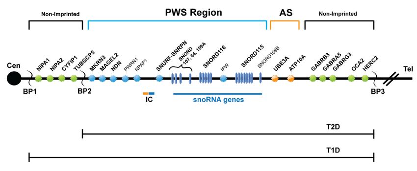
Figure 2. Summary of the genetic and expression map of chromosome region 15q11.2-q13
The Prader-Willi syndrome (PWS) region (shown in blue) has five paternal-only (PWS region) expressed unique-copy genes that
encode polypeptides (MKRN3, MAGEL2, NECDIN, and SNURF-SNRPN) and a family of six paternal-only expressed snoRNA genes
and IPW, a long non-coding RNA. Only UBE3A and ATP10A (shown in orange), related to Angelman syndrome (AS), have
preferential maternal-only expression, and this imprinted expression is limited to certain tissue specific regions (specifically brain). The
imprinting center (IC) has a bipartite structure with an AS (maternal, shown in orange) and a PWS (paternal, shown in blue)
component. The PWS-SRO has been localized to a 4.3-kb region which includes the promoter, CpG island, exon 1, and a small part of
intron 1 of bicistronic SNURF-SNRPN [Ohta et al 1999]. The AS-SRO lies approximately 35 kb proximal to exon 1 of SNURF-SNRPN
[Horsthemke & Buiting 2006]. The bipartite IC lies within the 2.5-Mb PWS/AS imprinted region. The cluster of GABA receptor genes
(GABRB3, GABRA5, and GABRG3), OCA2 (type II albinism) and HERC2 are not imprinted and have biparental expression (shown in
green). The jagged vertical lines denote the three common 5-6-Mb PWS and AS deletion breakpoints: BP1, BP2, and BP3. On rare
occasions there will be a distal breakpoint at BP4 or BP5. Between BP1 and BP2 lie four additional, non-imprinted genes: NIPA1,
NIPA2, CYFIP1, and GCP5. Type 1 deletions (T1D) extend from BP1 to BP3 and type 2 deletions (T2D) extend from BP2 to BP3. Note
that there are more copies of SNORD116 and SNORD115 than are shown and the map has not been precisely drawn to scale.
Hypogonadism. In both sexes, hypogonadism is present and manifests as genital hypoplasia, incomplete
pubertal development, and infertility in the vast majority. Genital hypoplasia is evident at birth and throughout
life.
• Males. The penis may be small, and most characteristic is a hypoplastic scrotum that is small, poorly
rugated, and poorly pigmented. Unilateral or bilateral cryptorchidism is present in 80%-90% of males.
• Females. The genital hypoplasia is often overlooked; however, the labia majora and minora and the clitoris
are generally small from birth.
The hypogonadism is usually associated with low serum concentration of gonadotropins and causes incomplete,
delayed, and sometimes disordered pubertal development. Precocious adrenarche occurs in approximately
15%-20%. Infertility is the rule, although a few instances of reproduction in females have been reported
[Akefeldt et al 1999; Schulze et al 2001; Vats & Cassidy, unpublished data]. Although the hypogonadism in PWS
has long been believed to be entirely hypothalamic in origin, recent studies have suggested a combination of
hypothalamic and primary gonadal deficiencies [Eldar-Geva et al 2009, Hirsch et al 2009, Eldar-Geva et al 2010,
Gross-Tsur et al 2012], a conclusion largely based on the absence of hypogonadotropism and abnormally low
inhibin B levels in some affected individuals of both sexes.
In one study of 84 individuals with PWS (half males, half females) ages 2-35 years [Crinò et al 2003], the
following were identified:Prader-Willi Syndrome 7
• Males. Cryptorchidism 100%, small testes 76%, scrotal hypoplasia 69%
• Females. Labia minora and/or clitoral hypoplasia 76%, primary amenorrhea 56%, spontaneous menarche
(mostly spotting) 44% of those older than age 15 years
• Both sexes. Premature pubarche 14%, precocious puberty 3.6% (1 male, 2 females)
Appetite and obesity. In contrast to the long-held view that there are only two distinct nutritional phases in
PWS (i.e., failure to thrive followed by hyperphagia leading to obesity) a multicenter study [Miller et al 2011]
found that the transition between nutritional phases is much more complex, with seven different nutritional
phases through which individuals with PWS typically progress (Table 2).
Table 2. Nutritional Phases in PWS
Phase Median Ages Clinical Characteristics
0 Prenatal - birth Decreased fetal movements & lower birth weight than sibs
1a 0-9 months Hypotonia with difficulty feeding & decreased appetite
1b 9-25 months Improved feeding & appetite; growing appropriately
2a 2.1-4.5 years Weight increasing without appetite increase or excess calories
2b 4.5-8 years Increased appetite & calories, but can feel full
3 8 years - adulthood Hyperphagic, rarely feels full
4 Adulthood Appetite no longer insatiable for some
Miller et al [2011]
The hyperphagia that occurs in PWS is believed to be caused by a hypothalamic abnormality resulting in lack of
satiety. Food-seeking behavior, with hoarding or foraging for food, eating of inedibles, and stealing of food or
money to buy food, are common. In most, gastric emptying is delayed, and vomiting is rare. Obesity results from
these behaviors and from decreased total caloric requirement. The latter is due to decreased resting energy
expenditure resulting from decreased activity and decreased lean body mass (primarily muscle) compared with
unaffected individuals. The obesity in PWS is primarily central (abdomen, buttocks, and thighs) in both sexes,
and interestingly, there is less visceral fat in obese individuals than would be expected for the degree of obesity.
Obesity and its complications are the major causes of morbidity and mortality (see Morbidity and mortality).
Several independent groups have shown that ghrelin levels are significantly elevated in hyperphagic older
children and adults with PWS before and after meals [Cummings et al 2002, DelParigi et al 2002, Haqq et al
2003b]. Ghrelin is a potent circulating orexigenic hormone that is produced mainly in the stomach. Circulating
ghrelin levels rise after fasting and are suppressed by food intake. The appetite-inducing effect acts through the
appetite regulating pathway in the hypothalamus. Ghrelin levels are lower in non-PWS obese individuals than in
lean controls, and they decrease with age [Scerif et al 2011].
A small study of nine non-hyperphagic children with PWS (age 17-60 months) found similar levels of circulating
ghrelin as in the eight control children matched for BMI, age, and sex [Erdie-Lalena et al 2006]. By contrast, in
two much larger and younger study cohorts of children and adolescents with PWS ghrelin levels were
significantly elevated in the PWS group at any age compared to controls [Feigerlová et al 2008, Kweh et al 2015].
In fact, the highest ghrelin levels in PWS were found in the youngest children. Thus, in these two large studies
the hyperghrelinemia occurred at an age long before the development of obesity and increased appetite in PWS.
Furthermore, several groups have now shown that pharmacologic reduction of ghrelin to normal levels in PWS,
using either short- or long-acting agents, did not affect the weight, appetite, or eating behavior in hyperphagic
individuals [Haqq et al 2003a, Tan et al 2004, De Waele et al 2008]. At this time there are no consistently
identified hormonal abnormalities to explain the hyperphagia, and the metabolic correlates of hyperphagia in
PWS remain uncertain.8 GeneReviews®
Endocrinologic concerns. Up to 25% of adults with PWS (particularly those with significant obesity) have type
2 diabetes [Butler et al 2002] with a mean age of onset of 20 years. In the last 15 years, earlier diagnosis and
education of parents, use of growth hormone therapy, and the frequency of group homes specific for PWS have
led to reduction in the development of morbid obesity (and as a result, in type 2 diabetes) among individuals
with PWS.
Central hypothyroidism, with a normal thyroid-stimulating hormone value and low free thyroxine level, has
been documented in up to 25% of individuals with PWS, with a mean age of diagnosis and treatment of two
years [Miller et al 2008, Diene et al 2010].
Central adrenal insufficiency (CAI) following overnight single-dose metyrapone tests was noted in 60% of
children with PWS in one study, suggesting that this may be the cause of the high incidence of sudden death in
this population [de Lind van Wijngaarden et al 2008]. It is known that introducing GH therapy can precipitate
adrenal crisis in individuals with incipient adrenal insufficiency by accelerating the peripheral metabolism of
cortisol, which may explain the correlation between the incidence of sudden death at the beginning of GH
treatment and CAI in individuals with PWS [Scaroni et al 2008]. However, subsequent studies have found
normal cortisol responses to low- and high-dose synacthen testing, as well as to insulin tolerance testing [Nyunt
et al 2010, Farholt et al 2011]; thus, whether CAI is a true issue for individuals with PWS remains uncertain at
this time and no consensus exists among endocrinologists as to whether evaluation for CAI should be
performed on every individual with PWS or only those with symptoms consistent with adrenal insufficiency.
Sleep abnormalities are well documented and include reduced REM (rapid eye movement) latency, altered sleep
architecture, oxygen desaturation, and both central and obstructive apnea [Festen et al 2006, Priano et al 2006].
Primary hypothalamic dysfunction is thought to be the cause of the alterations in sleep microstructure and
abnormalities in ventilation during sleep, with studies showing low levels of orexin and hypocretin in the
cerebrospinal fluid and decreased levels of acetyl-cholinergic neurons in the pedunculo-pontine tegmental
nucleus [Dauvilliers et al 2003, Nevsimalova et al 2005, Bruni et al 2010, Hayashi et al 2011]. Some individuals
with PWS have excessive daytime sleepiness, which resembles narcolepsy, with rapid onset of REM sleep and
decrease in non-REM sleep instability [Bruni et al 2010].
Behavior. A characteristic behavior profile with temper tantrums, stubbornness, controlling and manipulative
behavior, compulsivity, and difficulty with change in routine becomes evident in early childhood in 70%-90% of
individuals with PWS.
• Many of the behavioral characteristics are suggestive of autism; one study showed that 19% of 59
individuals with PWS versus 15% of age-, sex-, and IQ-matched controls satisfy diagnostic criteria for
autism [Descheemaeker et al 2006].
• In another study of 58 children, attention deficit/hyperactivity symptoms and insistence on sameness were
common and of early onset [Wigren & Hansen 2005].
• This behavior disorder has been reported to increase with age and body mass index (BMI) [Steinhausen et
al 2004], although it diminishes considerably in older adults [Dykens 2004].
• Psychosis is evident by young adulthood in 10%-20% of affected individuals, and is more frequent in those
with UPD [Boer et al 2002, Clarke et al 2002, Vogels et al 2004, Yang et al 2013].
Behavioral and psychiatric problems interfere most with the quality of life in adolescence and adulthood.
Growth. Short stature, if not apparent in childhood, is almost always present during the second decade in the
absence of growth hormone (GH) replacement, and lack of a pubertal growth spurt results in an average
untreated height of 155 cm for males and 148 cm for females. The hands and feet grow slowly and are generally
below the fifth centile by age ten years, with an average adult female foot size of 20.3 cm and average adult male
foot size of 22.3 cm. Growth charts for affected infants and children not treated with growth hormone have beenPrader-Willi Syndrome 9 published [Butler et al 2011, Butler et al 2015] and growth charts for growth hormone-treated children with PWS have been developed [MG Butler & DJ Driscoll, unpublished data]. Data from at least 15 studies involving more than 300 affected children [Burman et al 2001] document reduced GH secretion in PWS. GH deficiency is also seen in adults with PWS [Grugni et al 2006, Höybye 2007]. Dysmorphic features. Characteristic facial features (narrow bifrontal diameter, almond-shaped palpebral fissures, narrow nasal bridge, thin vermilion of the upper lip with down-turned corners of the mouth) may or may not be apparent at birth and slowly evolve over time. Hypopigmentation of hair, eyes, and skin is frequently found in patients with a deletion due to loss of a single copy of the gene OCA2. Ophthalmic issues. Strabismus is seen in 60%-70%. Skeletal findings. Hip dysplasia occurs in approximately 10%-20% [West & Ballock 2004, Shim et al 2010]. Bone fractures are a risk due to the increased frequency of osteopenia and osteoporosis. Scoliosis, present in 40%-80%, varies in age of onset and severity. Other. Rates of the following are increased: • Possibility of recurrent respiratory infections (in ≤50% of individuals) • Leg edema and ulceration (especially in the obese) • Skin picking • Altered temperature sensation • Decreased saliva flow • High vomiting threshold • Seizures (in 10%-20%) Morbidity and mortality. Mortality rate in PWS is higher than in controls with intellectual disability, with obesity and its complications being factors [Einfeld et al 2006]. Based on a population study, the death rate was estimated at 3% per year [Butler et al 2002], although a later study of the same population showed the rate to be decreasing to 1.25% per annum with improved management [Whittington et al 2015]. Two multicenter series of individuals who died of PWS have been reported [Schrander-Stumpel et al 2004, Stevenson et al 2004], and an extensive case and literature review of 64 cases of death in PWS was performed [Tauber et al 2008]. Respiratory and other febrile illnesses were the most frequent causes of death in children, and obesity-related cardiovascular problems and gastric causes or sleep apnea were most frequent in adults. Other causes of morbidity include diabetes mellitus, thrombophlebitis, and skin problems (e.g., chronic edema, infection from skin picking). A few individuals have been reported to have respiratory or gastrointestinal infections resulting in unexpected death; of these, three who died as a result were noted to have small adrenal glands [Stevenson et al 2004], although this is not a common finding. The report of central adrenal insufficiency (CAI) in 60% of tested individuals [de Lind van Wijngaarden et al 2008] suggests a possible explanation for some of these unexpected and sudden deaths. Other studies have not demonstrated a high incidence [Farholt et al 2011, Grugni et al 2013] of CAI in PWS. Acute gastric distention and necrosis have been reported in a number of individuals with PWS [Stevenson et al 2007a], particularly following an eating binge among those who are thin but were previously obese. It may be unrecognized because of high pain threshold and can be fatal. Choking, especially on hot dogs, has been reported as cause of death in approximately 8% of deaths in individuals with PWS [Stevenson et al 2007b].
10                                                                                                   GeneReviews®

Concern about the possible contribution of growth hormone (GH) administration to unexpected death has been
raised by reported deaths of individuals within a few months of starting GH therapy [Eiholzer 2005, Sacco & Di
Giorgio 2005]. The few reported deaths were mostly in obese individuals who had pre-existing respiratory or
cardiac disorders with evidence of upper airway obstruction and uncorrected tonsillar and adenoidal
hypertrophy. In the database of one pharmaceutical company, five of 675 children treated with GH died
suddenly of respiratory problems [Craig et al 2006]. In another study, the rate of death in affected individuals on
and off GH did not differ [Nagai et al 2005]. A study of the natural history of PWS in one region of the UK found
the overall death rate of individuals with PWS to be as high as 3% per year without GH therapy [Whittington et
al 2001]. Thus, the relationship of GH administration to unexpected death remains unclear. However, it is
advisable to obtain a sleep study before the initiation of GH therapy and again four to eight weeks after the
beginning of GH therapy to ensure that GH treatment has not caused or worsened sleep-disordered breathing
[Miller et al 2006b]. A long-term study of 48 treated children suggests that the benefits of treatment with GH
greatly exceed the risks [Carrel et al 2010].
Neuroimaging. In one study, all 20 individuals with PWS who were evaluated had brain abnormalities that were
not found in 21 sibs or 16 individuals with early-onset morbid obesity who did not have PWS [Miller et al 2007].
All had ventriculomegaly; 50% had decreased volume of brain tissue in the parietal-occipital lobe; 60% had
Sylvan fissure polymicrogyria; and 65% had incomplete insular closure. In another study, these authors reported
white matter lesions in some people with PWS [Miller et al 2006a]. A study of brain MRIs from 91 individuals
with PWS from another group showed reduced pituitary height in 49% and some neuroradiologic abnormality
in 67% [Iughetti et al 2008]. The implications of these findings are unknown.

Genotype-Phenotype Correlations
No phenotypic feature is known to correlate exclusively with any one of the three main molecular mechanisms
that result in PWS. However, some statistical differences in the frequency or severity of certain features between
the two largest molecular classes (deletion and UPD) are observed.
UPD
 • Post-term delivery is more common with UPD [Butler et al 2009].
 • Individuals with UPD are less likely to have the typical facial appearance, hypopigmentation, or skill with
   jigsaw puzzles [Dykens 2002]; they also have a somewhat higher verbal IQ and milder behavior problems
   [Dykens et al 1999, Roof et al 2000, Hartley et al 2005].
 • Individuals with UPD are more likely to have psychosis [Holland et al 2003, Yang et al 2013] and autism
   spectrum disorders [Veltman et al 2004, Whittington et al 2004b, Veltman et al 2005, Descheemaeker et al
   2006]. Studies suggest that as many as 62% of those with UPD develop atypical psychosis compared with
   16% of those with a deletion [Soni et al 2007].
Deletion
 • Individuals with a deletion showed a higher frequency of need for special feeding techniques, sleep
   disturbance, hypopigmentation, and speech articulation defects in a recent study of 91 children [Torrado
   et al 2007].
 • Individuals with the slightly larger, type 1 deletions (BP1 to BP3; see Figure 2) have been reported to have
   more compulsions and poorer adaptive behavior, intellectual ability, and academic achievement than those
   with type 2 deletions (BP2 to BP3) [Butler et al 2004, Hartley et al 2005]. Two other studies found much
   less clinically significant differences between individuals with these two deletion types [Milner et al 2005,
   Varela et al 2005]. Larger studies are needed to determine whether there are significant clinical differences
   between the two most frequent deletion classes.
Prader-Willi Syndrome                                                                                            11

Penetrance
Penetrance is complete.

Nomenclature
The term HHHO (hypogonadism, hypotonia, hypomentia, obesity) is no longer used.
The condition is sometimes called Willi-Prader syndrome or Prader-Labhart-Willi syndrome.

Prevalence
The estimated prevalence of PWS is 1:10,000 to 1:30,000 in a number of populations.

Genetically Related Disorders
Angelman syndrome (AS) is caused by loss of the maternally contributed PWS/AS region. It is clinically distinct
from PWS after age two years.
Maternally inherited duplication of the PWS/AS region causes intellectual disability, seizures, and autism [Boyar
et al 2001]. See 15q Duplication Syndrome and Related Disorders.

Differential Diagnosis
Many disorders can mimic parts of the PWS phenotype.
Craniopharyngioma and the results of its treatment show the greatest overlap with PWS. Damage to the
hypothalamus causes most of the same findings that characterize PWS, particularly when craniopharygioma
occurs at an early age. History and, if uncertain, DNA methylation analysis will distinguish craniopharyngioma
from PWS.
Hyperphagic short stature is an acquired condition related to psychosocial stress that includes growth hormone
insufficiency, hyperphagia, and mild learning disabilities [Gilmour et al 2001]. History and, if uncertain, DNA
methylation analysis should distinguish this disorder from PWS.
Hypotonia in infancy is also seen in the following conditions:
 • Neonatal sepsis
 • Central nervous system depression
 • Congenital myotonic dystrophy type 1, characterized by hypotonia and severe generalized weakness at
   birth, often with respiratory insufficiency and early death; intellectual disability is common. It is caused by
   expansion of a CTG trinucleotide repeat in DMPK.
 • Several myopathies and neuropathies, including some instances of spinal muscular atrophy (SMA) [Miller
   et al 1999, Richer et al 2001]. In these situations, poor respiratory effort may be present, a feature rarely
   seen in PWS. Molecular genetic testing, EMG/NCV, and/or muscle biopsy are often required to
   differentiate these conditions.
 • Angelman syndrome (AS), characterized by severe developmental delay or intellectual disability, severe
   speech impairment, gait ataxia and/or tremulousness of the limbs, and a unique behavior with an
   inappropriate happy demeanor that includes frequent laughing, smiling, and excitability. Microcephaly
   and seizures are also common. AS is caused by deficient expression or function of the maternally inherited
   UBE3A allele and may be diagnosed in 75%-80% of individuals with AS using DNA methylation analysis
   of chromosome 15. In infancy, hypotonia may be the only manifestation of AS. Affected individuals lack
   the characteristic sucking problems, hypogonadism, and facial appearance of individuals with PWS.
12                                                                                                  GeneReviews®

 • Fragile X syndrome, characterized by moderate intellectual disability in affected males and mild
   intellectual disability in affected females. Males may have a characteristic appearance (large head, long
   face, prominent forehead and chin, protruding ears), connective tissue findings (joint laxity), and large
   testes (postpubertally). Behavioral abnormalities, sometimes including autism spectrum disorder, are
   common. The diagnosis of fragile X syndrome rests on the detection of an alteration in FMR1 consisting
   of expansion of a triplet repeat and aberrant gene methylation. In infancy, hypotonia may be the only
   manifestation. Affected individuals lack the characteristic sucking problems, hypogonadism, and facial
   appearance of individuals with PWS.
 • WAC-related intellectual disability is typically characterized by variable degrees of global developmental
   delay and/or intellectual disability. Behavioral abnormalities including anxiety, attention deficit/
   hyperactivity disorder, and/or autism spectrum disorder are observed in the majority of older children
   and adults. Most affected infants have significant but nonspecific features at birth such as neonatal
   hypotonia and feeding problems. Some affected individuals come to medical attention with respiratory or
   vision problems. Facial features may be mildly dysmorphic, but are nonspecific. To date, 18 individuals
   have been identified with WAC-related ID. The diagnosis of WAC-related ID is established in a proband
   by identification of a heterozygous pathogenic variant in WAC on molecular genetic testing.
Developmental delay/intellectual disability and obesity with or without hypogonadism can be seen in the
following disorders:
 • Angelman syndrome (AS). Individuals with AS caused by paternal UPD 15 frequently have an elevated
   BMI for age. In one large study overweight was found in more than 70% and obesity in more than 40%
   [Lossie et al 2001].
 • Fragile X syndrome. A subset have been found with a "Prader-Willi-like" phenotype including
   hyperphagia and obesity [de Vries et al 1993].
 • Maternal uniparental disomy for chromosome 14, which also includes prenatal growth retardation,
   feeding problems, short stature, and precocious puberty [Cox et al 2004, Hosoki et al 2009]
 • Albright hereditary osteodystrophy (OMIM 103580), which also includes short stature, but lacks
   hypotonia and has different characteristic facial appearance (round face). Specific testing is possible by
   measurement of Gs receptor-coupling protein.
 • Bardet-Beidl syndrome (BBS), characterized by cone-rod dystrophy, truncal obesity, postaxial polydactyly,
   cognitive impairment, male hypogonadotropic hypogonadism, complex female genitourinary
   malformations, and renal dysfunction. Individuals with BBS have a different facial phenotype from those
   with PWS. Inheritance is typically autosomal recessive, although in fewer than 10% of individuals
   inheritance may be more complex.
 • Cohen syndrome, characterized by failure to thrive in infancy and childhood; truncal obesity in the teen
   years; early-onset hypotonia and developmental delays; microcephaly developing during the first year of
   life; moderate to profound psychomotor retardation; progressive retinochoroidal dystrophy and high
   myopia; neutropenia in many with recurrent infections and aphthous ulcers in some; a cheerful
   disposition; joint hypermobility; and characteristic facial features which are different from PWS. Cohen
   syndrome is caused by pathogenic variants in VPS13B. Inheritance is autosomal recessive.
 • Borjeson-Forssman-Lehmann syndrome (OMIM 301900), seen in males, characterized by severe
   cognitive deficit, epilepsy, hypogonadism, hypometabolism, marked obesity, infantile hypotonia and
   failure to thrive, and short stature. It can be distinguished by the severity of intellectual disability, the
   presence of nystagmus, and characteristic facial appearance with prominent superciliary ridges, ptosis,
   and deep-set eyes. Pathogenic variants in PHF6 are causative. Inheritance is X-linked. Heterozygous
   females who show manifestations of the disorder have skewed X-chromosome inactivation or a genomic
   deletion including PHG6.
 • Alstrom syndrome, characterized by cone-rod dystrophy, early-onset obesity, progressive sensorineural
   hearing impairment, dilated cardiomyopathy (>60%), the insulin resistance syndrome/type 2 diabetes
Prader-Willi Syndrome                                                                                            13

    mellitus associated with acanthosis nigricans, and developmental delay (~50%). Other endocrine
    abnormalities can include hypothyroidism and male hypogonadotropic hypogonadism. Urologic disorders
    of varying severity, characterized by detrusor-urethral dyssynergia, appear in females in their late teens.
    Severe renal disease is usually a late finding. Pathogenic variants in ALMS1 are found in 70%-80% of
    individuals of northern European descent, and about 40% of affected individuals worldwide.
Cytogenetic abnormalities with a similar phenotype include the following:
 • A "PWS-like phenotype" of syndromic obesity has been identified in individuals with an interstitial
   deletion of 6q16.2, which includes SIM1 [Varela et al 2005]. This deletion had been reported at least five
   times previously in syndromic obesity [Bonaglia et al 2008] as well a duplication [Desch et al 2015]. Also,
   pathogenic variants in SIM1 alone have been found in patients with severe obesity [Bonnefond et al 2013,
   Ramachandrappa et al 2013].
 • Several reports have associated a Prader-Willi-like phenotype with 1p36 deletion; additional findings
   include hypotonia, developmental delay, obesity, hyperphagia, and behavioral problems [Tsuyusaki et al
   2010, Stagi et al 2014].
 • Multiple reports describe deletions at 16p11.2 including SHB2B1, which is involved in leptin and insulin
   signaling [Maillard et al 2015].
 • Reports of other cytogenetic anomalies in individuals with a PWS-like phenotype have included
   dupXq27.2-ter and del10q26 [Lukusa & Fryns 2000, Ben-Abdallah-Bouhjar et al 2012, Rocha & Paiva
   2014].

Management
Management of the manifestations of Prader-Willi Syndrome (PWS) is age dependent and should include both
addressing of the consequences of PWS and anticipatory guidance. It is recommended that a team approach be
used, if possible. Several approaches to management have been published [Goldstone et al 2008, Cassidy &
Driscoll 2009, Cassidy & McCandless 2010, McCandless 2011, Cassidy et al 2012].

Evaluations Following Initial Diagnosis
To establish the extent of disease and needs in an individual diagnosed with PWS, the following evaluations are
recommended:
 • Consultation with a clinical geneticist and/or genetic counselor
 • Endocrinology consultation
 • Nutritional consultation
 • Assessment of newborns and young infants for sucking problems and failure to thrive
 • Regardless of age, measurement and plotting of height, weight, head circumference and body mass index
   (BMI) on either age-appropriate growth charts or charts developed for PWS [Butler et al 2011, Butler et al
   2015]
 • Assessment for hypothyroidism in children, especially those with prolonged failure to thrive, those with
   excess weight gain in the absence of increased food intake, and those with poor linear growth despite
   growth hormone treatment
 • Evaluation of respiratory status with a low threshold for performing a sleep study regardless of age. These
   studies are specifically recommended prior to the initiation of growth hormone therapy (GHT) and four
   to eight weeks after starting GHT, along with assessment of the size of tonsils and adenoids, particularly in
   the obese individual.
14 GeneReviews®
• Assessment of development of infants and of educational development of children including a speech
evaluation
• Assessment for the presence of behavioral problems and obsessive-compulsive features after age two years,
and for psychosis in adolescents and adults. If history reveals evidence of these problems, referral for more
detailed assessment is indicated.
• Assessment of males for the presence of cryptorchidism regardless of age
• Referral for ophthalmologic evaluation if strabismus is present, and for assessment of visual acuity by age
one year or at diagnosis if it is later
• Regardless of age, assessment for the presence of scoliosis clinically, and, if indicated, radiographically
Note: Very obese individuals cannot be adequately assessed for scoliosis clinically; x-rays are necessary to
establish the diagnosis.
Treatment of Manifestations
A team approach to management is recommended [Goldstone et al 2008, Cassidy & McCandless 2010].
Feeding, hyperphagia, and obesity. Special feeding techniques, including special nipples or gavage feeding, may
be necessary for the first weeks to months of life to assure adequate nutrition and avoid failure to thrive.
Individuals diagnosed with PWS typically do not need a G-tube since the feeding will improve with time.
When weight centiles begin increasing in nutritional phase 2a (typically between 18 and 36 months), a program
of a well-balanced, low-calorie diet, regular exercise, and close supervision to minimize food stealing should be
instituted to prevent obesity (i.e., BMI Z score ofPrader-Willi Syndrome 15 • A review of the results of one to two years of growth hormone treatment among 328 children documented in the database of one pharmaceutical company indicated improved height velocity, particularly in prepubertal children, but no change in BMI [Craig et al 2006]. • Significantly greater adult height was demonstrated in 21 individuals treated long term versus 39 untreated individuals without an increase in adverse side effects [Angulo et al 2007]. • Some improvements in cognition have been suggested with growth hormone therapy in individuals with PWS [Osório 2012, Siemensma 2012], but more work and longer studies need to be done. • Although there was initial concern about growth hormone treatment contributing to scoliosis in PWS, later studies showed no difference in frequency or severity in those treated compared to those who were not treated [Nagai et al 2006, Angulo et al 2007]. Decreased saliva production can be addressed with products developed for the treatment of dry mouth, including special toothpastes, gels, mouthwash, and gum. Therapies, education and behavior management. Early intervention in children before age three years, particularly physical therapy, may improve muscle strength and encourage achievement of developmental milestones. In older individuals, daily muscle training increases physical activity and lean body mass [Schlumpf et al 2006]. Initiate appropriate educational programming in children: • Begin speech therapy for language delay and articulation abnormalities in infancy and childhood. • Special education, either in an inclusion setting or in a self-contained classroom setting, is usually necessary during school age. An individual aide is helpful in assuring attendance to task. Social skills training groups have been beneficial. Behavioral disturbance should be addressed with behavioral management programs, including firm limit setting. While no medication is beneficial in managing behavior in all individuals with PWS, serotonin reuptake inhibitors have helped the largest proportion of affected teenagers and adults, particularly those with obsessive- compulsive symptoms [Brice 2000, Dykens & Shah 2003]. Psychosis is reported to respond well to selective serotonin reuptake inhibitors, but not to mood stabilizers [Soni et al 2007]. There are no well-designed studies of the effectiveness of treatment for psychosis in PWS [Ho & Dimitropoulos 2010]. Hypogonadism. Cryptorchidism may resolve spontaneously, even up to adolescence, but usually requires hormonal and surgical approaches; however, preservation of fertility is not an issue. Standard treatment is appropriate. Human chorionic gonadotropin treatment for infants with undescended testes should be considered as it can improve the size of the scrotal sac and improve surgical outcome [McCandless 2011; Angulo & Miller, unpublished data]. Replacement of sex hormones produces adequate secondary sexual characteristics but is somewhat controversial because of the possible role of testosterone replacement in behavior problems in males and the role of estrogen replacement in the risk of stroke as well as hygiene concerns related to menstruation in females. Daily use of the testosterone patch or gel may avert exacerbation of behavioral problems by providing a more even blood level than use of a slow-release depo-testosterone injection every month. Also, it was shown in the non-PWS adult population that depo- testosterone injections were associated with a greater risk of cardiovascular events, hospitalizations, and deaths compared with gels and patches [Layton et al 2015]. Concern about osteoporosis should be considered in deciding about hormone replacement. Recent reports of fertility in four women with PWS raise the issue of need for birth control [Akefeldt et al 1999; Schulze et al 2001; Vats & Cassidy, unpublished data].
16                                                                                                      GeneReviews®

Sleep issues. Disturbed sleep in children and adults should prompt a sleep study, as treatment may be available.
Treatment depends on the cause and may include tonsillectomy and adenoidectomy and/or CPAP or BiPAP, as
in the general population.
Excessive daytime sleepiness (unrelated to the degree of sleep apnea) is frequently seen in individuals with PWS.
Modafinil has been shown to be a safe and effective treatment for this condition [De Cock et al 2011].
Skin picking. One study demonstrated decreased skin picking with topiramate treatment in some individuals
[Shapira et al 2004]; other clinicians have reported anecdotally that approximately half of individuals with PWS
who skin pick benefit from low-dose (25-50 mg daily) topiramate.
A recent study of 35 individuals with PWS (age 5-39 years) using 450-1200 mg/day of N-acetylcysteine found a
high degree of success in reducing or eliminating skin picking [Miller & Angulo 2014].
Other
 • Management of strabismus is as for any infant.
 • Management of scoliosis, hip dysplasia, and complications of obesity is as in the general population.
Adulthood. For adults with PWS, the most successful living situation for behavior and weight management is a
group home specially designated for individuals with PWS, where diet and access to food are tightly restricted
and exercise is included in daily activities. Affected individuals generally require a sheltered employment
environment.
Issues of guardianship, wills, trusts, and advocacy should be investigated no later than adolescence.

Prevention of Primary Manifestations
Obesity may be prevented if the diet, exercise, and supervision program described in Treatment of
Manifestations is instituted. Early diagnosis allows the clinician to begin anticipatory guidance concerning the
natural history of PWS, and in particular the nutritional phases, informing the family about the risk of obesity
and the need to monitor weight increase and to restrict calories beginning around 18-36 months of age.
If started at a young age, growth hormone treatment, along with good dietary control, may retard obesity and
the high proportion of fat mass. It may also prevent development of the typical facial appearance and improve
motor milestones.

Prevention of Secondary Complications
Diabetes mellitus rarely occurs in the absence of obesity.
Calcium and vitamin D supplementation are probably beneficial, as low-calorie diets are often low in dairy
products and osteoporosis has been documented in the majority of older children and adults with PWS.
If osteoporosis develops, consider treatment with a bisphosphonate.
Although no formal study exists, individuals with PWS tend to be very sensitive to medications of all kinds.
Starting with lower doses is recommended.

Surveillance
Health supervision guidelines from the American Academy of Pediatrics (AAP) have been published
[McCandless 2011] (full text).
To assure appropriateness of exercise program and diet, including adequacy of vitamin and mineral intake and
monitor height, weight, and BMI (weight in kg/height in m2):
Prader-Willi Syndrome                                                                                                  17

 • Every month in infancy;
 • Every six months in the first decade of life;
 • At least annually thereafter.
Cryptorchidism can recur after orchidopexy; therefore, testicular position should be monitored.
Evaluate for the presence of diabetes mellitus by standard methods (e.g., obtaining glycosylated hemoglobin
concentration and/or glucose tolerance test) in anyone with significant obesity or rapid significant weight gain.
Test annually for hypothyroidism, including free T4 and TSH levels.
Obtain history of any sleep disturbance; if present, obtain a sleep study.
Monitor for development of scoliosis clinically or, in the presence of obesity, radiographically at least annually.
Perform bone densitometry by DEXA to evaluate for possible osteoporosis every two years in adulthood.
Obtain history for behavioral and psychiatric disturbance at least annually.

Evaluation of Relatives at Risk
See Genetic Counseling for issues related to testing of at-risk relatives for genetic counseling purposes.

Therapies Under Investigation
In the last few years there has been great interest by the pharmaceutical industry in testing treatments for the
major manifestations of PWS – particularly the hyperphagia, obesity, and behavioral problems. For a detailed
description of these studies, click here (pdf).
Search ClinicalTrials.gov in the US and EU Clinical Trials Register in Europe for access to information on
clinical studies for a wide range of diseases and conditions.

Genetic Counseling
Genetic counseling is the process of providing individuals and families with information on the nature, inheritance,
and implications of genetic disorders to help them make informed medical and personal decisions. The following
section deals with genetic risk assessment and the use of family history and genetic testing to clarify genetic status for
family members. This section is not meant to address all personal, cultural, or ethical issues that individuals may
face or to substitute for consultation with a genetics professional. —ED.

Mode of Inheritance
Prader-Willi syndrome (PWS) is caused by lack of expression of the paternally derived PWS/AS region of
chromosome 15q11.2-q13 by one of several genetic mechanisms.

Risk to Family Members
Parents of a Proband
 • The parents of the proband are unaffected.
 • Recommendations for genetic testing of the parents depend on the genetic mechanism of PWS in the
   proband (see Sibs of a Proband and Table 3).
 • Note: Germline mosaicism in the father is rare but has been observed in cases of 15q11.2 deletions
   [Fernández-Novoa et al 2001] and IC deletions [Buiting et al 2003, Wey et al 2005]. Recurrent meiotic
   nondisjunction of maternal chromosome 15 has also been observed [Harpey et al 1998].
18                                                                                                                 GeneReviews®

Sibs of a Proband
The risk to sibs of a proband with PWS depends on the genetic mechanism of PWS in the proband and the
molecular class (summarized in Table 3). The vast majority of families have a recurrence risk of less than 1%.
However, certain etiologies involve a recurrence risk as high as 50%, and a scenario with a risk of almost 100%
(i.e., a mother with a 15/15 Robertsonian translocation), though very unlikely, is theoretically possible.
For recurrence risk assessment. If the DNA methylation pattern is characteristic of maternal inheritance only,
the underlying genetic mechanism (deletion, UPD, or ID) and the specific genetic etiology (molecular class)
should be determined for genetic counseling purposes (see Figure 1 and Table 3).
It is typically most efficient to begin with a karyotype and FISH analysis for the 15q11.2-q13 deletion.
Simultaneous cytogenetic studies allow detection of a translocation or other anomaly involving proximal 15q.
With the increasing use of chromosomal microarray (CMA) in clinical genetics, arrays (particularly SNP arrays)
may replace FISH analysis for the identification of deletions in PWS. However, each technique has its
advantages.
 • CMA will precisely identify the deletion size, which is anticipated to become increasingly important for
   genotype-phenotype correlations in the future [Kim et al 2012]. In addition, in the non-deletion cases an
   SNP array will identify a high percentage of UPDs.
 • However, CMA will not detect the rare chromosome rearrangements (translocations and inversions)
   involving proximal 15q; these are detectable by simultaneous karyotype and FISH analysis and are
   important in recurrence risk determination.
 • For genetic counseling purposes, a chromosome analysis is advised in the proband to discern an
   interstitial de novo deletion from a balanced or unbalanced chromosome rearrangement involving the
   15q11.2 region.
 • CMA would also be indicated if an individual with PWS had a more severe phenotype than is typical in
   order to discern if there was a larger deletion present or an additional chromosome abnormality elsewhere
   in the genome.
If no deletion or other chromosome abnormality is detected, DNA polymorphism studies (requiring blood from
both parents and the proband) are conducted. Increasingly, clinical geneticists are using a CMA combined with
SNPs (i.e., oligo-SNP arrays) which will detect approximately 75% of UPDs by demonstrating long (>13.5 Mb)
contiguous stretches of homozygosity limited to the chromosome 15s [Papenhausen et al 2011; Butler &
Driscoll, unpublished data]. In these cases DNA polymorphism analysis is probably not necessary to diagnose
UPD.
If UPD is not detected, referral to a specialized laboratory for microdeletion analysis of the imprinting center
(IC) is indicated.

Table 3. Risks to Sibs of a Proband with PWS by Genetic Mechanism
                    Proportion of PWS by
Molecular Class 1                              Genetic Mechanism                              Risk to Sibs
                    Molecular Class
Ia                  65%-75%                    Interstitial 5- to 6-Mb 15q11.2-q13 deletion
Prader-Willi Syndrome 19
Table 3. continued from previous page.
Proportion of PWS by
Molecular Class 1 Genetic Mechanism Risk to Sibs
Molecular Class
IIIb 2% Epimutation - ID w/out IC deletion20 GeneReviews® Offspring of a Proband • With rare exception in females, individuals with PWS do not reproduce. • The risk to the child of an affected individual depends on the molecular class and the sex of the affected individual. • If the proband has PWS as the result of a deletion, the offspring have a 50% chance of having Angelman syndrome (AS) if the proband is female and PWS if the proband is male (the latter has never been reported) • If the proband has UPD, the offspring would be expected to be unaffected. A single report described a female with PWS caused by UPD who had a normal child [Schulze et al 2001]. • If the female proband has PWS as the result of an ID by an IC deletion, the offspring are at a 50% theoretic risk of having AS if the microdeletion extends into the AS SRO (never reported). • If the proband has a chromosome translocation, the offspring are theoretically at increased risk of having PWS or AS, depending on the sex of the proband (never reported). Other Family Members If a chromosome rearrangement (e.g., translocation or inversion) is identified in the proband and a parent, the sibs of the carrier parent should be offered genetic counseling and the option of genetic testing. Related Genetic Counseling Issues Family planning • The optimal time for determination of genetic risk, clarification of carrier status, and discussion of the availability of prenatal testing is before pregnancy. • It is appropriate to offer genetic counseling (including discussion of potential risks to offspring and reproductive options) to young adults who are affected, are carriers, or are at risk of being carriers. DNA banking is the storage of DNA (typically extracted from white blood cells) for possible future use. Because it is likely that testing methodology and our understanding of genes, allelic variants, and diseases will improve in the future, consideration should be given to banking DNA of affected individuals. Prenatal Testing Families who have a child with PWS (in whom the causative genetic mechanism has been identified) should be aware that prenatal testing for PWS is possible. Depending on the molecular mechanism, FISH, CMA, DNA methylation analysis, MS-MLPA, or DNA polymorphism studies for UPD may be appropriate. Note that only DNA methylation analysis (e.g., using MS-MLPA) at the 5' SNRPN locus will identify the imprinting defects [Kubota et al 1996, Glenn et al 2000, Ramsden et al 2010]. While prenatal detection of all three molecular classes of PWS is possible, availability may be limited. Laboratories offering PWS prenatal diagnosis by DNA methylation analysis typically accept only amniocytes (vs chorionic villi) for analysis because of the known hypomethylation of tissue derived from the placenta [Driscoll & Migeon 1990, Glenn et al 2000]. • Parents who have had one child with PWS caused either by deletion or UPD, and who do not have a chromosome rearrangement, have a low recurrence risk. • Parents who have had one child with PWS caused by an IC deletion, and in whom the father is a known carrier, have a high recurrence risk; DNA methylation analysis can be used in these cases. • Prenatal testing for an inherited translocation involving chromosome 15 and resulting in a deletion is relevant because of the theoretic 50% risk of PWS in the offspring. Pregnancies in which no family history of PWS exists. PWS may be a possibility in the following situations:
Prader-Willi Syndrome                                                                                             21

 • If a 15q11.2 deletion is suspected on cytogenetic studies from testing of cells obtained by CVS or
   amniocentesis, FISH or CMA is indicated. In this instance, parent-of-origin studies should be performed
   after confirmation of a deletion to determine if the deletion is maternally derived (fetus has AS) or
   paternally derived (fetus has PWS).
 • If a trisomy 15 or mosaic trisomy 15 is detected on testing of cells obtained by CVS, and if subsequent
   testing of cells obtained by amniocentesis reveals 46 chromosomes, the possibility of trisomy rescue
   leading to AS (paternal UPD) through loss of a maternal chromosome 15 or PWS (maternal UPD)
   through loss of a parental chromosome 15 can be considered. In this instance, parent-of-origin (UPD)
   studies or DNA methylation analysis on amniocytes should be considered [EUCROMIC 1999, Shaffer et al
   2001].
 • If an inherited or de novo translocation involving chromosome 15 is present or if a supernumerary
   chromosome derived from chromosome 15 is detected, FISH (to rule out a deletion) and parent-of-origin
   or DNA methylation studies (to rule out the possibility of UPD) are indicated.
Recently noninvasive prenatal tests (NIPT) using fetal cell-free DNA have become available for testing the major
trisomy conditions as well as the common microdeletion (including PWS and AS) conditions [Norton et al
2012]. If CMA is used in NIPT it will pick up 15q11.2 deletions, but it will not distinguish an AS deletion from a
PWS deletion and it will not pick up the UPD and ID mechanisms.
Preimplantation genetic diagnosis (PGD) may be an option for some families in which an IC deletion has been
identified. PGD can also be used in cases of familial translocation to rule out UPD.

Resources
GeneReviews staff has selected the following disease-specific and/or umbrella support organizations and/or registries
for the benefit of individuals with this disorder and their families. GeneReviews is not responsible for the
information provided by other organizations. For information on selection criteria, click here.
 • International Prader-Willi Syndrome Organisation (IPWSO)
    c/o BIRD Europe Foundation Onlus
    Via Bartolomeo Bizio 1
    Costozza 1-36023
    Italy
    Phone: +39 0444 555557
    Fax: +39 0444 555557
    Email: info@ipwso.org
    www.ipwso.org
 • My46 Trait Profile
    Prader-Willi Syndrome
 • Prader-Willi Syndrome Association (USA)
    8588 Potter Park Drive
    Suite 500
    Sarasota FL 34238
    Phone: 800-926-4797 (toll-free); 941-312-0400
    Fax: 941-312-0142
You can also read