Prenatal diagnosis of harlequin ichthyosis by ultrasonography: a case report
←
→
Page content transcription
If your browser does not render page correctly, please read the page content below
Case Report
                                                                                                                                     Page 1 of 5

Prenatal diagnosis of harlequin ichthyosis by ultrasonography: a
case report
Xiao-Jing Zhou1, Yu-Jie Lin2, Xi-Wei Chen1, Jia-Hua Zheng3, Ying-Jie Zhou1
1
Seven Section of Department of Gynaecology, the Second Hospital of Hebei Medical University, Shijiazhuang, China; 2Department of Ultrasound,
the Julu County Hospital, Xingtai, China; 3Department of Obstetrics and Gynecology, the Second Hospital of Hebei Medical University,
Shijiazhuang, China
Correspondence to: Ying-Jie Zhou. No. 215, He Ping Road (West), Xin Hua District, Shijiazhuang, China. Email: yingjiehero@163.com.

                Abstract: Autosomal recessive congenital ichthyosis is a genetically and phenotypically heterogeneous
                group of skin disorders, including harlequin ichthyosis (HI), lamellar ichthyosis, and bullous congenital
                ichthyosiform erythroderma. HI is the most phenotypically severe autosomal recessive congenital
                ichthyosis associated with the mutation of the adenosine triphosphate—binding cassette subfamily A
                member 12 (ABCA12) gene. The clinical manifestations include generalized hyperkeratotic plaques and
                deep fissures, ectropion, eclabium, and contractures. However, the severe HI may easily be misdiagnosed
                as epidermolysis bullosa or syndromic ichthyosis. Meanwhile, no consensus exists about the best used in
                clinical trials or clinical practice when more elaborate scoring systems have been proposed to evaluate skin
                xerosis, palmoplantar keratoderma, and disease extension an accurate prenatal diagnosis is necessary. Until
                the ABCA12 gene was identified as the pathogenic gene, prenatal diagnosis of HI had been performed by
                the invasive techniques of fetal skin biopsy. Now, advances in ultrasound technology and fetal DNA-based
                analysis have replaced it. The mortality rate is markedly high and prompt; prenatal diagnosis of neonate HI
                is critical for appropriate perinatal and postnatal management. It is also essential to prepare parents for future
                pregnancies and reduce the family’s physical and mental distress and financial burden. This report presents
                a rare case of harlequin ichthyosis diagnosed by the ultrasound and discusses the significance of prenatal
                ultrasound diagnosis and molecular diagnosis in the prenatal diagnosis of HI.

                Keywords: Harlequin ichthyosis; molecular diagnosis; prenatal ultrasound; prenatal diagnosis; case report

                Submitted Nov 10, 2020. Accepted for publication Jan 22, 2021.
                doi: 10.21037/atm-20-8223
                View this article at: http://dx.doi.org/10.21037/atm-20-8223

Introduction                                                                ichthyosis (HI), lamellar ichthyosis, and bullous congenital
                                                                            ichthyosiform erythroderma (2). HI is the most phenotypically
Inherited ichthyoses comprise of ichthyosis syndrome and
                                                                            severe ARCI associated with the mutation of the adenosine
non-syndromic ichthyosis. Ichthyosis syndromes are disorders
                                                                            triphosphate (ATP)—binding cassette subfamily A member
that can be observed in combination with other organ
involvements. Non-syndromic ichthyoses are disorders that                   12 (ABCA12) gene (3-6). The clinical manifestations include
can be observed only in the skin (1). These two syndromes                   generalized hyperkeratotic plaques and deep fissures,
are genetic skin disorders and exhibit typical features that                ectropion, eclabium, and contractures and affect 1 in 300,000
include hyperkeratosis, scaling, and generalized dry skin,                  live births (3)—onset at birth, often preterm babies. The risk
mainly because of mutations on one or over 30 different                     of death is extremely high during the neonatal period (2).
alleles involved in skin barrier formation (2). Autosomal                      This report presents a case of a pregnant woman who
recessive congenital ichthyosis (ARCI) is a genetically and                 underwent three adverse pregnancies at the ages of 21, 24,
phenotypically heterogeneous group of skin disorders, which                 and 25, respectively, with the recent pregnancy presenting
is one form of non-syndromic ichthyosis, including harlequin                with HI, revealed by ultrasound, and to discuss the

© Annals of Translational Medicine. All rights reserved.                  Ann Transl Med 2021;9(2):183 | http://dx.doi.org/10.21037/atm-20-8223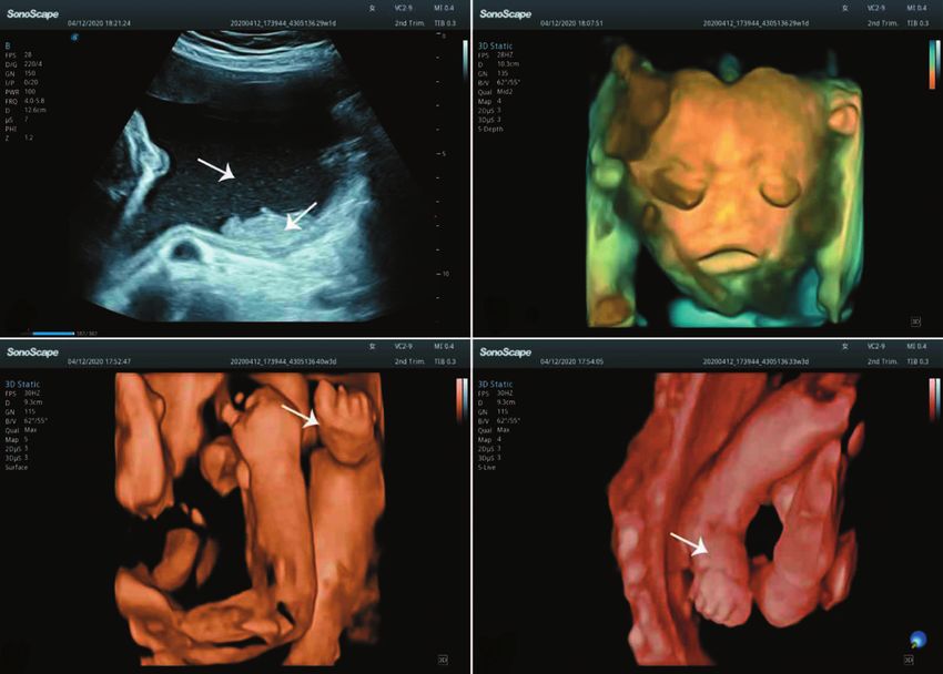
Page 2 of 5                                                                       Zhou et al. Prenatal diagnosis of HI by ultrasonography

                    A                                                  B

                    C                                                 D

Figure 1 Three-dimensional ultrasound. (A) The dense floating particles in amniotic fluid (arrows); (B) the harlequin phenotype (“clown-like
face”); (C) the contracted and edema hands (arrow); (D) the contracted and edema feet (arrow).

significance of prenatal ultrasound diagnosis and molecular                consanguineous marriage or contact with pesticides and
diagnosis in prenatal diagnosis of HI.                                     radioactive substances. However, repeated on admission,
   We present the following article in accordance with                     three-dimensional sonography construction revealed dense
the CARE reporting checklist (available at http://dx.doi.                  floating particles in amniotic fluid (“snowflake sign”). The
org/10.21037/atm-20-8223).                                                 bilaterally soft tissue in the anterior region of the eyeballs
                                                                           was thickened. The upper and lower lips were markedly
                                                                           thickened and everted, causing the mouth to be fixed in
Case presentation
                                                                           an O‑shape. No apparent nostrils and normal nasal shape
A 26-year-old multipara woman was referred to our hospital                 were observed. The fetus also had a fixed flexion deformity
with a suspected fetal abnormality in the third trimester                  of the extremities (Figure 1). Considering ichthyosis by
(28+5 weeks). On her admission, there were no symptoms or                  ultrasound, it is suggested to form a further prenatal
signs. Five years prior, she had undergone induced labor on                diagnosis. At 29+3 weeks, the labor was induced, and a dead
week 20 of pregnancy due to fetal abnormalities. Three-year                female infant was delivered. The premature infant exhibited
ago, she had a cesarean section at 32 weeks of pregnancy.                  typical characteristics of HI following the sonography (thick,
Unfortunately, the infant was premature, had deformities                   platelike scaling and deep fissures, extreme ectropion, and
(the whole skin was red, and the rest was unknown), and                    eclabium. The nose was not observed. The ears with the ear
died five days later (no fetal tissue examination). The                    canal were small and without auricles. Figure 2 shows the
present case was genetically tested for Down’s syndrome,                   palmoplantar involvement, synechiae of fingers, and toes.
neural tube defect, and trisomy 18 syndrome, and the                       With the patient and her family’s signature, a genetic study
results were all negative. Non-invasive DNA screening                      was performed with the dead infant’s lower extremities’
showed no chromosomal abnormalities. Meanwhile,                            gastrocnemius muscle tissue by whole-exome sequencing
there were no signs of hyperglycemia or hypertension                       (WES). Data analysis found that: The paternal variant was
during pregnancy, and the woman denied having a                            a missense mutation (c.1300C>T; p.Arg434*) present at

© Annals of Translational Medicine. All rights reserved.               Ann Transl Med 2021;9(2):183 | http://dx.doi.org/10.21037/atm-20-8223
Annals of Translational Medicine, Vol 9, No 2 January 2021 Page 3 of 5
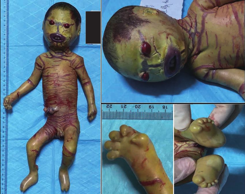
Figure 2 General view of harlequin ichthyosis fetal.
exon 12. According to the American Society of Medical in chromosome 2 (2q34) causes a change in the stratum
Genetics and Genomics (ACMG) guidelines, the mutation corneum’s lipid deposition, leading to skin barrier disruption
was pathogenic. According to the public database query, the and compensatory hyperkeratinization (4,7). In other words,
mutation may lead to autosomal recessive clown ichthyosis, HI is a lipid metabolism disorder (4). Skin development is
and the phenotype of the disease is consistent with the changed in the uterus, and hair canal hyperkeratosis occurs
disease. While the maternal variant was a deletion mutation in the second trimester (8). Historically, fetuses presenting
(c.3179+3_3179+6delAAGT) present at the intron 22 and with HI are born with a distinct clinical appearance and
according to the ACMG guidelines, the variation was have increased perinatal mortality (2). Hence, prenatal
judged to be an unknown variable. diagnosis of neonate HI is significantly essential for proper
All procedures performed in studies involving human perinatal and postnatal management, prepare parents for
participants were in accordance with the ethical standards future pregnancies, and reduce the physical and mental
of the Ethics Committee of the Second Hospital of Hebei distress and financial burden of the family.
Medical University (approval letter no. 2020-P019) and A correct diagnosis of HI is critical for genetic
with the Helsinki Declaration (as revised in 2013). Written counseling and adequate patient information about
informed consent was obtained from the patient. prognosis and therapeutic options. Given the patients
present with a harlequin phenotype precisely like the case,
one may surmise that it is easy to make a correct diagnosis
Discussion
of HI. However, the severe HI may easily be misdiagnosed
HI is a rare and severe ARCI genetic skin disorder. as epidermolysis bullosa or syndromic ichthyosis (9).
Reverend Oliver Hart reported the first case in 1750 (3). Meanwhile, no consensus exists about the best used in
Until 2005, the underlying mechanisms were not well clinical trials or clinical practice when more elaborate
understood. The mutation of the ABCA12 gene found scoring systems have been proposed to evaluate skin xerosis,
© Annals of Translational Medicine. All rights reserved. Ann Transl Med 2021;9(2):183 | http://dx.doi.org/10.21037/atm-20-8223Page 4 of 5 Zhou et al. Prenatal diagnosis of HI by ultrasonography
palmoplantar keratoderma, and disease extension (10,11). While the maternal variant (c.3179+3_3179+6delAAGT)
An accurate prenatal diagnosis is necessary. present at the intron 22 was judged to be an unknown
Until the ABCA12 gene was identified as the pathogenic variable. No related cases have been reported. The
gene, prenatal diagnosis of HI had been performed by mutation is close to the splicing site of "GT" and may affect
the invasive techniques of fetal skin biopsy (8,12). Now, mRNA’s transcriptional process. This may be supported
advances in ultrasound technology and fetal DNA-based by collecting more cases of similar gene mutations or
analysis have replaced it (3). In the present case, the functional verification at the RNA level. However, it is still
prenatal sonography analyzed the HI fetus images, and they difficult to predict its pathogenic potential. For example, a
were following the type of fetal HI, including eclabium, negligible point mutation in TGM1 may change the TGm-1
ectropion, contractures, and “snowflake sign” (dense 3D structure only when the skin temperature is elevated (20).
floating particles in amniotic fluid) (13,14). Moreover, Hence, genetic screening also should combine with detailed
three-dimensional ultrasound significantly improves anamnestic and clinical investigations.
the facial morphology analysis and may be conducive to In short, prenatal sonography and molecular diagnosis
prenatal diagnosis (15). It is worth noting these unusual are increasingly feasible in patients with HI and are essential
features are not detectable until the second trimester, for giving correct genetic advice. Combined with the pre-
except for early termination (3). Besides, fetal HI cannot implantation genetic diagnosis (PGD) to select and transfer
differentiate from fetal macroglossia and congenital tumor- an unaffected embryo will reduce the family’s physical and
like fetus angioma (13). They both had their tongues mental distress and financial burden.
extended from the mouth, causing the mouth to be fixed
in an O‑shape, like fetal HI. However, macroglossia is
Acknowledgments
always associated with genetic disorders (Down’s syndrome
and Beckwith-Wiedemann syndrome), and genetic testing Funding: None.
can differentiate it from HI (16). The thickened tongue in
congenital hemangioma fetal typically exhibits blood flow
Footnote
by color Doppler imaging (17). Therefore, HI ultrasound
diagnosis needs to be combined with disease characteristics Reporting Checklist: The authors have completed the CARE
and gene detection for differential diagnosis. reporting checklist. Available at http://dx.doi.org/10.21037/
A deep understanding of the HI genetic basis shows that atm-20-8223
genetic screening for candidate gene mutations associated
with HI may aid in prenatal diagnosis. The first DNA- Conflicts of Interest: All authors have completed the ICMJE
based prenatal diagnosis of HI was performed by direct uniform disclosure form (available at http://dx.doi.
sequence analysis of the ABCA12 mutation from amniotic org/10.21037/atm-20-8223). The authors have no conflicts
fluid cells, which showed the effectiveness of early DNA- of interest to declare.
based prenatal diagnosis (18). It is becoming a standard
technique for the early and accurate diagnosis of HI (19). Ethical Statement: The authors are accountable for all
The next generation sequencing (NGS) technology has aspects of the work in ensuring that questions related
been developed and applied to large-scale gene diagnosis, to the accuracy or integrity of any part of the work are
analyzing the whole coding part of a genome or even appropriately investigated and resolved. All procedures
the whole genome in a few days. If mutations cannot be performed in studies involving human participants were
identified, more genes will be analyzed, or whole-exome in accordance with the ethical standards of the Ethics
sequencing (WES) will be sequenced. Moreover, large-scale Committee of the Second Hospital of Hebei Medical
sequencing may lead to additional and coincidental findings. University (approval letter no. 2020-P019) and with the
However, exome sequencing may produce sequence variants Helsinki Declaration (as revised in 2013). Written informed
that cannot be interpreted as pathogenic or nonpathogenic. consent was obtained from the patient.
In the present case, a genetic study was performed
by WES and found the paternal variant was a missense Open Access Statement: This is an Open Access article
mutation (c.1300C>T; p.Arg434*) present at exon 12, and distributed in accordance with the Creative Commons
the pathogenicity of this mutation had been reported (4). Attribution-NonCommercial-NoDerivs 4.0 International
© Annals of Translational Medicine. All rights reserved. Ann Transl Med 2021;9(2):183 | http://dx.doi.org/10.21037/atm-20-8223Annals of Translational Medicine, Vol 9, No 2 January 2021 Page 5 of 5
License (CC BY-NC-ND 4.0), which permits the non- in the Treatment of Patients With moderate/severe
commercial replication and distribution of the article with Lamellar Ichthyosis: Results of a Randomized, Double-
the strict proviso that no changes or edits are made and the Blind, Multinational, Placebo-Controlled Phase II/III
original work is properly cited (including links to both the Trial. Br J Dermatol 2014;170:173-81.
formal publication through the relevant DOI and the license). 12. Akiyama M, Suzumori K, Shimizu H. Prenatal Diagnosis
See: https://creativecommons.org/licenses/by-nc-nd/4.0/. of Harlequin Ichthyosis by the Examination of Keratinized
Hair Canals and Amniotic Fluid Cells at 19 Weeks’
Estimated Gestational Age. Prenat Diagn 1999;19:167-71.
References
13. Rajpopat S, Moss C, Mellerio J, et al. Harlequin
1. Oji V, Traupe H. Ichthyosis: Clinical Manifestations Ichthyosis: A Review of Clinical and Molecular Findings
and Practical Treatment Options. Am J Clin Dermatol in 45 Cases. Arch Dermatol 2011;147:681-6.
2009;10:351-64. 14. Radner FPW, Marrakchi S, Kirchmeier P, et al. Mutations
2. Oji V, Tadini G, Akiyama M, et al. Revised Nomenclature in CERS3 Cause Autosomal Recessive Congenital
and Classification of Inherited Ichthyoses: Results of the Ichthyosis in Humans. PLoS Genet 2013;9:e1003536.
First Ichthyosis Consensus Conference in Sorèze 2009. J 15. Wortsman X, Aranibar L, Morales C. Postnatal 2- And
Am Acad Dermatol 2010;63:607-41. 3-dimensional Sonography of the Skin and Nail in
3. Ahmed H, O’Toole EA. Recent advances in the genetics Congenital Autosomal Recessive Ichthyosis Correlated
and management of harlequin ichthyosis. Pediatr Dermatol With Cutaneous Histologic Findings. J Ultrasound Med
2014;31:539-46. 2011;30:1437-9.
4. Akiyama M, Sugiyama-Nakagiri Y, Sakai K, et al. 16. Soejima H, Higashimoto K. Epigenetic and Genetic
Mutations in Lipid Transporter ABCA12 in Harlequin Alterations of the Imprinting Disorder Beckwith-
Ichthyosis and Functional Recovery by Corrective Gene Wiedemann Syndrome and Related Disorders. J Hum
Transfer. J Clin Invest 2005;115:1777-84. Genet 2013;58:402-9.
5. Washio K, Sumi M, Nakata K, et al. Case of Harlequin 17. Vohra N, Rochelson B, Smith-Levitin M. Three-
Ichthyosis With a Favorable Outcome: Early Treatment dimensional Sonographic Findings in Congenital
and Novel, Differentially Expressed, Alternatively Spliced (Harlequin) Ichthyosis. J Ultrasound Med 2003;22:737-9.
Transcripts of the ATP-binding Cassette Subfamily A 18. Akiyama M, Titeux M, Sakai K, et al. DNA-based Prenatal
Member 12 Gene. J Dermatol 2017;44:950-3. Diagnosis of Harlequin Ichthyosis and Characterization
6. Montalván-Suárez M, Esperón-Moldes US, Rodríguez- of ABCA12 Mutation Consequences. J Invest Dermatol
Pazos L, et al. A Novel ABCA12 Pathologic Variant 2007;127:568-73.
Identified in an Ecuadorian Harlequin Ichthyosis Patient: 19. Sugiura K, Akiyama M. Update on Autosomal Recessive
A Step Forward in Genotype-Phenotype Correlations. Congenital Ichthyosis: mRNA Analysis Using Hair
Mol Genet Genomic Med 2019;7:e608. Samples Is a Powerful Tool for Genetic Diagnosis. J
7. Akiyama M. The roles of ABCA12 in keratinocyte Dermatol Sci 2015;79:4-9.
differentiation and lipid barrier formation in the epidermis. 20. Raghunath M, Hennies HC, Ahvazi B, et al. Self-healing
Dermatoendocrinol 2011;3:107-12. Collodion Baby: A Dynamic Phenotype Explained by a
8. Akiyama M, Kim DK, Main DM, et al. Characteristic Particular transglutaminase-1 Mutation. J Invest Dermatol
Morphologic Abnormality of Harlequin Ichthyosis Detected 2003;120:224-8.
in Amniotic Fluid Cells. J Invest Dermatol 1994;102:210-3.
9. Takeichi T, Akiyama M. Inherited Ichthyosis: Non- (English Language Editor: J. Chapnick)
syndromic Forms. J Dermatol 2016;43:242-51.
10. Ezzedine K, Droitcourt C, Ged C, et al. Usefulness of a
Cite this article as: Zhou XJ, Lin YJ, Chen XW, Zheng JH,
Global Clinical Ichthyosis Vulgaris Scoring System for
Zhou YJ. Prenatal diagnosis of harlequin ichthyosis by
Predicting Common FLG Null Mutations in an Adult
ultrasonography: a case report. Ann Transl Med 2021;9(2):183.
Caucasian Population. Br J Dermatol 2012;167:1165-9.
doi: 10.21037/atm-20-8223
11. Vahlquist A, Blockhuys S, Steijlen S, et al. Oral Liarozole
© Annals of Translational Medicine. All rights reserved. Ann Transl Med 2021;9(2):183 | http://dx.doi.org/10.21037/atm-20-8223You can also read