PROTISTOLOGY CULTURE GUIDE - ATCC
←
→
Page content transcription
If your browser does not render page correctly, please read the page content below
PROTISTOLOGY
CULTURE GUIDE

      Credible leads to Incredible™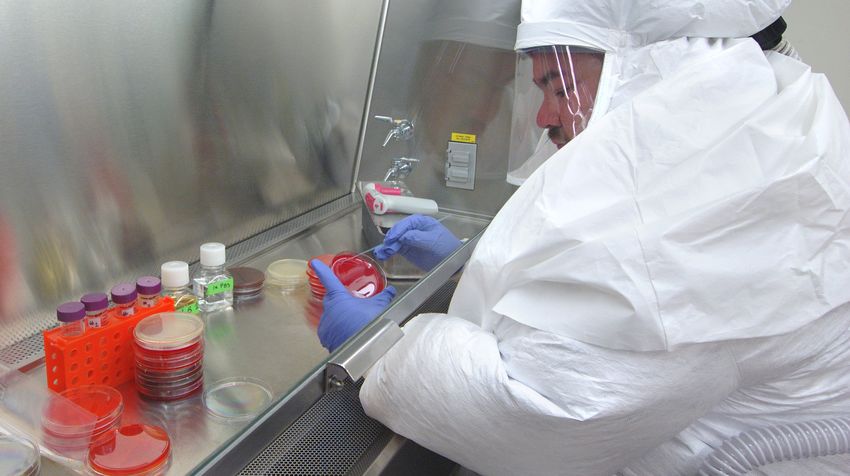
Page 2 Order online at www.atcc.org, call 800.638.6597, 703.365.2700, or contact your local distributor.
Table of Contents
Getting Started with an ATCC Protist Strain..... 4 Biosafety and Disposal........................................ 22
   Product Sheet................................................................. 4       Biosafety........................................................................ 22
   Preparation of Medium.................................................. 4              Disposal of Infectious Materials.................................. 22
   Opening Glass Ampoules.............................................. 4
   Initiating Frozen Cultures............................................... 6         Protist Authentication........................................ 23
   Initiating Freeze-Dried Cultures................................... 6                  Phenotypic Characterization....................................... 23
   Handling Live Protist Cultures Upon Arrival............... 6                           Genotypic Characterization........................................ 23

Protist Growth and Propagation...........................7 Protist Applications.............................................24
   Properties of Protists......................................................7          Bioassays........................................................................ 24
   Protist Metabolism......................................................... 8          Biofuel Production........................................................ 24
   Protist Propagation......................................................... 9         Fatty Acid Production.................................................. 24
   Propagation of Specific Protist Species...................... 12                       Diagnostics.................................................................... 24
   Cell Counts....................................................................16      Consumable Products.................................................. 24

Growth Media....................................................... 17 Glossary................................................................ 25
   Protist Culture Media.................................................... 17
                                                                                       References........................................................... 26
   Media Formulations ...................................................... 17
   Media Ingredients.......................................................... 17

Preservation..........................................................18
   Cryopreservation...........................................................18
   Special Hazards..............................................................21

                                   Order online at www.atcc.org, call 800.638.6597, 703.365.2700, or contact your local distributor.                                 Page 3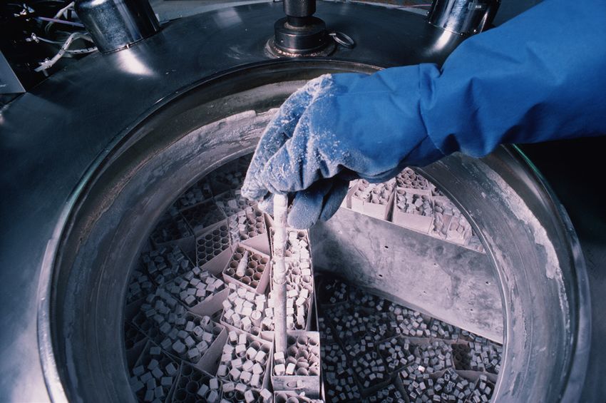
Getting Started with an ATCC Protist Strain
ATCC protist strains are shipped frozen on dry ice in plastic cryopreservation vials, as freeze-dried cultures in glass ampoules or serum
vials, or as live cultures in test tubes. Upon receipt of a frozen culture, immediately revive cells by thawing and transferring to an appro-
priate growth medium. If this is not possible, store frozen vials in liquid nitrogen vapor (below -130°C). Alternatively, the frozen material
can be stored between -70°C and -80°C for short periods (1 to 5 days); however, viability will decline at temperatures above -130°C. Upon
receipt of a freeze-dried culture, rehydrate cultures using the appropriate medium and incubation conditions specified on the product
sheet. Freeze-dried cultures can be stored safely at 4°C or colder. Live cultures should be immediately handled according to the recom-
mended protocols for that specific organism. Under no circumstances should live cultures be stored at refrigerated or frozen temperatures
as this may result in the death of the culture.
Product Sheet
ATCC protist strains typically have detailed information on the processing and expansion of materials, recommended media, as well as
recommended growth and propagation conditions. The complete information for a product and the product sheet can be found on the
product detail page for that specific material. Please note that ATCC will only warrant cultures that are handled according to the instruc-
tions provided on the ATCC product sheet. Product sheets, as well as additional information, can be found on the ATCC website or can be
requested from the ATCC Technical Service Department.
Preparation of Medium
In advance, prepare the appropriate medium and additional supplements necessary for algal or protozoan revival and growth. Additionally,
ensure that your incubator is set to maintain the optimal growth conditions of the strain and that any required light is available. Information
for the formulation and preparation of media is available on the ATCC website.
Opening Glass Ampoules
Overview
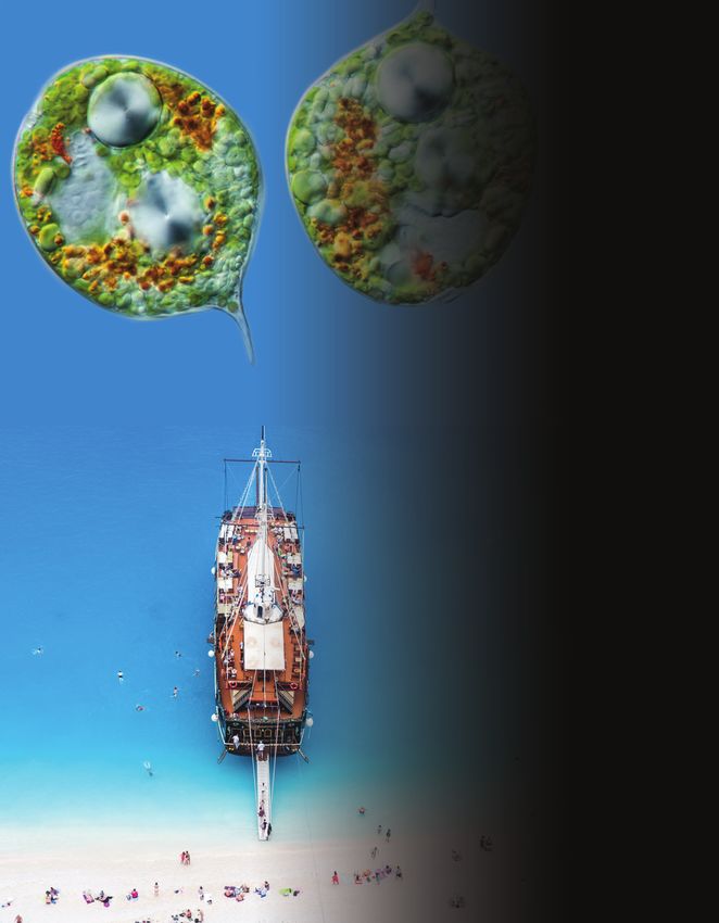
All cultures should be considered potentially hazardous and should be opened by individuals trained in microbiological techniques work-
ing in facilities with containment requirements appropriate for the biosafety level of the cultures. ATCC recommends that the handling
or opening of glass ampoules be performed in a biological safety cabinet. If this is not possible, wear protective clothing, gloves, a face
shield or safety goggles, and hold the vial away from your body. Ensure that all empty vials are sterilized before disposal.
A Opening Double-Vial Preparations
1 Heat the tip of the vial in a flame.
2 Add a few drops of water to the hot tip to crack the glass.
3 Strike the end of the vial with a file or pencil to remove the tip.
4 Remove the insulation and inner vial with sterile forceps. Gently raise the cotton plug.
B Opening Single-Vial Preparations
1 Recover the culture suspension from the glass ampoule by scoring the neck of the ampoule with a small, sterile file.
2 Disinfect the outside of the ampoule with freshly prepared 70% ethanol or dip it into a beaker of freshly prepared 70% ethanol.
3 Wrap the ampoule within several folds of a sterile towel or gauze to dry residual ethanol.
4 Hold the vial upright and snap the vial open within a laminar flow hood. Ensure that the gauze does not become too wet with
ethanol, or alcohol could be sucked into the culture when the vacuum is broken.
Page 4 Order online at www.atcc.org, call 800.638.6597, 703.365.2700, or contact your local distributor.
Tip
                    Insulator
                    Cotton plug
                    Outer vial (soft glass)                                                 Borosilicate glass
                    Inner vial
                    Freeze-dried pellet                                                     Freeze-dried cells
                    Cotton
                    Desiccant with indicator

                                                                      These preparations may be enclosed in a thin skin of
Heat the tip of the outer                                             cellulose; this skin must be removed (either with a sharp
vial in a flame                                                       blade or by soaking in water for a few minutes). Score the
                                                                      ampoule once briskly with a sharp file about one inch
                                                                      from the tip.

Squirt a few drops of water
on the hot tip to crack glass

                                                                      Disinfect the ampoule with alcohol-dampened gauze

Strike with file or
pencil to remove tip

                                                                      Wrap gauze around the ampoule, and break at the scored
                                                                      area. Care should be taken not to have the gauze too wet, or
                                                                      alcohol could be sucked into the culture when the vacuum is
                                                                      broken. Rehydrate material at once.

Remove insulation and inner vial with
forceps, gently raise cotton plug

                        Order online at www.atcc.org, call 800.638.6597, 703.365.2700, or contact your local distributor.          Page 5
Initiating Frozen Cultures
1 Prepare a sterile culture tube or flask that contains the recommended growth medium described in the product sheet. Ensure that
  the medium contains all necessary supplements or components and is equilibrated for temperature and pH.
2 Thaw the sample vial in a water bath that is set to 35°C. Thawing will be rapid; approximately 2 minutes or until all ice crystals have
  melted. Protists should not be thawed at room temperature or in the refrigerator.
3 Remove the vial from the water bath and decontaminate the outer surface using freshly prepared 70% ethanol. Follow strict aseptic
  conditions in a laminar flow hood for all further manipulations.
4 Unscrew the top of the vial and transfer the entire contents to a culture tube or flask containing the appropriate growth medium.
5 Incubate cultures under the appropriate temperature and atmospheric conditions as recommended on the product sheet.
6 Monitor the cultures by microscopic examination following
  initiation. Once growth has stabilized, cultures can be main-         NOTE 1
  tained as described on the product sheet. Please note that the        Following recovery from a freeze-dried or frozen state, some
  incubation period will vary between strains (See: NOTE 1).            protist strains may require an extended incubation period.
Initiating Freeze-Dried Cultures
1 Rehydrate freeze-dried strains by aseptically adding 0.5 mL of the recommended medium to the vial. For sensitive strains, rehydrate
  freeze-dried material using medium containing 12% sucrose.
2 Aseptically transfer the entire contents of the vial to a sterile culture tube or flask containing the appropriate growth medium.
3 Incubate the culture under the appropriate temperature and atmospheric conditions as recommended on the product sheet.
4 Examine cultures microscopically following initiation. Once growth has stabilized, cultures can be maintained as described on the
  product sheet. Please note that the incubation period will vary between strains (See: NOTE 1).
Handling Live Protist Cultures Upon Arrival
Immediately place the culture under the recommended atmospheric conditions and temperature and allow culture to equilibrate for 1-3
hours or as recommended in the product sheet for the specific organism.
Following equilibration, examine the culture to confirm its viability and healthy morphology, then handle as directed in the product sheet
for the specific item.
Examine cultures again after the recommended incubation period
                                                                         NOTE 2
(See: NOTE 2). The incubation period will vary between strains and is
listed on the product sheet. Follow the procedure for maintenance        Some live cultures need to be incubated for a few hours upon
of the culture as directed in the product sheet for the specific item.   receipt, then passaged as necessary. This will vary by taxon.

Page 6     Order online at www.atcc.org, call 800.638.6597, 703.365.2700, or contact your local distributor.
Protist Growth and Propagation
Properties of Protists
Protists are a diverse group of eukaryotic organisms that includes the protozoa and algae. These organisms are of clinical relevance as
many protozoan species are human pathogens, causing diseases such as malaria, giardiasis, and toxoplasmosis. Additionally, protists are
of ecological importance as they are the predominant primary producers in most aquatic and soil environments and many species func-
tion as bacterial consumers.
In 2005 the taxonomic classification of protists was revised to recognize six clusters of eukaryotes representing the basic groupings
similar to the traditional “kingdoms”. This taxonomic revision incorporated results obtained from ultrastructural studies and molecular
phylogenetic studies to characterize protists into “supergroups”. The six supergroups were named Rhizaria, Opisthokonta, Archaeplastidia,
Amoebozoa, Excavata, and Chromalveolata¹ (Table 1). A more recent revision of eukaryote classification published in 2012 retained most
of the super-groups proposed in 2005, although some have been assembled into still higher order groupings.²

Table 1: Protist Supergroups*
 Supergroup                          Characteristics                         Examples**                    Image

                                                                             Tubulinea
                                                                             Flabellinea
                                     Amoeboid locomotion using blunt,
                                                                             Stereomyxida
                                     oblong pseudopodia. Most species
 Amoebozoa                                                                   Acanthamoebidae
                                     are unicellular and are common to
                                                                             Entamoebida
                                     aquatic and soil habitats.¹
                                                                             Mastigamoebidae
                                                                             Eumycetozoa
                                                                                                           ATCC® 50924™ Vannella cirrifera

                                     Red and green algae, containing         Glaucophyta
 Archaeplastida                      photosynthetic plastid with chlo-       Rhodophyceae
                                     rophyll and pigments.¹                  Chloroplastida
                                                                                                           ATCC® 30929™ Dunaliella tertiolecta

                                     Species contain a plastid obtained      Cryptophyceae
 Chromalveolata                      by secondary endosymbiosis with         Haptophyta
                                     an ancestral archaeplastid.¹            Stramenopiles

                                                                                                           ATCC® 205082™ Tetrahymena elliotii

                                                                             Fornicata
                                     Unicellular eukaryotes that include     Parabasalia
                                     symbiotic, parasitic, and free-living   Preaxostyla
 Excavata
                                     forms. Many excavates lack classi-      Jakobida
                                     cal mitochondria.¹,³                    Heterolobosea
                                                                             Euglenozoa
                                                                                                           ATCC® 50286™ Trepomonas agilis

                                                                             Cercozoa
                                     Amoeboid in form with filose,
                                                                             Haplosporidia
 Rhizaria                            rediculose, or microtubule-sup-
                                                                             Foraminifera
                                     ported pseudopodia.¹,⁴
                                                                             Radiolaria
                                                                                                           ATCC® 50344™ Cercomonas longicauda

                                     Heterotrophic organisms having a         Mesomycetoza
 Opisthokonta                        single posterior cilium that is pres- Choanomonada
                                     ent in at least one life stage cycle.¹,⁵ Metazoa

                                                                                                           ATCC® 50964™ Monosiga gracilis

* Classification is based on the 2005 revision of the classification of unicellular eukaryotes by the International Society of Protistologists.¹
**Examples do not include a complete accounting of the full taxonomic diversity of the ATCC Protistology Collection but rather the most
 common protist groups housed by ATCC.

                               Order online at www.atcc.org, call 800.638.6597, 703.365.2700, or contact your local distributor.                 Page 7
In addition to characterization by taxonomic classification, at ATCC we find it helpful to categorize groups of protists based on how they
are handled (Table 2). This system of categorization allows us to generalize protocols and techniques for organisms with similar needs
based on the culture system used (i.e., whether the culture is xenic or axenic, whether liquid or solid media are used, the type of atmo-
sphere required, etc.).

Table 2: Types of Protist Cultures
 Type of Culture       Recommended Handling                               Examples*
 Algae                 Axenic culture grown in Erlenmeyer flasks or       Chlorella, Chlamydomonas, Dunaliella, Haematococcus, Chlorogonium, Scenedesmus,
                       test tubes. Some strains can be cultured on        Euglena
                       plates or slants.
 Free-living           Aerobic xenic culture, exclusively grown on        Xenic strains of Acanthamoeba, Naegleria, Hartmannella, Willaertia
 Amoebae               plates.
                       Aerobic xenic cultures grown both in flasks        Xenic strains of Vannella, Flabellula, Korotnevella, Paramoeba, Neoparamoeba,
                       and on plates.                                     Platyamoeba, Vexillifera
                       Monoxenic or axenic, bacteria-free culture,        Monoxenic or axenic, bacteria-free strains of Acanthamoeba, Naegleria,
                       exclusively grown in flasks. Few species are       Hartmannella, Willaertia, Vahlkampfia, Balamuthia
                       propagated in cell culture within flasks at a
                       5% CO₂ atmosphere.
 Ciliates              Xenic or axenic culture. Strains can be grown      Colpoda, Anophryoides, Chilodonella, Homalogastra, Pseudocohnilembus,
                       in both flasks and in tubes with loose caps.       Platyophrya, Opisthonecta
 Dinoflagellates       Axenic culture grown in flasks or tubes.           Axenic strains of Crypthecodinium, Amphidinium
 Free-living           Aerobic xenic culture grown in flasks.             Xenic strains of Cercomonas, Ancyromonas, Bodo
 Flagellates
                       Aerobic axenic culture grown in flasks.            Axenic strains of Diplonema, Rhynchopus
                       Microaerophilic, xenic culture grown in            Xenic strains of Hexamita, Mastigamoeba, Mastigella, Trepomonas
                       angled tubes.
 Non-motile            Non-motile parasites in xenic or axenic            Blastocystis strains only
 Anaerobic             culture, grown anaerobically in tubes.
 Parasites
 Non-Motile            Non-motile, non-photosynthetic parasites           Perkinsus, various thraustochytrids, Helicosporidium
 Parasites/            or saprophytes in axenic culture. Strains are
 Saprophytes           grown in flasks.
 Obligate              Parasites grown in cell culture free from          In vitro strains of Toxoplasma, Neospora, Encephalitozoon
 Intracellular         bacteria. Cultures are grown in flasks in a 5%
 Parasites             CO₂ atmosphere.
                       Parasites grown in vivo.                           Babesia microti, B. duncani, in vivo strains of Trypanosoma brucei, T. cruzi,
                                                                          Leishmania sp.
 Parasitic             Microaerophilic, parasitic strains in xenic or     Entamoeba, Dientamoeba
 Amoebae               axenic culture. Strains are grown in angled
                       tubes.
 Parasitic             Parasitic flagellates in monoxenic or axenic,    Leishmania, Trypanosoma, Crithidia, Leptomonas, Herpetomonas, Phytomonas
 Flagellates           bacteria-free culture. Protists are grown
                       upright in tubes or in some cases within flasks.
                       A small number of strains are grown in cell
                       culture within flasks at a 5% CO₂ atmosphere.
                       Microaerophilic, parasitic flagellates in axenic   Trichomonas, Tritrichomonas, Pentatrichomonas
                       culture, grown in angled tubes.

*Note that the examples included on this table are a list of the most common genera that fall into each category. The listed genera do not
 represent a complete accounting of the full taxonomic diversity of each type of culture. Further, the “protist culture type” is an internal
 ATCC categorization used for the purpose of generalizing culture protocols and handling techniques for similar organisms.

Protist Metabolism
In general, protists have three methods of nutrient acquisition: autotrophy, heterotrophy, and mixotrophy. Autotrophic organisms, such
as algae, require sunlight to obtain energy and organic material for carbon acquisition. These protist species use chlorophyll and various
other pigments to obtain energy through photosynthesis. In contrast, heterotrophic organisms require the consumption of nutrients
from other organisms for both energy and carbon. There are two forms of heterotrophy, phagotrophy, which is the engulfment of
matter, and osmotrophy, which is the absorption of dissolved nutrients through pinocytosis. For example, free-living amoebae, such as
Naegleria, directly consume bacteria through phagocytosis; whereas fungal-like protists, known as slime molds, consume organic matter
by absorbing it from the environment. Lastly, mixotrophic organisms, such as most dinoflagellates, can use either, or both, autotrophic
and heterotrophic forms of nutrient acquisition.

Page 8         Order online at www.atcc.org, call 800.638.6597, 703.365.2700, or contact your local distributor.
Protist Propagation
The following section includes general guidelines for the culture of
algae, bacteriovorus protists, and obligate intracellular parasites.
Algae
Algae are a very large and diverse group of autotrophic protists that
range in size from unicellular microalgae to large multicellular organ-
isms. To survive, algal species use chlorophyll and other pigments
to manufacture their own food source through a process termed
photosynthesis. The types of pigments used for photosynthesis
differ among the algal divisions, resulting in colorful organisms that
can appear green, red, brown, yellow, and golden-brown. In addi-
tion to variations in pigmentation, algal species also differ in the
type of carbohydrate compounds stored for energy and the type
of flagella used for motility. These differences can be used to help
classify algal species.
In the laboratory, algae are commonly grown as axenic cultures in Image of Prototheca filamenta alga courtesy of Dr. W. Kaplan, M.S.
either liquid media or on agar media. Strains are commonly propa- Sudman, CDC
gated in aerobic/ventilated culture vessels under a 14 hour light/10
hour dark cycle to promote photosynthesis. ATCC recommends the following procedure for the in vitro cultivation of algae:
Propagation
1 Rehydrate freeze-dried preparations by aseptically adding 0.5 mL of ice-cold medium to the freeze-dried inner shell vial. For frozen
   preparations, thaw the contents of the ampoule by placing the vial in a 35°C water bath until thawed (approximately 2 minutes).
   Immerse the ampoule just sufficiently to cover the material. Do not agitate the ampoule.
2 Aseptically transfer the entire contents to a 16 x 125 mm screw-capped test tube containing 5 mL of the recommended medium.
   Alternatively, add the entire contents to the surface of a 20 x 100 mm Petri plate containing 20 mL of the recommended medium.
   Some algae, and particularly freeze-dried preparations, do not recover well in broth media. For these cultures, agar media is
   recommended.
3 Incubate broth cultures horizontally at a 15° slant with the
   cap screwed on loosely at 25°C under a 14 hour light /10 hour        NOTE 3
   dark cycle. For agar-based cultures, wrap the plate culture in       Some green algae may lose their color shortly after recovery
   Parafilm and incubate upright.                                       from the frozen state. In most cases, the natural coloration will
4 Subculture as necessary. This may be every 14-21 days in broth        return after a period of time with normal incubation.
   media (See: NOTE 3).
Bacterivorous Protists
Xenic, bacterized protist cultures require the presence of bacte-
ria as a food source. Typically, the bacterial strain will survive the
preservation process better than most protists, and will be present
in the culture when it is re-initiated from preserved material. The
concentration of the bacterial food source can affect the growth
of many protist species or may make it difficult to observe the
protist strain. For these cultures, control over bacterial density may
enhance protist propagation. The following procedures regarding
propagation and bacterial control can be applied to the majority of
free-living, aerotolerant, xenic protist cultures:
Propagation
1 Rehydrate freeze-dried preparations by aseptically adding 0.5
   mL of ice-cold medium to the freeze-dried inner shell vial. For
   frozen preparations, thaw the contents of the ampoule by
   placing the vial in a 35°C water bath until thawed (approxi-
   mately 2 minutes). Immerse the ampoule just sufficiently to          Image of Acanthamoeba polyphaga. Photo courtesy of Catherine
   cover the material. Do not agitate the ampoule.                      Armbruster and Margaret Williams.
2 Transfer the entire contents to 10 mL of the appropriate ATCC
   medium in a T-25 tissue culture flask. The bacterial flora present in the shipped culture will support growth, but growth may be
   improved by using medium that has been bacterized with Enterobacter aerogenes approximately 24 hours before inoculation with the
   protist. (See: NOTE 4, Table 3)
3 Subculture every 14-21 days. To subculture, aseptically scrape
   the bottom of the flask and agitate the flask to distribute the
                                                                          NOTE 4
   cells evenly. Aseptically transfer 0.25 to 0.5 mL to a T-25 flask      All three bacterial strains listed in Table 3 can be used almost
   containing 9 mL of bacterized medium.                                  interchangeably with the vast majority of free-living, bacterio-
4 Incubate the flask at 25°C with the cap screwed on tightly.             vorus protist strains

                              Order online at www.atcc.org, call 800.638.6597, 703.365.2700, or contact your local distributor.    Page 9
Table 3: Bacterial Strains Used as a Food-Source for Free-Living Bacteriovorus Protists
ATCC® Number Species
27889™ Klebsiella pneumoniae subsp. pneumoniae
13048™ Enterobacter aerogenes
700831™ Klebsiella pneumoniae subsp. pneumoniae
For some xenic protist cultures, better growth may be obtained if bacterial density is reduced to some degree. This phenomenon is
not uncommon among aerobic, free-living flagellate, ciliate, and amoeba cultures, particularly where mixed bacteria flora are present.
Microaerophilic protists in xenic culture may also exhibit reduced or inhibited growth if bacterial density becomes excessive, primarily
as a result of mixed bacterial flora native to the culture. Some degree of control over bacterial density in these cultures can be exerted
through the addition of antibiotics or by a reduction in the quantity or strength of whichever component in the growth medium is promot-
ing excessive bacterial growth. Any alteration to growth media should be tested empirically to determine the optimal culture conditions.
Controlling bacterial density in xenic cultures
Dilute Cerophyl® media to reduce bacterial density by mixing
NOTE 5
it 1:1 or 1:2 with a suitable buffer medium, such as ATCC media
formulations 2348 or 1323 for freshwater cultures or ATCC ATCC medium formulations can be found on the ATCC website
medium formulation 1405 for marine cultures (See: NOTE 5). at www.atcc.org.
Media containing rice starch can sometimes promote exces-
sively dense bacterial growth. Manual reduction in the amount of rice starch in the medium prior to inoculation with bacteria can
help modulate subsequent bacterial growth. Rice starch may be removed from media by light centrifugation.
Further control of bacterial growth may require use of antibiotics such as penicillin/streptomycin; however, the appropriate concen-
trations should be determined empirically.
Free-living amoebae such as Naegleria, Hartmannella, or Acanthamoeba in xenic/bacterized culture on agar media covered with dense bacte-
rial growth can be challenging to observe or cultivate successfully. Especially for the inexperienced, it can be quite difficult to discern
amoebae amidst the dense growth of mixed bacterial flora. To achieve optimal culture conditions, it can be helpful to either reduce the
overall bacterial density or to separate amoebae from the dense bacteria around them.
Reducing bacterial density on agar media
An amoeba culture containing mixed bacterial flora may be washed with a suitable buffer solution to remove most of the bacteria, poten-
tially enabling remaining amoebae to grow to greater numbers.
1 Flood a 20 x 100 mm agar petri plate containing the amoeba culture with 4 to 5 mL of a balanced saline solution. For freshwater
strains, use Page's amoeba saline (ATCC medium formulation 1323) or PBS; for marine strains, use an artificial seawater wash.
2 Gently rub the plate surface with a spread bar to carefully dislodge adherent amoebae, and then transfer suspended amoebae and
bacteria to a 15 mL plastic centrifuge tube.
3 Centrifuge the culture at approximately 400 to 500 x g for 5 to 6 minutes.
4 Remove all but 0.5 to 1.0 mL of the supernatant and then resuspend pelleted amoebae. Make a wet mount slide to confirm presence
of amoebae or cysts. Transfer the resuspended culture material onto several fresh, uninoculated agar plates and spread over the
plate surface using a spread bar.
5 Allow all plates to air dry, then seal with parafilm and incubate as recommended. Observe cultures daily for presence of newly-emer-
gent amoebae; these may be further isolated from problematic bacterial growth as described below.
Separating amoebae from undesirable bacteria on agar media
A fresh lawn of a single, preferred bacterial species may be less dense than the mixed bacterial flora on the parent culture plate, offering
a better opportunity for amoebae to proliferate in a more visible way.
1 Prepare a lawn of either E. aerogenes or K. pneumoniae bacte-
ria on several fresh agar plates, and allow the plates to dry NOTE 6
completely. Instead of using an inoculating loop to transfer cells, a small
2 Transfer the amoeba culture with an inoculation loop to a loca- block of agar may be cut from the parent culture plate using a
tion on one of the freshly-prepared agar plates containing the sterilized fine-point scalpel or Pasteur pipette tip. The agar block
lawn of a single bacterial species. Attempt to transfer from a is then physically transferred to one of the freshly-prepared
region on the source plate that contains amoeba tophozoites subculture plates and inverted onto the surface of the bacterial
or cysts if possible; otherwise, if amoebae or cysts are not lawn. Gently slide the agar block along the lawn a short distance
apparent, blind transfers can be made to multiple subculture to transfer amoebae from the block to the new bacterial lawn.
plates. (See: NOTE 6).
3 Amoebae that successfully transfer to a new agar plate may
proliferate more readily and subsequently migrate to a region
of the plate containing only the preferred bacterial species. A second transfer of amoebae from this new region will result in a
monoxenic culture of amoebae and a single bacterial food source when inoculated onto a fresh agar plate pre-inoculated with a
lawn of the preferred bacterium.
Page 10 Order online at www.atcc.org, call 800.638.6597, 703.365.2700, or contact your local distributor.Intracellular Parasites
Several protist species, such as Toxoplasma gondii and the fungus
Encephalitozoon cuniculi, are considered obligate intracellular NOTE 7
parasites. These organisms require a living host cell for in vitro All procedures pertaining to the manipulation of cells and
propagation. Below are general ATCC procedures that describe how dispensing of growth media into flasks should be performed in
to prepare a host cell line and propagate an intracellular parasite a biological safety cabinet.
culture (See: NOTE 7, Table 4).
Table 4: Cell Lines Used in the In Vitro Propagation of Intracellular Parasites
ATCC® Number Description Application
CCL-34™ MDCK Encephalitozoon sp., Vittaforma corneae
CCL-37™ RK13 Encephalitozoon sp.
CCL-75™ Human lung fibroblasts Encephalitozoon sp.
CCL-81™ Vero Toxoplasma gondii, Encephalitozoon sp., Trypanosoma cruzi
CCL-163™ BALB/3T3 fibroblasts Trypanosoma cruzi
CRL-1634™ Human foreskin fibroblasts Toxoplasma gondii
In vitro propagation
Preparation of the Host Cell Line
1 Thaw a vial of the host cell culture by placing the ampoule in a water bath set at 35°C (approximately 2 minutes). Immerse the vial
just sufficiently to cover the frozen material. Do not agitate the vial.
2 Sterilize the outer surface of the vial using freshly prepared 70% ethanol.
3 Aseptically transfer the entire thawed contents of the ampoule to a T-25 tissue culture flask containing 10.0 mL of the recom-
mended medium. Be sure that your medium is properly buffered with sodium bicarbonate to ensure proper cellular growth in a 5%
CO₂ incubator.
4 If flasks with plug seal caps are used, outgas each flask for 30-45 seconds with a 95% air, 5% CO₂ gas mixture. Tightly cap the flask.
5 Incubate the flask horizontally in a in a 35-37°C, 5% CO₂ incubator.
6 Observe the culture regularly using an inverted microscope to determine the health and confluency of the cells. When the cells form
a monolayer completely covering the bottom surface of the flask, the culture may be passaged or expanded.
Transferring the Host Cell Line
1 When the cell line reaches the desired density, remove the medium and replace it with an appropriate volume of Phosphate
Buffered Saline (PBS) (ATCC® 30-2200™) to cover the cell layer. Incubate the flask at 35°C to 37°C for 3-5 minutes (or up to 30
minutes for cell lines that do not detach easily) to remove serum and/or other potential trypsin inhibitors present.
2 Remove all the PBS and replace it with a small volume of 1X
Trypsin-EDTA solution (ATCC® 30-2101™) sufficient to leave NOTE 8
a thin film of the solution over the entire bottom surface of
the flask. For a T-25 flask, 1-2 mL of 1X Trypsin-EDTA solution Trypsin-EDTA solution must be fresh for proper detachment.
should be sufficient. (See: NOTE 8). Some cell lines will not detach easily or completely, in which
case the addition of other enzymes may be necessary.
3 Incubate the flask horizontally at 35°C to 37°C until the
desired detachment results are obtained. Cells may be
observed using an inverted microscope to confirm they have become rounded and detached.
4 Neutralize the Trypsin-EDTA solution by dispensing an appropriate volume of the growth medium with FBS. For medium with 10%
FBS, add at least 5 mL for every 1 mL of trypsin used. For medium with 5% FBS, add at least 10 mL for every 1 mL of trypsin used.
5 Resuspend the cells by aspirating and expelling the fluid gently with a pipette and transfer an appropriate volume to each of the
desired number of destination flasks.
6 Aseptically dispense suitable fresh growth medium with the defined concentrations of supplements to each destination flask at an
appropriate volume. If flasks with plug seal caps are used, outgas each destination flask for 30-45 seconds with a 95% air, 5% CO₂ gas
mixture and tightly cap the flask.
7 Incubate the flask horizontally at 35-37°C in a 5% CO₂ incubator.
Propagation
1 Thaw a frozen ampoule containing the protist strain by placing it in a 35°C water bath such that the lip of the ampoule remains
above the water line. Thawing time is approximately 2 minutes. Do not agitate the ampoule. Do not leave ampoule in water bath
after it is thawed. Further, protist cultures should not be thawed at room temperature or in the refrigerator.
2 Aseptically transfer the entire thawed contents of the ampoule to a T-25 tissue culture flask containing a fresh monolayer of the
propagation host cells and 10 mL of the recommended medium.
3 Incubate in a 35-37°C, 5% CO₂ incubator. If a flask with a plug seal cap is used, outgas the flask for 10 seconds with a 95% air, 5% CO₂
gas mixture. Tightly cap the flask.
4 Observe the culture daily under an inverted microscope for the presence of intracellular parasites inside the cells.
Order online at www.atcc.org, call 800.638.6597, 703.365.2700, or contact your local distributor. Page 115 To maintain the culture, remove the medium from a fresh confluent monolayer of the host cell line in a T-25 tissue culture flask and
replace it with 10 mL of the recommended medium.
6 Transfer the protist culture by removing the old medium from the flask containing the organism and centrifuge at 1300 x g for 10
min.
7 Remove all but 0.5 mL of the supernatant and resuspend the cell pellet. Transfer the resuspended pellet to the fresh flask contain-
ing the host cell line. Outgas the flask for 10 seconds with a 95% air, 5% CO₂ gas mixture if using a plug seal cap.
8 Incubate at 35-37°C in a 5% CO₂ incubator with the cap screwed on tightly.
Parasites requiring In vivo Propagation
For the propagation of parasitic protists that lack an in vitro culture system such as certain species of Babesia, in vivo cultivation using
an appropriate host is required. The required host species and inoculation route can vary significantly depending on the species. Below
is a general ATCC procedure for in vivo cultivation of parasites. For specific details regarding the recommended preparation of a protist
strain and the method of inoculation, please review the product sheet for that organism.
Propagation
1 Prior to thawing the frozen ampoule, prepare the host animal under the recommended conditions.
2 Once the host animal is prepared, thaw the frozen ampoule in a 35°C water bath such that the lip of the ampoule remains above
the water line. Thawing time is approximately 2 minutes. Do not agitate the ampoule. Do not leave ampoule in water bath after it is
thawed. Further, protist cultures should not be thawed at room temperature or in the refrigerator.
3 Immediately after thawing, aseptically remove the contents of the ampoule with a syringe and inoculate an uninfected, animal host.
Follow the recommended protocol for maintenance in vivo described on the product sheet; this procedure will vary significantly
between protist species. Note that the course of infection may also vary depending on the virulence of the parasite strain and its
recovery from the frozen state.
4 Monitor parasitemia and passage as needed.
Propagation of Specific Protist Species
The following section includes general guidelines for the culture of some of the most commonly ordered protist species from ATCC.
Additional information regarding the recommended media used in the cultivation of these species is found on the ATCC website as well
as on the product sheet.
Trichomonas vaginalis
T. vaginalis is a single-celled, flagellated protozoan known to cause
the human sexually transmitted disease, trichomoniasis. This protist
is oval in shape with four posterior flagella and a fifth flagellum
wrapped around the surface of the organism. A conserved feature
of T. vaginalis is the production of adherence factors that allow for
epithelial attachment and colonization. ATCC recommends the
following procedure for the propagation of T. vaginalis:
1 Thaw a frozen ampoule by placing it in a 35°C water bath until
thawed (approximately 2 minutes). Immerse the ampoule just
sufficiently to cover the frozen material. Do not agitate the
ampoule.
2 Immediately after thawing, aseptically transfer the entire
Image of Trichomonas vaginalis
contents to a screw-capped test tube containing either 9.0
mL of completed TYM media adjusted to pH 6.0 or 13.0 mL of
completed LYI media adjusted to pH 6.0. Incubate the tube at 35°C in the upright position in the case of TYM media, or horizontally
at a 15° slant in the case of LYI media.
3 When the culture reaches peak density, place the tube on ice for 10 minutes.
4 Gently invert the culture tube 10 times and aseptically transfer a 0.1-0.4 mL aliquot to a screw-capped test tube containing an
appropriate volume of the recommended medium as described in step 2.
5 Screw the cap on tightly and incubate the culture at 35ºC.
6 Transfer the culture every 3-4 days as described in steps 3-5. The transfer interval will depend on the quantity of the inoculum and
the quality of the medium. This should be empirically determined by examining the culture on a daily basis, until the growth cycle
has stabilized. Do not allow the culture to overgrow as protist death occurs soon after reaching peak density.
Page 12 Order online at www.atcc.org, call 800.638.6597, 703.365.2700, or contact your local distributor.Acanthamoeba castellanii
A. castellanii is a freshwater amoeba known to cause keratitis and
encephalitis in humans. Cells are small, can form cysts, and are oval
to triangular in shape. In addition to their role as a human patho-
gen, A. castellanii is also a host to various bacterial strains, including
Legionella pneumophila. ATCC recommends the following procedure
for the propagation of axenic strains of A. castellanii:
1 To thaw a frozen ampoule, place it in a 35°C water bath such
that the lip of the ampoule remains above the water line.
Thawing time is approximately 2 minutes. Do not agitate the
ampoule. Do not leave ampoule in water bath once thawed.
Protists should not be thawed at room temperature or in the
refrigerator.
2 Immediately after thawing, aseptically transfer the entire
contents to either a 16 x 125 mm plastic test tube or a T-25
tissue culture flask containing the recommended medium (5.0
mL in a test tube, 10.0 mL in a flask). Image of Acanthamoeba sp. courtesy of Janice Haney Carr , CDC
3 Screw the cap on tightly and incubate the tube or flask at 25°C.
4 When the culture reaches peak density, vigorously agitate the culture.
5 Transfer approximately 0.25 mL to a fresh tube or flask of the recommended medium, freshly prepared (5.0 mL in a test tube, 10.0
mL in a flask).
6 Screw the cap on tightly and incubate at 25°C (incubate test tubes horizontally at a 15° slant).
7 The amoebae will form an almost continuous sheet of cells on the bottom surface of the flask or test tube. Repeat steps 4-6 at
10-14 days intervals.
For propagation of xenic/bacterized A. castellanii strains, ATCC recommends the following:
1 To thaw a frozen ampoule, place it in a 35°C water bath such that the lip of the ampoule remains above the water line. Thawing time
is approximately 2 minutes. Do not agitate the ampoule. Do not leave ampoule in water bath once thawed. Protists should not be
thawed at room temperature or in the refrigerator.
2 Immediately after thawing, aseptically transfer the entire contents to an agar plate of the recommended medium. Distribute the
material evenly over the plate using a spread bar and allow to dry.
3 Wrap the entire edge of the plate with parafilm and incubate upright at 25°C. Amoeba trophozoites should be evident within 2-5
days.
4 When the amoeba culture reaches peak density, transfer amoebae or cysts to a fresh plate of the recommended medium with an
inoculation loop. Agar plates pre-inoculated with a lawn of food-source bacteria will promote faster growth of amoebae.
5 Seal the agar plate with parafilm and incubate upright at 25°C.
6 The amoebae will form an almost continuous sheet of cells on the surface of the agar. Repeat steps 4-5 at 7-10 day intervals.
Giardia intestinalis
G. intestinalis is a flagellated protozoan that is known to cause giardiasis in humans and other mammals. These organisms enter the host
intestine through the consumption of soil, food, or water that has been contaminated with the feces of an infected individual. During
infection, G. intestinalis attaches to the intestinal epithelium and reproduces by binary fission. ATCC recommends the following proce-
dure for the propagation of G. intestinalis:
1 To thaw a frozen ampoule, place it in a 35°C water bath until thawed (approximately 2 minutes). Immerse the ampoule just suffi-
ciently to cover the frozen material. Do not agitate the ampoule. Protists should not be thawed at room temperature or in the
refrigerator.
2 Immediately after thawing, aseptically transfer the entire contents to a screw-capped test tube containing 13 mL of medium.
Commonly use media include Keister's Modified TYI-S-33 (ATCC medium 2695) and LYI Giardia medium (ATCC medium 2155). Incubate
the tube on a 15º horizontal slant at 35ºC.
3 When the culture has reached or is near peak density, place the tubes on ice for 10 minutes.
4 Gently invert the culture tube 10 times and aseptically transfer a 0.1-0.4 mL aliquot to a screw-capped test tube containing 13 mL
of the recommended medium.
5 Incubate the culture horizontally at a 15° slant at 35ºC with the cap on tightly.
6 Transfer the culture every 3-4 days as described in steps 3-5. The transfer interval will depend on the size of the inoculum and the
quality of the medium. This should be empirically determined by examining the culture on a daily basis until the growth cycle has
stabilized. Do not allow the culture to overgrow. The culture crashes soon after reaching peak density.
Order online at www.atcc.org, call 800.638.6597, 703.365.2700, or contact your local distributor. Page 13Trypanosoma cruzi T. cruzi is a vector-borne parasitic euglenoid trypanosome that causes Chagas disease in humans. Within the human body, T. cruzi can be found in two forms: The trypomastigote, which is the infec- tious stage in the human bloodstream, and the amastigote, which is the reproductive stage that occurs within host cells. The epimas- togote stage is found naturally in the insect vector and is the life stage most commonly cultured in vitro. ATCC recommends the following procedure for the propagation of T. cruzi strains adapted to in vitro cultivation in LIT medium: 1 To thaw a frozen ampoule, place it in a 35°C water bath until thawed (approximately 2 minutes). Immerse the ampoule just sufficiently to cover the frozen material. Do not agitate the ampoule. Protist cultures should not be thawed at room temperature or in the refrigerator. Image of Trypanosoma cruzi courtesy of Dr. A.J. Sulzer, CDC 2 Immediately after thawing, aseptically transfer the entire contents to a T-25 tissue culture flask containing 10 mL of medium. Incubate the tube at 25ºC. 3 When the culture is at or near peak density, agitate the flask by inverting several times. 4 Aseptically transfer a 0.1 mL aliquot to a T-25 tissue culture flask containing 10 mL of the recommended medium, freshly prepared. 5 Screw the cap on tightly and incubate the flask at 20°C to 25°C. 6 Subculture every 7-14 days. Entamoeba histolytica E. histolytica is a parasitic protozoan that is transmitted among humans and primates through the consumption of contaminated water, solids, and foods. Upon ingestion of E. histolytica cysts, amoeba trophozoites are extruded into the digestive tract, eventually causing amoebic dysentery or the formation of amoebic liver abscesses. ATCC recommends the following procedure for the propagation of axenic strains of E. histolytica that are distributed as frozen preparations: 1 To thaw a frozen ampoule, place in a 35°C water bath until thawed (approximately 2 minutes). Immerse the ampoule just sufficiently to cover the frozen material. Do not agitate the ampoule. Protist cultures should not be thawed at room temperature or in the refrigerator. 2 Immediately after thawing, aseptically transfer the entire contents to a glass screw-capped tube containing 13 mL of medium. Axenic strains are commonly cultured in LYI medium (ATCC medium 2154) while xenic strains are cultured in TYGM-9 medium(ATCC medium 2154, 2463, and 2692). Screw the cap on tightly and incubate horizontally at a 15° slant at 35°C. 3 When the culture is at or near peak density, ice the culture tube for 10 min. 4 Gently invert culture 20 times to dislodge adherent amoeba. 5 Aseptically transfer a 0.1 and 0.25 mL aliquot to freshly prepared (no older than 7-10 days) tubes of the recommended medium. 6 Screw caps on tightly and incubate horizontally at a 15° slant at 35°C. 7 Subculture as described in steps 3-6 when many trophozoites are observed (typically every 2-4 days). The transfer interval will depend on the quantity of the inoculum and the quality of the medium. This should be empirically determined by examining the culture on a daily basis until the growth cycle has stabilized. Do not allow the culture to overgrow. The culture crashes soon after reaching peak density. For propagation of xenic/bacterized E. histolytica strains distributed as growing cultures, ATCC recommends the following: 1 Upon receipt, immediately incubate culture horizontally at a 15° slant at 35°C for at least three hours before observing the NOTE 9 culture. Amoeba trophozoites should be evident attached to Growth of xenic/bacterized E. histolytica is often better if the the tube wall amidst rice starch and bacteria. growth medium is inoculated with bacteria at least 24 hrs before 2 Regardless of the state of the culture, remove 5.5 mL of fluid inoculating with amoebae. Also, growth can be improved by and centrifuge at 500 x g for 5 minutes. controlling bacterial density with addition of antibiotics to the 3 Inoculate 2 fresh tubes of the recommended medium with culture. In general, addition of penicillin G at 75 U/mL and strep- 0.25 mL of supernatant (preinoculated bacterized culture tomycin at 75 µg/mL to the medium may be necessary if the tubes allow for better growth) (See: NOTE 9). Xenic strains are bacterial density in the culture is excessive. Antibiotics, if used, commonly cultured in 8 mL TYGM-9 medium (ATCC medium should be added at least 24 hours after medium is inoculated 1171). with bacteria 4 Divide the remainder of the supernatant in two equal aliquots in 16 x 125 mm screw-capped tubes and bring the volumes up to 8 mL with fresh medium. 5 Ice the parent culture 5 minutes, invert 20 times and transfer 0.5 and 1.0 mL aliquots to the test tubes containing the equal volumes of supernatant. 6 Re-feed the parent culture by centrifuging it at 200 x g for 5 min, aspirate most of the supernatant (leaving approximately 1.0-1.5 mL), and resuspend the pellet with fresh growth medium up to 8 mL. 7 Incubate all cultures (including the tubes of bacterized growth medium) horizontally at a 15° slant at 35°C. Page 14 Order online at www.atcc.org, call 800.638.6597, 703.365.2700, or contact your local distributor.
8 Observe the culture daily and transfer when many trophozoites are observed (i.e., early stationary phase): Ice a test tube culture
  at or near peak density for 10 minutes, invert 20 times and aseptically transfer a 0.1 and 0.3 mL aliquot to a fresh tube of recom-
  mended medium.
9 Screw cap on tightly and incubate horizontally at a 15° slant at 35°C.
Babesia microti
B. microti is a blood parasite that causes the hemolytic disease, babesiosis. This organism is spread between hosts through the saliva of the
Ixodes scapularis tick. Upon infection, B. microti enters erythrocytes where it is able to develop and eventually reproduce. ATCC recom-
mends the following procedure for the in vivo cultivation of B. microti:
1 To thaw a frozen ampoule, place it in a 35°C water bath such that the lip of the ampoule remains above the water line. Thawing time
  is approximately 2 minutes. Do not agitate the ampoule. Do not leave ampoule in water bath after it is thawed. Further, protist
  cultures should not be thawed at room temperature or in the refrigerator.
2 Immediately after thawing, aseptically inoculate entire
  infected blood suspension intraperitoneally into a hamster             NOTE 10
  using a 1.0 mL syringe equipped with a 27 gauge 1/2 inch
  needle. Note that the course of infection may vary depend-             Hamsters may be primed for faster infection by treatment with
  ing on the virulence of the parasite strain and its recovery from      Cortisone (2mg/day/hamster) 1-3 days prior to inoculation
  the frozen state (See: NOTE 10).
3 Monitor the infection at 1-2 day intervals by examination of blood films stained with 5% Giemsa solution.
4 Count the number of infected red blood cells (RBC) versus the total number of red cells under oil immersion and determine the %
  parasitemia: % parasitemia = infected rbc / total rbc X 100. A minimum of 500 red blood cells should be counted. (Note that a red
  blood cell infected with multiple parasites is counted as a single infected cell.)
5 When the level of parasitemia is ≥ 10% the strain should be passaged. Normally this would occur 1-3 weeks post-inoculation, but
  the rate of infection may vary considerably. (Note that the level of parasitemia before the host will succumb will vary with the strain
  used. Monitoring on a daily basis will alert the researcher as to when the strain should be passaged.)
6 Passage the strain by removing blood from the infected hamster using an appropriate cardiac puncture procedure.
7 Inject 0.5 mL of the infected blood suspension into each uninfected hamster.
8 Monitor parasitemia and passage as needed.
Toxoplasma gondii
T. gondii is an obligate, intracellular protozoan known to cause the disease toxoplasmosis. Infection in humans and mammals can occur by
the consumption of raw or undercooked meat containing T. gondii cysts, through the ingestion of products contaminated with oocysts,
or by transplacental transmission. ATCC recommends the following procedure for the propagation of T. gondii:
Preparation of the Host Cell Line
1 To establish a cell culture from the frozen state place an ampoule in a water bath set at 35°C (2-3 min). Immerse the vial just suffi-
   ciently to cover the frozen material. Do not agitate the vial.
2 Immediately after thawing, aseptically remove the contents of the ampoule and inoculate into 10.0 mL of fresh media in a T-25
   tissue culture flask.
3 Incubate in a 35-37°C, 5% CO₂ incubator. If a flask with a plug seal cap is used, outgas the flask for 10 seconds with a 95% air, 5% CO₂
   gas mixture. Tightly cap the flask.
4 Change the medium 1-2 times per week.
Transferring the Host Cell Line
1 When the cell line forms a confluent layer, remove the medium and replace it with 3 mL of Phosphate Buffered Saline (PBS) (ATCC®
   cat. 30-2200™). Incubate the flask at 35-37°C for 3-5 minutes (or up to 30 minutes for cell lines that do not detach easily) to remove
   serum and/or other potential trypsin inhibitors present.
2 Remove all the PBS and replace it with 2 mL of 0.25% (w/v) trypsin dissolved in Hank's Balanced Salt Solution (ATCC® cat. 30-2101™).
3 Gently distribute the trypsin over the monolayer, remove the trypsin, and place the flask at 35-37°C for 5-10 min. or until the desired
   detachment results are obtained.
4 Add 2 mL of the recommended growth medium and detach any cells still adherent by alternately aspirating the medium into a
   pipette and discharging the contents over the monolayer.
5 Distribute the cell suspension in 0.5 mL aliquots to 4 T-25 flasks containing 10 mL fresh growth medium.
6 Incubate in a 35-37°C, 5% CO₂ incubator. If a flask with a plug seal cap is used, outgas the flask for 10 seconds with a 95% air, 5% CO₂
   gas mixture. Tightly cap the flask.
Propagation
1 Thaw a frozen ampoule by placing it in a 35°C water bath such that the lip of the ampoule remains above the water line. Thawing
   time is approximately 2 to 3 minutes. Do not agitate the ampoule. Do not leave ampoule in water bath after it is thawed.
2 Aseptically transfer the entire thawed contents of the ampoule to a T-25 tissue culture flask containing a fresh monolayer of the
   propagation host cells and 10 mL of the recommended medium.
3 Incubate in a 35-37°C, 5% CO₂ incubator. If a flask with a plug seal cap is used, outgas the flask for 10 seconds with a 95% air, 5% CO₂
   gas mixture. Tightly cap the flask.
4 Observe the culture daily for the presence of intracellular parasites inside the cells.

                             Order online at www.atcc.org, call 800.638.6597, 703.365.2700, or contact your local distributor.      Page 15
5 To maintain the culture, remove the medium from a fresh confluent monolayer of the host cell line in a T-25 tissue culture flask and
replace it with 10 mL of the recommended medium.
6 Transfer the Toxoplasma culture by removing the old medium from the flask, and centrifuge at 1300 x g for 10 min.
7 Remove all but 0.5 mL of the supernatant and resuspend the cell pellet. Transfer the resuspended pellet to the fresh flask contain-
ing the host cell line. Outgas the flask for 10 seconds with a 95% air, 5% CO₂ gas mixture if using a plug seal cap.
8 Incubate in a 35-37°C CO₂ incubator with the cap screwed on tightly.
Cell Counts
Cell counts are often necessary to monitor growth rates of protists and establish new cultures. Hemocytometers (also spelled hemacytom-
eters) are commonly used to estimate protist cell density and determine viability. A hemocytometer is a thick glass slide with two counting
chambers, one on each side. Each counting chamber has a mirrored surface with a 3 x 3 mm grid of 9 counting squares. The chambers have
raised sides that will hold a coverslip exactly 0.1 mm above the chamber floor. Each of the 9 counting squares holds a volume of 0.0001 mL.
Hemocytometers are excellent for determining culture viability and estimating density, but are not precise for determining actual cell
numbers due to the relatively low number of cells actually counted. Although rarely used with protist cultures, an automated counter will
generate the most reliable data, particularly when used in combination with the viability data from a hemocytometer.
Count cells as follows:
1 Clean, thoroughly dry, and assemble the hemocytometer with
the cover slip.
2 Transfer a small amount of the culture suspension to the edge
1 2
of each of the two counting chambers. Allow the culture to be
drawn into the counting chamber by capillary action.
3 Place the hemocytometer over the objectives of an inverted
microscope and view the cells at 100x magnification.
4 Focus on the squares of each of the four corners, labeled 1, 2,
3, and 4 in the adjacent figure.
5 Record the number of cells in each square. Average the number
of cells, and multiply by the dilution factor. If the cells have not
been diluted, this factor will be 10⁴ cells/mL. Any dilution of
the sample after it was removed from the cell suspension, such
as using vital stain, needs to be included in the calculation.
For example, if the four counts are 60, 66, 69, and 75, the concentra-
tion would be 68 x 10⁴ protists/mL for the sample that was loaded
3 4
into the hemocytometer. For best results, adjust the concentra-
tion of the suspension so that 50 to 100 cells are in each of the four
counting squares.
The growth rate and culturing requirements of protists can vary
drastically between species. ATCC recommends that growth rates
be determined empirically. For more information on how to culture
a particular strain, see the product sheet for details or contact ATCC
Technical Services at Tech@atcc.org.
Page 16 Order online at www.atcc.org, call 800.638.6597, 703.365.2700, or contact your local distributor.Growth Media
Protist Culture Media
Protist culture media are often mixtures of proteins, trace elements, amino acids, and carbohydrates. The requirements for these compo-
nents vary among algal and protozoa strains. In addition to supplying nutrients, medium assists in the maintenance of pH. The pH can be
sustained through one or more buffering systems such as MOPS or potassium phosphate. ATCC uses numerous types of media in order
to provide the optimal growth conditions for each protist. Each medium is tested by ATCC for microbial growth promotion. The recom-
mended media for all strains is indicated on the product sheet and online at www.atcc.org.
Keister’s Modified TYI-S-33 Giardia Medium (ATCC medium formulation 2695, ATCC® PRA-2695™) is a complex growth medium used for
the axenic cultivation of Giardia and Spironucleus. This medium is prepared as a mixture of casein digest, yeast extract, bovine bile, salts,
Diamond’s Vitamin Solution, and heat-inactivated bovine serum. It is provided freeze-dried, complete with 10% bovine serum.
LYI Entamoeba Medium (ATCC medium formulation 2154, ATCC® PRA-2154™) is a growth medium used for the axenic cultivation of
Entamoeba, Hypotrichomonas, Mastigamoeba, Pentatrichomonas, Tetratrichomonas, Trichomitus, Trichomonas, and Tritrichomonas. This
medium is prepared as a mixture of neutralized liver digest, yeast extract, glucose, salts, vitamins, and heat-inactivated bovine serum. It
is provided freeze-dried, complete with 10% bovine serum.
LYI Giardia Medium (ATCC medium formulation 2155, ATCC® PRA-2155™) is a growth medium used for the axenic cultivation of Giardia
and Spironucleus. This medium is prepared as a mixture of neutralized liver digest, yeast extract, glucose, salts, vitamins, and heat-inac-
tivated bovine serum. It is provided freeze-dried, complete with 10% bovine serum.
Modified PYNFH Medium (ATCC medium formulation 1034, ATCC® 327-X™) is a growth medium used for the axenic cultivation of small,
free-living amoebae including Hartmanella and Naegleria, as well as insect trypanosomes including Crithidia, Herpetomonas, Leptomonas,
Phytomonas, and Tetrahymena. This medium is prepared as a mixture of peptone, yeast extract, yeast nucleic acids, folic acids, salts, and
heat-inactivated fetal bovine serum. It is provided freeze-dried, complete with 10% fetal bovine serum.
TYGM-9 Medium (ATCC medium formulation 1171, ATCC® PRA-1171™) is a growth medium used for the cultivation of bacterized strains
of Entamoeba, Dientamoeba, and Endolimax. This medium is prepared as a mixture of yeast extract, gastric mucin, salts, heat-inactivated
bovine serum, tween 80, and rice starch. It is provided in liquid form, complete with 3% bovine serum.
Media Formulations
The formulations for media used by ATCC can be found on the ATCC website at www.atcc.org. Please note that the majority of ATCC micro-
bial media are not currently available for sale from ATCC.
Media Ingredients
Diamond’s Vitamin Solution
Diamond’s Vitamin Solution is used to supplement protist growth media with vitamins and nutrients that stimulate growth, reduce the
biosynthetic burden of in vitro cultures, and prolong cell viability.
Neutralized Liver Digest
A standardized digest of liver is used as a source of nutrients or as a supplement in microbial culture media. This reagent contains rela-
tively high levels of iron and can be used as a source of nitrogen and amino acids.
Peptone
Peptone is a water-soluble protein derivative used in culture media. This reagent is prepared via the partial hydrolysis of an animal protein
by an enzyme or acid. Not all microbial species can use free atmospheric nitrogen. Many species require either organic or inorganic fixed
nitrogen⁶. Peptone is used in media as an organic source of nitrogen and is often used in serum-free medium. The nutritional value of
peptone is dependent on the amino acid content that supplies the essential nitrogen. The starting material for peptone can range from
animal to plant; these can include meat, soybean, casein, and whey⁷.
Serum
Sera can serve as a source of growth factors, proteins, vitamins, hormones, carbohydrates, lipids, amino acids, minerals, and trace elements.
The exact composition of sera is unknown and varies from lot to lot, although consistency from one lot to another has improved in recent
years. Sera from bovine and fetal bovine sources are often used to support the growth of some protist strains in culture. For protist growth
media, serum is often heat-inactivated at 56°C for 30 minutes to ensure the inactivation of blood components that may inhibit propagation.
Tryptose
Tryptose is an enzymatic digest of protein commonly used in microbial culture media to provide uniformity for the cultivation of fastid-
ious organisms. It is commonly used in media formulations as a source of vitamins and amino acids.
Yeast Extract
Yeast extract is prepared as a water-soluble extract of autolyzed Saccharomyces cerevisiae yeast cells. During autolysis, endogenous yeast
digestive enzymes break down protein content into peptides and amino acids, which can be used by protists as a source of nitrogen.
Additionally, yeast extract provides an essential source of water-soluble B-complex vitamins, carbohydrates, and free glutamic acid⁸.
Order online at www.atcc.org, call 800.638.6597, 703.365.2700, or contact your local distributor. Page 17You can also read