Quality control of B-lines analysis in stress Echo 2020 - Digital ...
←
→
Page content transcription
If your browser does not render page correctly, please read the page content below
Washington University School of Medicine
Digital Commons@Becker
Open Access Publications

2018

Quality control of B-lines analysis in stress Echo
2020
Julio E. Perez
Washington University School of Medicine in St. Louis

et al

Follow this and additional works at: https://digitalcommons.wustl.edu/open_access_pubs

Recommended Citation
Perez, Julio E. and et al, ,"Quality control of B-lines analysis in stress Echo 2020." Cardiovascular Ultrasound.16,. 20. (2018).
https://digitalcommons.wustl.edu/open_access_pubs/7170

This Open Access Publication is brought to you for free and open access by Digital Commons@Becker. It has been accepted for inclusion in Open
Access Publications by an authorized administrator of Digital Commons@Becker. For more information, please contact engeszer@wustl.edu.
Scali et al. Cardiovascular Ultrasound (2018) 16:20
https://doi.org/10.1186/s12947-018-0138-7

 RESEARCH                                                                                                                                          Open Access

Quality control of B-lines analysis in stress
Echo 2020
Maria Chiara Scali17,40, Quirino Ciampi1,2*, Eugenio Picano1, Eduardo Bossone18, Francesco Ferrara18, Rodolfo Citro3,
Paolo Colonna4, Marco Fabio Costantino5, Lauro Cortigiani6, Antonello D’. Andrea7, Sergio Severino7,
Claudio Dodi8, Nicola Gaibazzi9, Maurizio Galderisi10, Andrea Barbieri11, Ines Monte12, Fabio Mori13,
Barbara Reisenhofer14, Federica Re15, Fausto Rigo16, Paolo Trambaiolo19, Miguel Amor20, Jorge Lowenstein21,
Pablo Martin Merlo21, Clarissa Borguezan Daros22, José Luis de Castro e Silva Pretto23,
Marcelo Haertel Miglioranza24, Marco A. R. Torres25, Clarissa Carmona de Azevedo Bellagamba25,
Daniel Quesada Chaves26, Iana Simova27, Albert Varga28, Jelena Čelutkienė29, Jaroslaw D. Kasprzak30,
Karina Wierzbowska-Drabik30, Piotr Lipiec30, Paulina Weiner-Mik30, Eva Szymczyk30, Katarzyna Wdowiak-Okrojek30,
Ana Djordjevic-Dikic31, Milica Dekleva32, Ivan Stankovic33, Aleksandar N. Neskovic33, Angela Zagatina34,
Giovanni Di Salvo35, Julio E. Perez36, Ana Cristina Camarozano37, Anca Irina Corciu38, Alla Boshchenko39,
Fabio Lattanzi40, Carlos Cotrim41, Paula Fazendas42, Maciej Haberka43, Bozena Sobkowic44, Wojciech Kosmala45,
Tomasz Witkowski45, Piotr Gosciniak46, Alessandro Salustri47, Hugo Rodriguez-Zanella48, Luis Ignacio Martin Leal40,
Alexandra Nikolic49, Suzana Gligorova50, Madalina-Loredana Urluescu51, Maria Fiorino52, Giuseppina Novo53,
Tamara Preradovic-Kovacevic54, Miodrag Ostojic49,54, Branko Beleslin31, Bruno Villari2, Michele De Nes1,
Marco Paterni1, Clara Carpeggiani1 and on behalf of Stress Echo 2020 study group of the Italian Society of
Echocardiography and Cardiovascular Imaging (SIECVI)

  Abstract
  Background: The effectiveness trial “Stress echo (SE) 2020” evaluates novel applications of SE in and beyond coronary
  artery disease. The core protocol also includes 4-site simplified scan of B-lines by lung ultrasound, useful to assess
  pulmonary congestion.
  Purpose: To provide web-based upstream quality control and harmonization of B-lines reading criteria.
  Methods: 60 readers (all previously accredited for regional wall motion, 53 B-lines naive) from 52 centers of 16 countries
  of SE 2020 network read a set of 20 lung ultrasound video-clips selected by the Pisa lab serving as reference standard,
  after taking an obligatory web-based learning 2-h module (http://se2020.altervista.org). Each test clip was scored for B-
  lines from 0 (black lung, A-lines, no B-lines) to 10 (white lung, coalescing B-lines). The diagnostic gold standard was the
  concordant assessment of two experienced readers of the Pisa lab. The answer of the reader was considered correct if
  concordant with reference standard reading ±1 (for instance, reference standard reading of 5 B-lines; correct answer 4, 5,
  or 6). The a priori determined pass threshold was 18/20 (≥ 90%) with R value (intra-class correlation coefficient) between
  reference standard and recruiting center) > 0.90. Inter-observer agreement was assessed with intra-class correlation
  coefficient statistics.
  (Continued on next page)

* Correspondence: qciampi@gmail.com
1
 CNR, Institute of Clinical Physiology, Biomedicine Department, Pisa, Italy
2
 Cardiology Division, Fatebenefratelli Hospital, Benevento, Italy
Full list of author information is available at the end of the article

                                          © The Author(s). 2018 Open Access This article is distributed under the terms of the Creative Commons Attribution 4.0
                                          International License (http://creativecommons.org/licenses/by/4.0/), which permits unrestricted use, distribution, and
                                          reproduction in any medium, provided you give appropriate credit to the original author(s) and the source, provide a link to
                                          the Creative Commons license, and indicate if changes were made. The Creative Commons Public Domain Dedication waiver
                                          (http://creativecommons.org/publicdomain/zero/1.0/) applies to the data made available in this article, unless otherwise stated.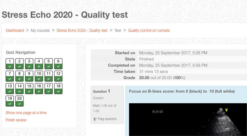
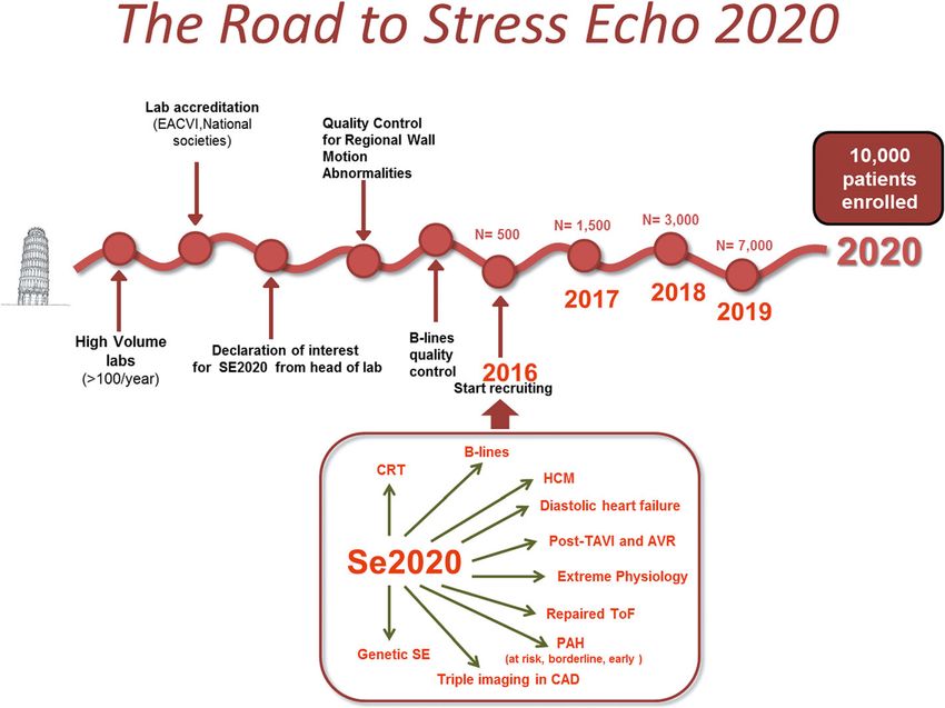
Scali et al. Cardiovascular Ultrasound (2018) 16:20                                                                   Page 2 of 9

  (Continued from previous page)
  Results: All 60 readers were successfully accredited: 26 (43%) on first, 24 (40%) on second, and 10 (17%) on third
  attempt. The average diagnostic accuracy of the 60 accredited readers was 95%, with R value of 0.95 compared to
  reference standard reading. The 53 B-lines naive scored similarly to the 7 B-lines expert on first attempt (90 versus 95%,
  p = NS). Compared to the step-1 of quality control for regional wall motion abnormalities, the mean reading time per
  attempt was shorter (17 ± 3 vs 29 ± 12 min, p < .01), the first attempt success rate was higher (43 vs 28%, p < 0.01), and
  the drop-out of readers smaller (0 vs 28%, p < .01).
  Conclusions: Web-based learning is highly effective for teaching and harmonizing B-lines reading. Echocardiographers
  without previous experience with B-lines learn quickly.
  Keywords: Certification, Lung comets, Quality control, Stress echocardiography, Wall motion

Background                                                        dyspnea is linked to acute backward heart failure [15].
Stress echocardiography (SE) has some advantages over             B-lines assessment must be properly standardized and
competing imaging techniques, including low cost, port-           quality-controlled prior to dissemination and use for
ability, radiation-free nature and versatility. Its major         clinical and scientific purposes. The present report was
limitation is the dependence upon operator’s expertise,           part of the larger SE2020 study and focuses on the edu-
which may impact on the quality and consistency of                cational aspects of LUS-SE, describing the results of the
diagnostic results [1, 2]. This limitation is magnified           upstream quality control and harmonization of B-lines
when the technique is used for scientific purposes in a           reading criteria across 52 SE2020 centers.
multi-center trial such as Stress Echo 2020 (SE2020)
study, designed to provide effectiveness data in 10,000           Methods
patients from > 100 laboratories in a variety of condi-           The Pisa lab coordinated the quality control assessment
tions ranging from coronary artery disease to heart fail-         for B-lines of all investigators who expressed their
ure (with preserved or depressed ejection fraction),              intention to participate in the study (Fig. 1). The coord-
hypertrophic cardiomyopathy, repaired congenital heart            inating center was in the National Research Council, In-
disease, valvular heart disease and extreme physiology            stitute of Clinical Physiology in Pisa, Italy. The candidate
[3]. To achieve harmonization, one possible approach is           centers included 52 centers (each with at least one certi-
the use of the core lab which analyses centrally images           fied reader) from 16 countries (Argentina, Brazil,
sent from all recruiting sites. This approach is typically        Bulgaria, Costa Rica, Hungary, Italy, Lithuania, Mexico,
the preferred choice in a clinical trial and minimizes the        Poland, Portugal, Romania, Qatar, Russia, Serbia, UK,
sources of measurement variability [4, 5]. The core lab           USA). The selection criterion was that all readers had
option was discarded in SE 2020 for two reasons. First,           already passed the quality control for RWMA reading
it was too costly and logistically demanding. Second, it          (step 1 in the “Road to SE 2020”). The B-lines reading
would provide efficacy data under ideal conditions, but           was the step 2 in the “Road to SE 2020”. The complete
our aim was to obtain effectiveness data realistically gen-       list of participants in the SE2020 consortium (as per
erated when the technique is deployed in the clinical             January 20th, 2018) is reported in the Appendix. The
arena, populated by real patients, real doctors and real          study protocol was reviewed and approved by the insti-
problems [6]. A feasible approach to ensure consistency           tutional ethics committee as a part of the SE 2020 study
in data acquisition and interpretation in this challenging        (1487-CE Lazio-1, July 20, 2016). The study was funded
setting is to develop an upstream reading quality control         with institutional funding of the Italian National Re-
for prospective centers willing to enter the study [7, 8].        search Council and with travel grants of the Italian Soci-
In SE2020, this approach has already been implemented             ety of Echocardiography and Cardiovascular Imaging
for regional wall motion abnormalities (RWMA), which              with dedicated sessions during national meetings. No
remains the diagnostic cornerstone of SE [9]. However, a          fort from industry was asked for or received.
separate quality control needs to be performed for other             An obligatory web-based educational platform was de-
aspects of contemporary SE practice, such as B-lines ob-          veloped to facilitate the training process. Participating car-
tained with lung ultrasound (LUS) [10]. Also known as             diologists were invited by email to join the platform,
ultrasound lung comets, B-lines are a sign of accumula-           which was protected by user-specific passwords. The plat-
tion of extra-vascular lung water [11] and can acutely in-        form includes files and videos with detailed instructions
crease during stress [12–14]. Their presence and/ or              on how to start the training and allows downloading and
increase during stress places the patient in a higher risk        uploading of external files. The sequence of the certifica-
subset for any level of RWMA [13] and indicates that              tion process and web-based learning has already been
Scali et al. Cardiovascular Ultrasound (2018) 16:20                                                                                        Page 3 of 9

 Fig. 1 The road to accreditation for the aspiring recruiting centers. After the first essential step of RWMA, the reader completes the second step
 (B-lines) and starts enrolling with dual imaging (RWMA and B-lines)

detailed and follows the same template used for RWMA                         patients requiring less depth and obese patients need-
[9]. We decided to have this platform mandatory and not                      ing greater depth to visualize the pleural line. A B-line
optional as in the step-1 for RWMA, since in case of                         was defined with 4 constant criteria: vertical, laser-like,
B-lines the technique is relatively young and recent ad-                     hyperechoic reverberation; arises from the pleural line
vances in acquisition (with 4-site scan mode) and report-                    extending to the bottom of the screen without fading;
ing were adopted in the SE2020 platform [16].                                moves synchronously with lung sliding; and erases the
                                                                             A-lines, which are a part of the normal lung pattern as
Study population of readers                                                  a horizontal, multiple reverberation artefact, equidistant
Sixty readers from 52 different centers initially asked to                   from one another below the pleura, at exact multiples
enter the SE2020 study, had passed the RWMA test for                         of the transducer-pleural line distance [17]. Detailed
quality control and therefore were allowed to enter the                      description of the scanning procedure and scanning
step-2 of SE2020.                                                            sites is also available in a 2-min movie from our labora-
  All participants were clinical cardiologists and expert                    tory on YouTube (The incredible ULCs – ultrasound
echocardiographers with ongoing high volume (> 100                           lung comets. Available at http://www.youtube.com/
tests per year) SE activity and the years of experience in                   watch?v=7y_hUFBHStM. Accessed: July 10, 2018). LUS
SE ranged from 5 to 31 years (mean value 18 years). All                      scanning was performed with the cardiac probe in the
were certified by national and/or international societies .                  supine position at rest and soon after stress (with the
                                                                             patient again resuming the supine position). The 4-site
Lung ultrasound acquisition                                                  simplified scan of the lung was used [16]. We analyzed
To acquire lung ultrasound (LUS) images adopted for                          the anterior and lateral hemithoraces, scanning along
quality control test, we used commercially available ultra-                  the anterior axillary (AA) and midaxillary (MA) lines
sound machines (IE 33, Philips, Medical Systems, Andover,                    on the third intercostal space (Fig. 2).
Massachusetts, USA with a 2.5–3.5 MHz phased-array sec-
tor scan probe; Vivid E9, GE Healthcare, USA, manufac-                       Web-based learning module
tured in Horten, Norway, equipped or standard M5S                            The 2-h web-based training module (http://se2020.alter
transducer with second harmonic technology; Mylab Eight                      vista.org) consisted of five sequential learning blocks: a-
platform Esaote, Genova, Italy). The depth was adjusted ac-                  Selected readings of 3 recent review or original articles
cording to the body habitus of the patient, with thin                        summarizing the evidences supporting the use of B-lines
Scali et al. Cardiovascular Ultrasound (2018) 16:20 Page 4 of 9
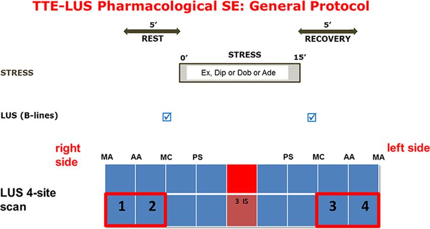
Fig. 2 The Stress-LUS general protocol. LUS for B-lines are assessed at baseline and at the end of stress, after the acquisition for RWMA. The
adopted protocol is the 4-site simplified scan
during stress and the adopted scan technique and scor- (white lung with coalescing B-lines). The diagnostic gold
ing criteria [8, 10, 13]; b- A power-point file of 25 slides standard was the reading of Pisa lab. The answer of the
summarizing key points and specific literature support- reader was considered correct if concordant with refer-
ing the proposed reading policy illustrating tips and ence standard reading ±1 (for instance, reference stand-
tricks highlighting the most frequent problems in B-lines ard reading of 5 B-lines; correct answer 4, 5, or 6). The a
interpretation with special focus on the technicalities of priori determined pass threshold was 18/20 (≥ 90%) with
the 4-site simplified scanning technique; c- A theory R value of intra-class correlation coefficient > .90.
self-assessment test with five questions with four an- The LUS images were selected to represent the garden
swers each (only one correct) preliminary to video-clip variety of stress testing modes, responses, results and
reading; d- Short (< 15 s) video-clips of examinations image quality. They came from six different laboratories
with the same format of official test reading, with 5 min (Benevento, Lucca, Pisa, Porto Alegre, Rome, St Peters-
per reading with countdown clock, and one possible an- burg) in three countries (Brasil, Italy, Russian Federation),
swer (from 0 to 10) for each video-clip (Fig. 3). and showed the full spectrum of responses (from 0, n = 7;
An expert trainer (QC or MCS) remained available to to 10, n = 1). All images were considered readable, with
all readers for e-mail or phone contact to provide assist- quality ranging from average-to-good (n = 16) to excellent
ance with any issue concerning the training. (n = 4) in the assessment of the reference standard reading.
At all times there was the possibility of face-to-face The stress employed was exercise in 17 subjects, high dose
discussion (via Skype) to address issues requiring special accelerated dipyridamole (0.84 mg/kg over 6 min) in 2 and
clarification with the principal investigator. After com- dobutamine (40 mcg/kg/min) in 1. The projection selected
pleting the web-based module the reader could take the was the third intercostal space between left mid-axillary
test (maximum three attempts). After each attempt, the and anterior axillary lines in 4; third intercostal space be-
sequence of videos was mixed. tween right mid-axillary and anterior axillary lines in 4;
third intercostal space between right anterior-axillary and
Reading sessions and pass threshold mid-clavicular lines in 4; third intercostal space between
We selected 20 cases of 10 patients (with rest and stress left anterior-axillary and mid-clavicular lines in 8.
images) in which the presence and number of B-lines
was documented by unanimous decision of 2 experi- After the pass or fail response
enced observers (EP and QC). The privacy of patients The response was pass (≥ 90% accuracy) or fail. With
during acquisition, storage, and transmission of the SE pass, the reader received a certificate of accreditation
study was protected. All images were anonymized, and and could start recruiting with a written informed con-
the identity of patients or the study condition (rest or sent signed by each patient and after clearance by the
stress) was not disclosed at any time to the readers. Each local ethical committee. With fail, the unsuccessful
SE study was structured in a single video-clip of 10– reader could retake the test after 1 month. After the sec-
15 s, with either resting or stress images. Each test clip ond fail, the reader could undergo training in a recom-
was scored from 0 (black lung, A-lines, no B-lines) to 10 mended center and try again after 1 year.Scali et al. Cardiovascular Ultrasound (2018) 16:20 Page 5 of 9
Fig. 3 The screenshot of the test-match step during B-lines quality control. There are 5 still frames (or videos) with B-lines and the trainee has to
choose among 5 possible answers, ranging from 0 (left lower panel) to 9 (upper middle panel)
Statistical analysis was very high (R = 0.95, p < .0001). In the 20 peripheral
Each reader was evaluated against the gold standard of readers who repeated the test a second time at least
reference standard reading for assessment of individual 3 months after accreditation, the Spearman correlation co-
accuracy (in %). The intra-class correlation coefficient efficient was also very high (R = 0.97, p < .0001).
was calculated, for each reader, in the whole series of 20
paired measurements made by the peripheral reader and Discussion
the reference reader. Intra-observer agreement was A user-friendly web-based learning is highly effective for
tested in 20 peripheral readers who volunteered to re- training B-lines also for echocardiographers without pre-
peat the measurement session after at least 3 months vious exposure to B-lines. After a limited learning effort,
from the first reading. A p value < 0.05 was considered the accuracy of B-lines reading is comparable between
significant. very experienced and freshly trained readers. B-lines
with 4-site simplified scan of the lung has a very high
Results success rate in acquisition and analysis. It has been em-
Of the initial 60 readers who started, 53 were B-lines naive bedded as an integral part of dual imaging SE adopted
(without previous exposure to B-lines). All 60 readers as the core protocol in SE2020 for all forms of physical
were successfully accredited (Fig. 4): 26 (43%) on first, 24 and pharmacological stress for all patients, from coron-
(40%) on second, and 10 (17%) on third attempt. The 53 ary artery disease to heart failure.
B-lines naive scored similarly to the 7 B-lines expert on
first attempt (90 versus 95%, p = NS). Compared to the Comparison with previous studies
step-1 of quality control for regional wall motion abnor- The American College of Chest Physicians has defined
malities [6], the mean reading time per attempt was the knowledge and technical elements required for com-
shorter (17 ± 3 vs 29 ± 12 min, p < .01), the first attempt petence in lung ultrasound [18]. There have been a
success rate was higher (43 vs 28%, p < 0.01), and the number of prior lung ultrasound education papers,
drop-out of readers smaller (0 vs 28%, p < .01). The aver- showing that a limited training of a few hours can im-
age diagnostic accuracy of the 60 accredited readers was prove the capability of execution and interpretation of
95%. Considering the final attempt of the 60 readers, the LUS even in medical students without previous exposure
Spearman correlation coefficient between the expert refer- to ultrasound [19, 20]. In the present study we are deal-
ence reading and the reading of each peripheral reader ing with a specific and limited aspect of LUS of specialScali et al. Cardiovascular Ultrasound (2018) 16:20 Page 6 of 9
Fig. 4 The test results of a reader passed with full marks (20/20)
interest for cardiologists, i.e. the detection of B-lines. modules are currently under construction within the
There is a lack of a specific training and certification framework of “SE 2020” to cover the entire spectrum of
pathway in cardiology, and as a result training and per- key aspects of SE diagnosis, from coronary flow velocity
formance of LUS varies widely among different institu- reserve to left ventricular volumes and pulmonary
tions. An approach similar to the one adopted in the hemodynamics [3].
present study was developed in Pisa for centers recruit- A key aspect in the evaluation of SE results is the
ing in the LUST study [21]. However, this study differs adoption of an undisputed diagnostic “gold standard”.
from the previous one under some aspects: first, it was The lack of a universally acceptable gold standard makes
focused on LUS-SE, not on resting LUS; the adopted the assessment of reading performance difficult. From
scan scheme was the simplified 4-site scan, easier to do, the library of images arriving from all the world and
to teach and to learn than the previously adopted 28-site stored in our data bank, we selected cases meeting the
scan; and the quality control procedures required some conditions of unanimous reading of the two most expe-
prior reading and slide presentation to facilitate a stan- rienced readers from the reference lab. This is a far from
dardized learning [9]. perfect gold standard, yet a reasonable, and perhaps the
Our findings are consistent with a large body of litera- only possible, one.
ture showing that stable web applications are increas- We restricted our validation phase to participants in
ingly used for improving medical image interpretation the SE2020 study, who had a substantial reading experi-
skills regardless of time and space and without the need ence and certification in RWMA as a prerequisite. This
for expensive imaging equipment or a patient to scan reader pool may have been especially knowledgeable and
[22]. With the adopted web-based approach, the educa- motivated, thereby justifying the excellent learning re-
tional path is standardized, shared, and - after validation sults. However, 53 of them were B-lines naive, and there-
and refinement - prospectively available in open source, fore probably the selection criteria of our readers did
and exploitable for scientific purposes and clinical edu- not affect the generalizability of results.
cation. The use of enabling technologies makes the ac- We adopted a simplified 4-site scan for acquisition of
creditation process faster, smoother and cheaper, and B-lines at rest and during stress. This approach intro-
coupled with the open-source platform grants an unpre- duces a substantial abbreviation compared to other pro-
cedented opportunity for continuing education, also fos- tocols such as the 28-region scan originally adopted in
tered by endorsement and governance by the scientific the Pisa laboratory in the first application of LUS in
society supporting the study. heart failure patients [23] and also recommended by an
international consensus in 2012 [24]. Over the years,
Study limitations simplified 8-zone and 4-zone lung imaging protocols
We focused on the assessment of B-lines, which is a par- were proposed [25, 26], with comparable information
ticularly simple aspect of LUS diagnosis [10, 11]. Similar between the 2 protocols as shown by Platz et al. [25].
harmonization and accreditation issues are present for Scali et al. showed that the simplified 4-site scan allows
other aspects of SE diagnosis. Separate and parallel training to complete the assessment of B-lines in 20 s (instead ofScali et al. Cardiovascular Ultrasound (2018) 16:20 Page 7 of 9
the 3 min required by the 28- region scan). There is a Giovanni di Salvo35, Julio E. Perez 36, Ana Camarozano 37, Anca Corciu38, Alla
linear, close correlation between the 28-site and the Boshcenko39, Fabio Lattanzi40, Carlos Cotrim41, Paula Fazendas42, Maciej
Haberka43, Bozena Sobkowicz44, Wojciech Kosmala 45, Tomasz Witkowski45,
4-site B-lines score [16]. Therefore, there is no signifi- Piotr Gosciniak 46, Alessandro Salustri 47, Hugo Rodriguez Zanella48, Alexandra
cant loss of information when going from 28- to 4-site Nikolic49, Suzana Gligorova 50, Madalina-Loredana Urluescu 51, Maria Fiorino
52
scan, but a substantial simplification and time saving, , Giuseppina Novo53, Tamara Preradovic-Kovacevic54, Miodrag Ostojic33, 54,
Dario Gregori55.
vital for SE imaging, when there are so many things to 1
Institute of Clinical Physiology, National Research Council, Pisa; 2Cardiology
see and so little time available. Division, Fatebenefratelli Hospital, Benevento, Italy; 3Cardiology Department
and Echocardiography Lab, University Hospital “San Giovanni di Dio e Ruggi
d’Aragona”, Salerno, Italy; 4Cardiology Hospital, Policlinico of Bari, Italy;
Clinical implications 5
Cardiology Department, San Carlo Hospital, Potenza, Italy; 6Cardiology
B-lines are a useful adjunct to mainstream SE based on Department, San Luca Hospital, Lucca, Italy; 7Cardiology Department,
RWMA [27, 28], but its impact may be limited by the Monaldi Hospital, Second University of Naples, Italy; 8Casa di Cura Figlie di
San Camillo, Cremona; 9Cardiology Department, Parma University Hospital,
relatively few centers currently using it in their routine Italy; 10Department of Advanced Biomedical Sciences, Federico II University
SE practice, and the lack of standardization in acquisi- Hospital, Naples, Italy; 11Cardiology Department, Modena University Hospital,
tion, scoring and reporting [29]. After a web-based mod- Modena, Italy; 12Cardio-Thorax-Vascular Department, Echocardiography lab,
“Policlinico Vittorio Emanuele”, Catania University, Italy; 13Cardiology
ule and certification, the approach is better harmonized Department, Careggi Hospital, Florence, Italy; 14Cardiology Division,
and the accumulation of clinical practice also allows the Pontedera-Volterra Hospital, ASL Toscana 3 Nord-Ovest, Italy; 15Cardiology
rapid growth of scientifically unique data. To achieve Department, San Camillo-Forlanini Hospital, Roma, Italy; 16Cardiology
Department, Ospedale dell’Angelo Mestre-Venice, Italy; 17Cardiology
this goal, simplification is essential, and the 4-site simpli- Department, Nottola Hospital, Siena, and Cardiothoracic Department,
fied scan is ideal for LUS rest and stress testing. University of Pisa, Italy; 18Cardiology Department, Ospedale Santa Maria
However, the SE technique does not tolerate improvisa- Incoronata dell’ Olmo, Cava de’ Tirreni, Salerno, Italy; 19Department of
Cardiology, Sandro Pertini Hospital, Rome, Italy; 20Cardiology Department,
tion, and an accurate standardization of terminology, stan- Ramos Mejia Hospital, Buenos Aires, Argentina; 21Cardiodiagnosticos,
dards of execution, and interpretation criteria is required Investigaciones Medicas, Buenos Aires, Argentina; 22Cardiology Division,
before a center is allowed to enter its experience in the Hospital San José, Criciuma, Brasil; 23Hospital Sao Vicente de Paulo e
Hospital de Cidade, Passo Fundo, Brasil; 24Cardiology Institute of Rio Grande do
common data bank. Similarly to what has been said for Sul, Porto Alegre, Brasil; 25Hospital de Clinicas de Porto Alegre - Universidade
meta-analysis [30], multicenter SE studies are like a bouil- Federal do Rio Grande do Sul, Porto Alegre, Brasil; 26Hospital San Vicente de
labaisse: no matter how much seafood (or recruiting cen- Paul, Heredia, Costa Rica; 27Acibadem City Clinic Cardiovascular Center,
University Hospital, Sofia, Bulgaria; 28Institute of Family Medicine, University of
ters) is added, one tainted fish (an unreliable center Szeged, and Department of Internal Medicine, Elisabeth Hospital,
generating inconsistent reading) will spoil the pot. Hodmezovasarhely, Hungary; 29Centre of Cardiology and Angiology,
Vilnius University Hospital Santaros Klinikos, Faculty of Medicine, Vilnius
University, State Research Institute for Innovative Medicine, Vilnius,
Conclusion Lithuania; 30Chair of Cardiology, Bieganski Hospital, Medical University,
Web-based learning is highly effective for teaching and Lodz Poland; 31Cardiology Clinic, Clinical Center of Serbia, Medical
harmonizing B-lines reading, with an enormous saving School, University of Belgrade, Serbia; 32Clinical Hospital Zvezdara Belgrade,
Serbia; 33Department of Cardiology, Clinical Hospital Center Zemun, Faculty of
of time and resources versus the conventional hands-on Medicine, University of Belgrade, Serbia; 34Cardiology Department, University
approach of teaching and learning ultrasound tech- Clinic, Saint Petersburg, Russian Federation; 35Pediatric Cardiology Department,
niques. Echocardiographers without previous experience Brompton Hospital, London, UK, Division of Cardiology; 36Washington University
School of Medicine, Barnes-Jewish Hospital, St. Louis, Missouri, USA; 37Hospital
with B-lines learn quickly. de Clinicas UFPR, Medicine Department, Federal University of Paranà, Curitiba,
Brasil; 38Department of Cardiology, IRCCS Policlinico San Donato Clinic, Milan,
Abbreviations Italy; 39Cardiology Research Institute, Tomsk National Tomsk National Research
CAD: Coronary artery disease; LUS: Lung Ultrasound; RWMA: Regional wall Medical Center of Russian Academy of Sciences; 40Cardiothoracic Department,
motion abnormalities; SE: Stress echocardiography; TE: Transthoracic University of Pisa, Italy; 41Heart Center, Hospital da Cruz Vermelha, Lisbon, and
echocardiography Medical School of University of Algarve, Faro, Portugal; 42Cardiology
Department, Hospital Garcia de Orta, Almada, Portugal; 43Department of
Acknowledgments Cardiology, School of Health Sciences, Medical University of Silesia,
The study was partially funded with the project Aging of the National Katowice, Poland; 44Department of Cardiology, Medical University of
Research Council. Białystok, Poland; 45Department of Cardiology, Wroclaw Medical University,
On behalf of the Stress Echo 2020 Study Group of the Italian Society of Wroclaw, Poland; 46Department of Cardiology, Provincial Hospital, Szczecin,
Cardiovascular Echography (as per December 20, 2017). Eugenio Picano1, Poland; 47Hamad Medical Corporation, Heart Hospital, Doha, Qatar;
Maria Grazia Andreassi1, Clara Carpeggiani1, Michele De Nes1, Marco Paterni1, 48
Instituto Nacional de Cardiologia Ignacio Chavez, Mexico City, Mexico;
Lorenza Pratali1,Quirino Ciampi2, Bruno Villari2, Eduardo Bossone3, Rodolfo 49
Institute for Cardiovascular Diseases Dedinje, Belgrade, Serbia;
Citro3, Francesco Ferrara3, Paolo Colonna4, Marco Fabio Costantino5, Lauro 50
Cardiology Division Ospedale Casilino, Roma Italy; 51Cardiology
Cortigiani6, Antonello D’Andrea7-1, Claudio Dodi8, Nicola Gaibazzi9, Maurizio Department, County Hospital Sibiu, Invasive and Non-Invasive Center for
Galderisi10, Andrea Barbieri11, Ines Monte12, Fabio Mori13, Iacopo Olivotto13, Cardiac and Vascular Pathology in Adults - CVASIC Sibiu, Faculty of
Barbara Reisenhofer 14, Federica Re15, Fausto Rigo16, Maria Chiara Scali17,41, Medicine Sibiu, Romania; 52Cardiology Division Ospedale Civico Di Cristina
Sergio Severino7-2, Paolo Trambaiolo19, Miguel Amor20, Jorge Lowenstein21, Benfratelli Palermo; 53Cardiology Division, University Hospital, Palermo, Italy;
Pablo Martin Merlo21, Clarissa Borguezan Daros22, José Luis de Castro e Silva 54
University Clinical Center, Banja Luka, Republic of Srpska, Bosnia and
Pretto23, Marcelo H. Miglioranza24, Marco A.R. Torres25, Daniel Quesada Herzegovina; 55Department of Biostatistics, University of Padua, Padua, Italy.
Chaves26, Melissa Rodriguez Israel26, Iana Simova27, Albert Varga 28, Gergely
Agoston 28, Attila Palinkas 28, Jelena Čelutkienė29, Jaroslaw D. Kasprzak30,
Karina Wierzbowska-Drabik30, Ana Djordjevic-Dikic31, Branko Beleslin 31, Milica Funding
Dekleva32, Aleksandar N. Neskovic33, Ivan Stankovic33, Angela Zagatina34, Institutional funding from CNR Institute of Clinical Physiology.Scali et al. Cardiovascular Ultrasound (2018) 16:20 Page 8 of 9
Availability of data and materials Klinikos, Faculty of Medicine, Vilnius University, State Research Institute for
Data sharing not applicable to this article as no data-sets were generated or Innovative Medicine, Vilnius, Lithuania. 30Chair of Cardiology, Bieganski
analyzed during the current study. Hospital, Medical University, Lodz, Poland. 31Cardiology Clinic, Clinical Center
See the stress echo 2020 website at: http://se2020.altervista.org/index.php/en/. of Serbia, Medical School, University of Belgrade, Belgrade, Serbia. 32Clinical
(user name: reviewer; temporary password: N4ppGVgu70). Hospital Zvezdara Belgrade, Belgrade, Serbia. 33Department of Cardiology,
See the quality control content and modalities at: https://stressecho2020.mo Clinical Hospital Center Zemun, Faculty of Medicine, University of Belgrade,
odlecloud.com/login/index.php. (user name: reviewer; temporary password: Belgrade, Serbia. 34Cardiology Department, University Hospital, Saint
N4ppGVgu70). Petersburg, Russian Federation. 35Pediatric Cardiology Department, Brompton
Hospital, London, UK. 36Washington University School of Medicine,
Authors’ contributions Barnes-Jewish Hospital, St. Louis, MO, USA. 37Hospital de Clinicas UFPR,
EP is the study chairman, designed the protocol, organized the content of web- Medicine Department, Federal University of Paranà, Curitiba, Brazil.
38
based training and drafted the manuscript; QC is the principal investigator of Department of Cardiology, IRCCS Policlinico San Donato Clinic, Milan, Italy.
39
SE2020, helped to organize the structure of training, contributed to developing Cardiology Research Institute, Tomsk National Research Medical Center of
the web-based training, critically revised the manuscript for an intellectually Russian Academy of Sciences, Tomsk, Russia. 40Cardiothoracic Department,
important contribution and approved the submitted version; McS is the project University of Pisa, Pisa, Italy. 41Heart Center, Hospital da Cruz Vermelha,
leader of B-lines subproject in SE2020; MdN is the computer scientist who Lisbon and Medical School of University of Algarve, Faro, Portugal.
42
developed the website (SE 2020) and the web-based training material; MP is Cardiology Department, Hospital Garcia de Orta, Almada, Portugal.
43
the computer scientist who organized and governed the quality control access, Department of Cardiology, School of Health Sciences, Medical University of
results, and data analysis; all other authors contributed to study design, Silesia, Katowice, Poland. 44Department of Cardiology, Medical University of
undertook the quality control up to certification, are active members of SE 2020 Białystok, Białystok, Poland. 45Department of Cardiology, Wroclaw Medical
consortium and critically revised the manuscript for an intellectually important University, Wroclaw, Poland. 46Department of Cardiology, Provincial Hospital,
contribution and approved the submitted version. RC and PC also coordinated Szczecin, Poland. 47Hamad Medical Corporation, Heart Hospital, Doha, Qatar.
48
the involvement of SIECVI (Società Italiana di Ecocardiografia e Cardiovascular Instituto Nacional de Cardiologia Ignacio Chavez, Mexico City, Mexico.
49
Imaging). CC is responsible for data quality control and reader’s certification. Institute for Cardiovascular Diseases, Dedinje, Belgrade, Italy. 50Cardiology
Division Ospedale Casilino, Rome, Italy. 51Cardiology Department, County
Ethics approval and consent to participate Hospital Sibiu, Invasive and Non-Invasive Center for Cardiac and Vascular
The study protocol was reviewed and approved by the institutional ethics Pathology in Adults - CVASIC Sibiu, Faculty of Medicine, Sibiu, Romania.
52
committee as a part of the SE 2020 study (1487-CE Lazio-1, July 20, 2016). Cardiology Division Ospedale Civico Di Cristina Benfratelli, Palermo, Italy.
53
Cardiology Division, University Hospital, Palermo, Italy. 54University Clinical
Consent for publication Center, Banja Luka, Republic of Srpska, Bosnia and Herzegovina.
All the authors have read and approved the manuscript and accorded the
consent for pubblication. Received: 8 March 2018 Accepted: 3 August 2018
Competing interests
The authors declare that they have no competing interest.
References
1. Pellikka PA, Nagueh SF, Elhendy AA, Kuehl CA, Sawada SG. American Society
Publisher’s Note of Echocardiography recommendations for performance, interpretation, and
Springer Nature remains neutral with regard to jurisdictional claims in application of stress echocardiography. J Am Soc Echocardiogr. 2007;20:1021–4.
published maps and institutional affiliations. 2. Sicari R, Nihoyannopoulos P, Evangelista A, Kasprzak J, Lancellotti P,
Poldermans D. European Association of Echocardiography Stress
Author details echocardiography expert consensus statement: European Association of
1
CNR, Institute of Clinical Physiology, Biomedicine Department, Pisa, Italy. Echocardiography (EAE) (a registered branch of the ESC). Eur J
2
Cardiology Division, Fatebenefratelli Hospital, Benevento, Italy. 3Cardiology Echocardiogr. 2008;9:415–37.
Department and Echocardiography Lab, University Hospital “San Giovanni di 3. Picano E, Ciampi Q, Citro R, et al. Stress echo 2020 : The international Stress
Dio e Ruggi d’Aragona”, Salerno, Italy. 4Cardiology Hospital, Policlinico of Bari, Echo study in ischemic and non-ischemic heart disease Cardiov Ultras 2017 ;
Bari, Italy. 5Cardiology Department, San Carlo Hospital, Potenza, Italy. Jan 18 15 (1): 3. DOI: https://doi.org/10.1186/s12947-016-0092-1
6
Cardiology Department, San Luca Hospital, Lucca, Italy. 7Cardiology 4. Gottdiener JS, Bednarz J, Devereux R, Gardin J, Klein A, Manning WJ, et al.
Department, Echocardiography Lab, Monaldi Hospital, Second University of American Society of Echocardiography. American Society of
Naples, Naples, Italy. 8Casa di Cura Figlie di San Camillo, Cremona, Italy. Echocardiography recommendations for use of echocardiography in clinical
9
Cardiology Department, Parma University Hospital, Parma, Italy. trials. J Am Soc Echocardiogr. 2004;17:1086–119.
10
Department of Advanced Biomedical Sciences, Federico II University 5. Galderisi M, Henein MY, D' hooge J, Sicari R, Badano LP, Zamorano JL,
Hospital, Naples, Italy. 11Cardiology Department, Modena University Hospital, Roelandt J. Recommendations of the European Association of
Modena, Italy. 12Cardio-Thorax-Vascular Department, Echocardiography lab, Echocardiography. How to use echo-Doppler in clinical trials: different
Policlinico Vittorio Emanuele, University of Catania, Catania, Italy. 13Cardiology modalities for different purposes. Eur J Echocardiogr. 2011;12:339–53.
Department, Careggi Hospital, Florence, Italy. 14Cardiology Division, 6. Feinstein AR. Diagnostic and spectral markers. Philadelphia: Clinical
Pontedera-Volterra Hospital, ASL Toscana 3 Nord-Ovest, Florence, Italy. epidemiology. Saunders; 1985. p. 597–631.
15
Cardiology Department, San Camillo-Forlanini Hospital, Rome, Italy. 7. Picano E, Landi P, Bolognese L, Chiarandà G, Chiarella F, Seveso G, et al.
16
Cardiology Department, Ospedale dell’Angelo Mestre-Venice, Venice, Italy. Prognostic value of dipyridamole echocardiography early after
17
Cardiology Department, Nottola Hospital, Siena, Italy. 18Cardiology uncomplicated myocardial infarction: a large-scale, multicenter trial. The
Department, Ospedale santa Maria Incoronata dell’Olmo, cava de’ Tirreni, EPIC study group. Am J Med. 1993;95:608–18.
Salerno, Italy. 19Department of Cardiology, Sandro Pertini Hospital, Rome, 8. Picano E, Mathias W Jr, Pingitore A, Bigi R, Previtali M. Safety and tolerability
Italy. 20Cardiology Department, Ramos Mejia Hospital, Buenos Aires, of dobutamine-atropine stress echocardiography: a prospective, multicentre
Argentina. 21Cardiodiagnosticos, Investigaciones Medicas, Buenos Aires, study. Echo Dobutamine international cooperative study group. Lancet.
Argentina. 22Cardiology Division, Hospital San José, Criciuma, Brasília, Brazil. 1994;344:1190–2.
23
Hospital Sao Vicente de Paulo e Hospital de Cidade, Passo Fundo, Brazil. 9. Ciampi Q, Picano E, Paterni M, Daros CB, Simova I, de Castro e Silva Pretto
24
Cardiology Institute of Rio Grande do Sul, Porto Alegre, Brazil. 25Hospital de JL, D'Andrea A, Scali MC, Gaibazzi N, Severino S, Djordjevic-Dikic A, Kasprzak
Clinicas de Porto Alegre - Universidade Federal do Rio Grande do Sul, Porto J, Zagatina A, Varga A, Lowenstein J, Merlo P, Amor M, Celeutkiene J, Perez
Alegre, Brazil. 26Hospital San Vicente de Paul, Heredia, Costa Rica. 27Acibadem JE, Di Salvo G, Galderisi M, Mori F, Costantino MF, Massa L, Dekleva M,
City Clinic Cardiovascular Center, University Hospital, Sofia, Bulgaria. Chavez D Q, Trambaiolo P, Citro R, Colonna P, Rigo F, Torres MAR, Monte I,
28
Institute of Family Medicine, University of Szeged, Szeged, Hungary. Stankovic I, Neskovic A, Cortigiani L, Re F, Dodi C, D'Andrea A, Villari B,
29
Centre of Cardiology and Angiology, Vilnius University Hospital Santaros Arystan A, De Nes M, Carpeggiani C, on behalf of Stress Echo 2020. QualityScali et al. Cardiovascular Ultrasound (2018) 16:20 Page 9 of 9
control of regional wall motion analysis in stress Echo 2020. Int J Cardiol 27. Picano E, Scali MC. The lung water cascade in heart failure.
2017;249479 - 485. Echocardiography. 2017;34:1503–7.
10. Picano E, Frassi F, Agricola E, Gligorova S, Gargani L, Mottola G. Ultrasound 28. Picano E, Scali MC. Stress echo, carotid arteries and more: its versatility for
lung comets: a clinically useful sign of extravascular lung water. J Am Soc our imaging times. Editorial comment JACC img. 2017; https://doi.org/10.
Echocardiogr. 2006;19:356–63. 1016/j.jcmg.2017.01.023.
11. Picano E, Pellikka PA. Ultrasound of extravascular lung water: a new 29. Picano E, Pellikka PA. Stress echo applications beyond coronary artery
standard for pulmonary congestion. Eur Heart J. 2016;37:2097–104. disease. Eur Heart J. 2014;35:1033–40.
12. Agricola E, Picano E, Oppizzi M, Pisani M, Zangrillo A, Margonato A. 30. Messerli FH. Meta-analysis and bouillabaisse. Ann Intern Med. 1996;125:519.
Assessment of stress-induced pulmonary interstitial edema by chest
ultrasound during exercise echocardiography and its correlation with left
ventricular function. J Am Soc Echocardiogr. 2006;19:457–63.
13. Scali MC, Cortigiani L, Simionuc A, Gregori D, Marzilli M, Picano E. The
added value of exercise-echocardiography in heart failure patients:
assessing dynamic changes in extravascular lung water. Eur J Heart Failure.
2017;19:1468–78.
14. Simonovic D, Coiro S, Carluccio E, Girerd N, Deljanic-Ilic M, Ambrosio G.
Exercise elicits dynamic changes in extravascular lung water and
hemodynamic congestion in heart failure patients with preserved ejection
fraction. Research letter. Eur J Heart Fail. 2018;21. https://doi.org/10.1002/
ejhf.1228. [Epub ahead of print]
15. Lancellotti P, Pellikka PA, Budts W, Chaudhry FA, Donal E, Dulgheru R,
Edvardsen T, Garbi M, Ha JW, Kane GC, Kreeger J, Mertens L, Pibarot P,
Picano E, Ryan T, Tsutsui JM, Varga A. The clinical use of stress
echocardiography in non-ischaemic heart disease: recommendations from
the European Association of Cardiovascular Imaging and the American
Society of Echocardiography. Eur Heart J Cardiovasc Imaging. 2016;17:1191–229.
16. Scali MC, Zagatina A, Simova I, Zhuravskaya N, Ciampi Q, Paterni M, Marzilli
M, Carpeggiani C. Picano E. B-lines with Lung Ultrasound: the optimal scan
technique at rest and during stress Ultrasound Med Biol. 2017;43:2558–66.
17. Picano E, Scali MC, Ciampi Q, Lichtenstein D. Lung ultrasound for the
cardiologist. JACC imaging. 2018;12:381–90.
18. Mayo PH, Beaulieu Y, Doelken P, et al. American College of Chest
Physicians/La Société de Réanimation de Langue Française statement on
competence in critical care ultrasonography. Chest. 2009;135:1050–60.
19. Beaulieu Y, Laprise R, Drolet P, Thivierge RL, Serri K, Albert M, Lamontagne
A, Belliveau M, Denault AY, Patenaude JV. Bedside ultrasound training using
web-based e-learning and simulation early in the curriculum of residents.
Critical Ultrasound Journal. 2015;7:1.
20. Sun Lim J, Lee S, Ho Do H, Ho Oh K. Can Limited Education of Lung
Ultrasound Be Conducted to Medical Students Properly? A Pilot Study
BioMed Research International Volume 2017, Article ID 8147075, 6 pages
doi https://doi.org/10.1155/2017/8147075
21. Gargani L, Sicari R, Raciti M, Serasini L, Passera M, Torino C, Letachowicz K,
Ekart R, Fliser D, Covic A, Balafa O, Stavroulopoulos A, Massy ZA, Fiaccadori
E, Caiazza A, Bachelet T, Slotki I, Shavit L, Martinez-Castelao A, Coudert-Krier
MJ, Rossignol P, Kraemer TD, Hannedouche T, Panichi V, Wiecek A,
Pontoriero G, Sarafidis P, Klinger M, Hojs R, Seiler-Mußler S, Lizzi F,
Onofriescu M, Zarzoulas F, Tripepi R, Mallamaci F, Tripepi G, Picano E,
London GM, Zoccali C. Efficacy of a remote web-based lung ultrasound
training for nephrologists and cardiologists: a LUST trial sub-project.
Nephrology Dialysis Transplantation. 2016;31:1982–8.
22. Lindseth F, Hallan ML, Tonnessen MS, Smistad E, Vapenstad C. MIIP: a web-
based platform for medical image interpretation training and evaluation
focusing on ultrasound. Proceedings Volume 10138, Medical Imaging 2017:
Imaging Informatics for healthcare Research and Applications; 10138W; doi:
10.117/12.2254158.
23. Jambrik Z, Monti S, Coppola V, Agricola E, Mottola G, Picano E. Usefulness of
ultrasound lung comets as a nonradiologic sign of extravascular lung water.
Am J Cardiol. 2004;93:1265–70.
24. Volpicelli G, Elbarbary M, Blaivas M, et al. International Liaison Committee on
Lung Ultrasound for International Consensus Conference on Lung
Ultrasound International evidence-based recommendations for point-of-care
lung ultrasound. Intensive Care Med. 2012;38:577–91.
25. Platz E, Pivetta E, Merz AA, Peck J, Rivero J, Cheng S. Impact of device
selection and clip duration on lung ultrasound assessment in patients with
heart failure. Am J Emerg Med. 2015;33:1552–6.
26. Ohman J, Harjola VP, Karjalainen P, Lassus J. Assessment of early treatment
response by rapid cardiothoracic ultrasound in acute heart failure: cardiac
filling pressures, pulmonary congestion and mortality. Eur Heart J Acute
Cardiovasc Care. 2018;7:311–20.You can also read