Radiological Comparison of the Biomechanical Characteristics of Short Segment Posterior Instrumentation (Moss-Miami System), with and without ...
←
→
Page content transcription
If your browser does not render page correctly, please read the page content below
Original Article                                                             Nepal Journal of Neuroscience 2022;19(1):31-37

Radiological Comparison of the Biomechanical
Characteristics of Short Segment Posterior
Instrumentation (Moss-Miami System), with and
without Transverse Connector in Unstable Thoracolumbar
Fracture
Anand Doshi1 , Prashant Khandelwal2 , Sejal Mehta3 , Sudhir Sudumbrekar4
1,2,3,4
       Department of Neurosurgery, Bharati Vidyapeeth (DU) Medical College and Hospital, Pune
Date of submission: 22nd July 2021               Date of acceptance: 21st January 2022         Date of publication: 15th March 2022

Abstract
Introduction: Wedge compression fracture was most common type of fracture (66.6 %) and first Lumbar vertebra
(L1) was the most common fracture site (38 %). The choice of surgical approach and instrumentation depends on
fracture type, injury level and degree of neural injury. This paper evaluated efficacy of transpedicular screw fixation
by Moss Miami instrumentation with transverse connector for surgical treatment in traumatic unstable thoraco-
lumbar fractures.
Materials and Method: Fifty-five cases were prospectively studied over three years. Comparison was done between
two groups- Group 1 without transverse connector and Group 2 with transverse connector. Radiologically evaluation
was done in both groups pre-operatively, tenth day, six weeks and six months interval. All X-rays were assessed by
lateral and dynamic antero- posterior views for measuring kyphotic (cobb’s) angle and inter-rod angle.
Results: In both groups post-operative Mean Cobb’s angle level were increased and remained same until 06 months.
Variation in Group 2 with transverse connector was slightly higher in initial period (i.e.) after 10 days and then
it remains minimal from 6 weeks to 6 months. But in Group 1 without transverse connector the change in angle
variation was minimal which worsen over 6 weeks and it continues to remain worse at 6 months, suggestive of
parallelogram effect.
Conclusion: Transverse connector does play role in preventing parallelogram effect of implants and provides
additional stability to the construct. This is first study done in vivo which is done to study the role of transverse
connector and evaluate any parallelogram effect in spinal instrumentation.
Key words: Transverse connector, Thoraco-lumbar fracture, parallelogram effect

                                                                                           Introduction

                                                                      F
Access this article online
                                                                          racture-dislocation of the spine is a serious injury
Website: https://www.nepjol.info/index.php/NJN
                                                                          that is prevalent in physically active individuals. The
DOI: https://doi.org/10.3126/njn.v19i1.38548                          management and evaluation of this injury has changed
HOW TO CITE                                                           tremendously over the last decade.
Doshi A, Khandelwal P, Mehta S, Sudumbrekar S. Radiological                Boucher was the first to get credit for pedicle screw
Comparison of the Biomechanical Characteristics of Short
Segment Posterior Instrumentation (Moss- Miami System), with          fixation of the spine in 1959. Significant advances by
and without Transverse Connector, in Unstable Thoraco-Lumbar          Magrel and dick, Steffee, Luque, Cotrel and Dubousset
Fracture. NJNS. 2022;19(1):31-7.                                      and the others in biomechanical design and placement
                                                                      technique have led to a rapid increase in the use of implants
Address for correspondence:
Dr. Prashant Khandelwal                                               in spinal fixation. In 1996, Odgers et al. was the first to
Department of Neurosurgery, Bharati Vidyapeeth (DU) Medical           publish pedicle screw insertion under image guidance.1
College and Hospital, Pune                                                 Spinal stability is defined as the ability for the
E-mail: drpash72@gmail.com                                            vertebrae to maintain their relationship and limit their
Copyright © 2022 Nepalese Society of Neurosurgeons (NESON)            relative displacements during physiologic postures and
                                                                      loads.2 Unstable thoraco-lumbar spine injuries requires
ISSN: 1813-1948 (Print), 1813-1956 (Online)
                                                                      stabilization to obtain and maintain spinal stability,
             This work is licensed under a Creative Commons           facilitate neurological recovery and relieve pain in
             Attribution-Non Commercial 4.0 International License.    incomplete neurological deficit as well as rehabilitation

                                   Nepal Journal of Neuroscience, Volume 19, Number 1, 2022                              31
Doshi et al

of the patient starting from mobilization to ambulation       surgical procedures example decompression, reduction
where possible.3 In the majority of comparison studies        of fracture, fracture dislocation, or dislocation etc were
between surgical and non-surgical treatment, it appears       added to this operative procedure and carried out as per
that neurologic recovery is enhanced in surgically            standard technique, when required.
treated patients. The choice of surgical approach and             Surgery was done under general anaesthesia through
instrumentation requires a thorough understanding of          posterior midline approach. Standard Pedicle entry
fracture type, injury level and degree of neural injury.4     point and directions were followed. Intra-operative
Moss Miami posterior spinal instrumentation is a spinal       confirmation under c-arm guidance was carried out.
instrumentation introduced, which is a hybrid system          Transverse connector was used in alternate patients; hence
using pedicular screws and rods.                              two Groups were formed. Comparison was done between
     The use of transverse connector in this posterior        the two groups- Group 1 without transverse connector
stabilization has a controversial role. Krag MH et al.        and Group 2 with transverse connector. Radiologically
found that pedicle fixation techniques may fail during        evaluation was done in both groups pre-operatively, tenth
axial loading.5 This is in part due to a tendency towards     day, six weeks and six months interval. All X-rays were
the development of a parallelogram-like translational         assessed by lateral and dynamic antero- posterior views
deformity. Toeing-in of the screws and the use of             for measuring kyphotic (Cobb’s) angle and inter-rod
transverse connectors help to prevent this mechanism of       angle. Four X-ray shoots were taken- Antero-posterior
construct failure.                                            view (Right bending, Left bending, Neutral) and Lateral
     The aim of our study is radiological comparison of the   view. The Difference between Neutral and Right bending
biomechanical characteristics of short segment posterior      angles calculated on X-ray were considered as ‘Right
instrumentation (Moss- Miami System), with and without        bending angle difference’. Similarly difference between
transverse connector, in unstable thoraco-lumbar fractures.   Neutral and Left bending angles calculated on X-ray were
Transverse connector have additional stabilization role in    considered as ‘Left bending angle difference’.
Moss Miami pedicle screw system, and it does help in              Traditional Cobb’s beta angle: On the lateral X-ray
preventing parallelogram effect.                              view the angle is derived from the slope of selected vertebral
                                                              end‐plates, which also provides objective measurement
              Materials and Methods                           of frontal plane spinal deformity. Perpendiculars were
                                                              extended from lines drawn through superior endplate
    A prospective observational study been carried out        of one vertebra above the cranial implanted vertebrae
in our neurosurgical department over a period of three        and inferior endplate of one vertebra below the caudal
years from Dec 2016 to Dec 2019 after ethical committee       implanted vertebrae. The resulting angle was measured
approval.                                                     from the intersection of the two perpendiculars.
    The inclusion criteria were:
    1. Traumatic insult to the thoracolumbar spine of         Standardization of x-rays
         less than five days duration                             Tube to plate distance kept at 100 cm
    2. Without significant neurological deficit                   Exposure made on arrested expiration
    3. Unstable fracture/subluxation or dislocation               X-ray machine: Konika Minolta AERO DR (Model-
    Criteria for the instability of the thoracolumbar         X 70)
injuries during the study:                                        Goniometry used for calculating angle of lateral
    a) Loss of vertebral body height by more than 50%         bending- which was kept standard for all patients at 40
    b) Kyphotic deformity                                     degrees. All statistical analysis were done using SPSS
    c) Involvement of two or more of the Denis’ three         (Statistical package for social science) software program,
         columns.                                             version 18. The mean and standard deviations were
                                                              computed for continuous variables. Percentages were
     Patient data included demographics, mode of injury,      used for categorical variables.
duration of trauma, and pre injury ambulatory status.
The deformity, disability, distal neurovascular status and                             Results
other associated injuries were recorded. The neurological
status was assessed according to American Spinal Injury            Out of sixty-five patients with unstable thoraco-lumbar
Association Score.                                            spinal fractures, ten patients were lost for subsequent
     All laboratory tests and a thorough radiological         follow up and were hence excluded from the study. Hence
investigation including CT scans were done to define the      only fifty-five cases were analysed for final result. Most of
fracture morphology. All the surgeries were performed         the patients were in years of age group 36-45 years (24%).
on routine basis by a single consultant. Other additional     The mean age was 41 years (15-67 years).

      32                       Nepal Journal of Neuroscience, Volume 19, Number 1, 2022
Radiological Comparison of the Biomechanical Characteristics of Short Segment Posterior ...
Out of 55 total number of cases, 47 (85%) were males after 10th post-operative day and remained same until 6
and 8 (15%) were females at the ratio of 5.9 males: 1 months as shown in table 1 and Graph 3. Within the post-
female. operative time period the mean Cobb’s (beta) angle level
70% of the patients were laborers, 15% students 10% were similar after 10 days to 6 months with an increase
were involved in household activities, & 5% other. Out of of maximum 3.3 degrees in Group 2 (with transverse
55 patients 71 % (39 patients) sustained injury due to fall connector) also mean Cobb’s (beta) angles were similar
from height whereas 29 % (16 patients) sustained injury with a decrease of 0.9 degrees. This statistical analysis
by Road traffic accidents. Wedge compression fractures was carried out paired t test (p value < 0.001).
(63.6 %) was the most common type of injury followed by
Unstable burst fracture (30.9 %) and translational injuries
(5.5 %).
L1vertebra was the most common level of injury.
Out of 55 patients, 21 patients (38 %) had L1 vertebral
fracture. Out of 55 total number of cases, 26 (47.3 %) had
mild neurological involvement and 29 (52.7 %) had no
neurological involvement.
Graph 1 and Graph 2 shows the variation is more
stable from 10 days to 6 months in Group 2 as compared
to Group 1. The variation in Group 2 was slightly higher
in initial period (i.e.) after 10 days and then it remains
minimal from 6 weeks to 6 months. But in Group 1 the
change in angle variation was minimal which worsen over
6 weeks and it continues to remain worse at 6 months.
This statistical analysis was carried out using Independent
t test (p value < 0.001)
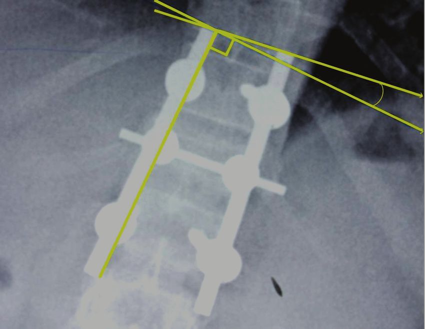
In Group 1 the mean Cobb’s (beta) angle level Figure 1: Radiograph of Dorso-Lumbar spine in
increased up to 22 degrees after 10th post-operative day Anteroposterior -Left lateral bending at sixth post-
and remained same until 6 months. In Group 2 the mean operative week showing measurement of the Left lateral
Cobb’s (beta) angle level increased up to 17.7 degrees bending angle.
Graph 1. Graphical representation showing trend in variation of alpha (α) angle after Right bending in group 1 and 2.
Nepal Journal of Neuroscience, Volume 19, Number 1, 2022 33Doshi et al
Graph 2. Graphical representation showing trend in variation of alpha (α) angle after Left bending in group 1 and 2.
Graph 3. Graphical representation showing change in Cobbs (Beta) angle during post-operative period in both Group 1
and Group 2 patients
34 Nepal Journal of Neuroscience, Volume 19, Number 1, 2022Radiological Comparison of the Biomechanical Characteristics of Short Segment Posterior ...
Cobbs (beta) angle
Cobbs (beta) angle Cobbs (beta) Cobbs (beta) angle
after 10th post-
Group pre-operatively in angle at 6 weeks in at 6 months in
operative day in
supine (pre_opkd) supine (latsu6w) supine(latsu6m)
supine (latsu10d)
Mean 142.500 164.548 166.619 163.62
1 N 21 21 21 21
Std. Deviation 16.4187 15.9122 14.5146 15.571
Mean 145.191 162.853 164.647 166.21
2 N 34 34 34 34
Std. Deviation 14.9585 13.2828 11.9370 11.908
Mean 163.500 165.400 165.22
Total N 55 55 55
Std. Deviation 14.2228 12.8857 13.344
Table 1: Correlation of two groups with respect to Mean and Standard deviation of Cobb’s (Beta) angle in supine position
at given time intervals
Discussion L1 junction.11,12,13 During the surgery we did not observe
any major intraoperative complications. However, Post-
The efficacy of transverse connector is controversial operatively six patients developed complications- one had
and conflicting reports has been published in the literature. bed sore and five developed urinary tract infection.
There are concerns regarding the efficacy of these devices, The improvements were observed in the radiological
as several other studies have failed to demonstrate any parameters (kyphotic deformity) measured pre and post-
appreciable clinical benefits despite the significant operatively. A good correction of kyphosis was achieved
additional cost of each supplementary transverse by surgery with 22 degree and 17.7 degree in Group 1
connector.6 and Group2 respectively. None of the patients developed
Many surgeons all over the world have used transverse significant kyphotic collapse till six months follow-up.
rod connectors to increase the axial and torsional stability Kim et al shows the average preoperative kyphotic angle
of the instrumented spine. There were research studies of all patients was 19.7 degree (+/-8.9), and corrected to
done in vitro which evaluated increased torsional stability 5.5 degree (+/-7.0) post-operatively, and progressed to 8.2
of the construct and common fatigue failure sites with use degree (+/-6.3) at the latest follow-up (14.6 months).14 Jin-
of transverse connector.7,8 Ho Hwang et al shows the mean preoperative, immediate
In cervical spine in vitro studies showed adding postoperative and final kyphosis angles at the fracture site
Transverse connector to C1 Lateral mass- C2 pars can were respectively 20.8 degrees (+/- 6.4), 8.2 degrees (+/-
effectively decrease the axial rotation and enhance the 4.8), and 15.2 degrees (+/- 6.0).15
stability of C1-C2 segment.9 Spinal rotation instability and its role in spinal fusions
We used McAfee’s system to classify the fractures after is difficult to measure. Numerous in vitro biomechanical
radiological evaluation. The most common fracture pattern studies have evaluated the use of transverse connector as
in our study was wedge compression (63.6 %), as revealed adjuvants to posterior constructs. Dick et al analysed various
in the CT scan. The second most common pattern was designs of transverse connector in sawbones and Lynn et
unstable burst fracture (30.9 %), followed by translational al explored the effect of adding Transverse connector to
injuries (5.5 %). The CT reconstruction characteristically pedicle screw-rod constructs for thoracolumbar fractures
showed the mal-alignments. Unstable burst fractures and, in animal cadaveric models.16,17 Both studies concluded
in particular, translational injuries were associated with that transverse connector increased the torsional rigidity
severe neurological involvement. Kim et al. also reported and rotational stiffness of constructs with transverse
a high degree of neurological involvement in patients with connector. But Lynn et al studied lateral bending also,
posterior element involvement – i.e. burst fractures and which states that construct with one transverse connector
rotational injuries.10 In the present study 38 % showed was significantly stiffer than that without any transverse
clustering of the spinal injuries at the first lumbar vertebra connector at 0.5 inch of displacement.17 Lim et al predicted
(L1) level and 47.3 % had mild neurological involvement. improvement in both axial rotation and lateral bending
Other studies show clustering of thoraco-lumbar trauma when transverse connector is used. This study was also
around D12 and L1. Weyns et al. showed 60% injuries done in animal models in vitro.18
over D12- L1, Viale et al. 55% and Carl et al. 82% at D12- Our study states that transverse connector does help
Nepal Journal of Neuroscience, Volume 19, Number 1, 2022 35Doshi et al
in preventing parallelogram effect between the vertical 2. Leone A, Guglielmi G, Cassar-Pullicino VN,
rods especially while lateral bending and thus increases Bonomo L. Lumbar intervertebral instability: a
the stability of the implant. review. Radiology. 2007;245(1):62-77. https://doi.
Variation in Group 2 with transverse connector was org/10.1148/radiol.2451051359
slightly higher in initial period (i.e.) after 10 days and 3. Alhelal F. Design, development, manufacturing
then it remains minimal from 6 weeks to 6 months. But in and biomechanical testing of Stand-alone cage for
Group 1 without transverse connector the change in angle posterior lumbar interbody fusion.
variation was minimal which worsen over 6 weeks and 4. Rajasekaran S, Kanna RM, Shetty AP. Management
it continues to remain worse at 6 months, suggestive of of thoracolumbar spine trauma. Indian journal
parallelogram effect. of orthopaedics. 2015;49(1):72-82. https://doi.
This is first study done in vivo which is done to org/10.4103/0019-5413.143914
study the role of transverse connector and evaluate any 5. Krag MH, Weaver DL, Beynnon BD, Haugh LD.
parallelogram effect in spinal instrumentation. We haven’t Morphometry of the thoracic and lumbar spine
studied torsional stability of instrumented spine as done in related to transpedicular screw placement for surgical
other studies. spinal fixation. Spine. 1988;13(1):27-32. https://doi.
org/10.1097/00007632-198801000-00007
Conclusion 6. Wood KB, Wentorf FA, Ogilvie JW, Kim
KT. Torsional rigidity of scoliosis constructs.
Fractures and fracture dislocations of the spine Spine. 2000;25(15):1893-8. https://doi.
are serious injuries and are occurring mostly among org/10.1097/00007632-200008010-00006
productive age group people. Therefore, their absence 7. Stambough JL, Sabri EH, Huston RL, Genaidy
from the work can cost a lot to the patient, the patient’s AM, Al-Khatib F, Serhan H. Effects of cross-
family and the nation as a whole. So early treatment linkage on fatigue life and failure modes of
and rehabilitation leading to early return to work can stainless steel posterior spinal constructs. Journal
minimize the loss. The management and evaluation of of spinal disorders. 1998;11(3):221-6. https://doi.
these types of injuries have changed dramatically over the org/10.1097/00002517-199806000-00008
last decade due to improvement of imaging technologies 8. Serhan H, Slivka M. Spinal implant transverse rod
and spinal instrumentation. The results showed that Moss connectors: A delicate balance between stability and
Miami pedicle screw system with transverse connector fatigue performance. InSpinal Implants: Are We
provides stable, reliable, segmental construct, helps in Evaluating Them Appropriately? 2003 Jan. ASTM
immediate rehabilitation of patients. There was significant International.
improvement in Kyphotic deformity after instrumentation. 9. Li T, Ma C, Du YQ, Qiao GY, Yu XG, Yin YH.
Transverse connector does play role in preventing The Role of Transverse Connectors in C1-C2
parallelogram effect of implants and provides additional fixation for Atlantoaxial Instability: Is It Necessary?
stability to the construct. We claim that this could be the A Biomechanical Study. World Neurosurgery.
first study done in vivo which is done to study the role 2020;140:e212-8. https://doi.org/10.1016/j.
of transverse connector and evaluate any parallelogram wneu.2020.04.247
effect in spinal instrumentation. 10. Kim NH, Lee HM, Chun IM. Neurologic injury
and recovery in patients with burst fracture of the
Acknowledgement thoracolumbar spine. Spine. 1999;24(3):290-3.
https://doi.org/10.1097/00007632-199902010-00020
We are thankful to XXXXXXX and XXXXX for 11. Weyns F, Rommens PM, Van Calenbergh F, Goffin
reviewing the article. J, Broos P, Plets C. Neurological outcome after
surgery for thoracolumbar fractures. A retrospective
Funding: None study of 93 consecutive cases, treated with dorsal
Conflict of interest: None instrumentation. Eur Spine J Off Publ Eur Spine
Soc Eur Spinal Deform Soc Eur Sect Cerv Spine
Res Soc. 1994;3(5):276–81. https://doi.org/10.1007/
References
BF02226579
12. Viale GL, Silvestro C, Francaviglia N, Carta F,
1. Walker CT, Kakarla UK, Chang SW, Sonntag VK.
Bragazzi R, Bernucci C, Maiello M. Transpedicular
History and advances in spinal neurosurgery: JNSPG
decompression and stabilization of burst fractures of
75th anniversary invited review article. Journal of
the lumbar spine. Surgical neurology. 1993;40(2):104-
Neurosurgery: Spine. 2019;31(6):775-85. https://doi.
11. https://doi.org/10.1016/0090-3019(93)90119-L
org/10.3171/2019.9.SPINE181362
36 Nepal Journal of Neuroscience, Volume 19, Number 1, 2022Radiological Comparison of the Biomechanical Characteristics of Short Segment Posterior ...
13. Carl AL, Tromanhauser SG, Roger DJ. Pedicle 7. PMID: 20166366
screw instrumentation for thoracolumbar burst 16. Dick JC, Zdeblick TA, Bartel BD, Kunz DN.
fractures and fracture-dislocations. Spine. 1992;17(8 Mechanical evaluation of cross-link designs in rigid
Suppl):S317-24. https://doi.org/10.1097/00007632- pedicle screw systems. Spine. 1997;22(4):370-5.
199208001-00018 https://doi.org/10.1097/00007632-199702150-00003
14. Kim KS, Oh SH, Huh JS, Noh JS, Chung BS. Dorsal 17. Lynn G, Mukherjee DP, Kruse RN, Sadasivan
short-segment fixation for unstable thoracolumbar KK, Albright JA. Mechanical stability of
junction fractures. Journal of Korean Neurosurgical thoracolumbar pedicle screw fixation: the effect of
Society. 2006;40(4):249-55. PMid:23826483 crosslinks. Spine. 1997;22(14):1568-72. https://doi.
15. Hwang JH, Modi HN, Yang JH, Kim SJ, Lee SH. org/10.1097/00007632-199707150-00007
Short segment pedicle screw fixation for unstable 18. Lim TH, Eck JC, An HS, Hong JH, Ahn JY, You
T11-L2 fractures: with or without fusion? A three-year JW. Biomechanics of transfixation in pedicle screw
follow-up study. Acta Orthop Belg. 2009;75(6):822- instrumentation. Spine. 1996;21(19):2224-9. https://
doi.org/10.1097/00007632-199610010-00009
Nepal Journal of Neuroscience, Volume 19, Number 1, 2022 37You can also read