Radiologically suspected COVID 19 associated organizing pneumonia responding well to corticosteroids: A report of two cases and a review of the ...
←
→
Page content transcription
If your browser does not render page correctly, please read the page content below
EXPERIMENTAL AND THERAPEUTIC MEDICINE 24: 453, 2022

            Radiologically suspected COVID‑19‑associated organizing
                pneumonia responding well to corticosteroids: A
                report of two cases and a review of the literature
        CHRISTOS SIAFARIKAS1*, CHRISTOS STAFYLIDIS2*, ANASTASIOS TENTOLOURIS1*, STAMATIA SAMARA3,
         IRENE ELIADI3, SOTIRIA MAKRODIMITRI3, DEMETRIOS A. SPANDIDOS4, NIKOLAOS MATHIOUDAKIS5,
                 PANAGIOTIS KARAMICHALOS6, PETROS PAPALEXIS7,8, SERAFEIM CHLAPOUTAKIS9,
           PAGONA SKLAPANI10, NIKOLAOS TRAKAS11 and VASILIKI EPAMEINONDAS GEORGAKOPOULOU3

    1
  First Department of Propedeutic and Internal Medicine, and 2First Department of Internal Medicine, Laiko General Hospital,
    Medical School, National and Kapodistrian University of Athens; 3Department of Infectious Diseases‑COVID‑19 Unit,
Laiko General Hospital, 11527 Athens; 4Laboratory of Clinical Virology, School of Medicine, University of Crete, 71003 Heraklion;
      5
        Renal Transplantation Unit, and 6Department of Pathophysiology, Laiko General Hospital; 7Unit of Endocrinology,
First Department of Internal Medicine, Laiko General Hospital, National and Kapodistrian University of Athens, 11527 Athens;
8
  Department of Biomedical Sciences, University of West Attica, 12243 Athens; 9Department of Thoracic Surgery, Agios Savvas,
       Hospital, 11522 Athens; 10Department of Cytology, Mitera Hospital, 15123 Athens; 11Department of Biochemistry,
                                         Sismanogleio Hospital, 15126 Athens, Greece

                                         Received April 3, 2022; Accepted May 11, 2022

                                                   DOI: 10.3892/etm.2022.11379

Abstract. Organizing pneumonia (OP) is a type of diffuse             pneumonia; thus, the optimal time for administration, the dose
interstitial lung disease, which may be induced in the context       and treatment duration have not yet been determined. The
of several clinical conditions, such as drug reactions, infec‑       present study presents two cases of patients with COVID‑19,
tions, autoimmune diseases and cancer. Coronavirus disease           who exhibited clinical deterioration following the initial phase
2019 (COVID‑19)‑associated OP has been reported as a                 of infection and with radiological characteristics of OP who
late‑stage consequence of the infection or a histological form of    received corticosteroids and had a favorable outcome. The
COVID‑19‑associated pneumonia. Autopsies and postmortem              early diagnosis of COVID‑19‑associated OP may lead to
lung biopsies have demonstrated that the majority of patients        targeted treatment, decreased requirements for ventilatory
with COVID‑19‑associated pneumonia develop secondary OP,             support and an improved survival rate.
and COVID‑19‑associated pneumonia and OP have common
radiological features. The diagnosis of COVID‑19‑associated          Introduction
OP should be suspected in patients with severe acute respira‑
tory syndrome coronavirus 2 infection who exhibit clinical           Since December, 2019, the new coronavirus disease 2019
deterioration despite optimal care, or who have aggravating          (COVID‑19) has become a worldwide hazard. The multi‑organ
symptoms following an initial recovery. The use of cortico‑          manifestations of COVID‑19 have been well‑established. The
steroids is a typical treatment for OP. However, to date, at         most common manifestation is COVID‑19‑associated pneu‑
least to the best of our knowledge, there are a few reports          monia. Although the majority of cases of COVID‑19 are mild,
regarding the role of corticosteroids in COVID‑19‑associated         in more severe cases, acute lung damage may be followed by
                                                                     interstitial lung disease, pulmonary fibrosis and chronic lung
                                                                     function impairment (1,2).
                                                                         Organizing pneumonia (OP) is a type of diffuse interstitial
                                                                     lung disease, which is histopathologically characterized by
Correspondence to: Dr Vasiliki Epameinondas Georgakopoulou,          inflammatory debris in the distal airway containing myofi‑
Department of Infectious Diseases‑COVID‑19 Unit, Laiko General
                                                                     broblasts, fibroblasts and inflammatory cells embedded in a
Hospital, 17 Agiou Thoma Street, 11527 Athens, Greece
                                                                     matrix of connective tissue, and interstitial inflammation of
E‑mail: vaso_georgakopoulou@hotmail.com
                                                                     the surrounding lung parenchyma (3). OP may develop in the
*
    Contributed equally                                              context of various clinical conditions, such as reactions to
                                                                     medication, infections, connective tissue disorders and solid
Key words: organizing pneumonia, coronavirus disease 2019,           organ or hematologic malignancies (4). The term cryptogenic
corticosteroids, chest computed tomography, respiratory infections   OP is used for the primary disease, in which no cause is
                                                                     identified (5). Typical radiological features of OP are peribron‑
                                                                     chovascular and peripheral ground glass opacities (GGOs) or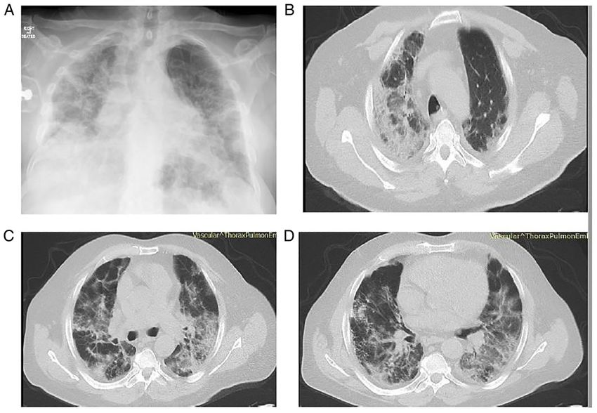
2 SIAFARIKAS et al: COVID-19-ASSOCIATED ORGANIZING PNEUMONIA
consolidations. These lesions may be migratory and accom‑
panied by nodules, masses and interstitial opacities. Another
radiological finding consistent with OP is the reversed halo
sign, a central GGO surrounded by a consolidation halo.
According to previous research, OP is considered to be in the
spectrum of manifestations of acute lung injury (6).
Research on viral‑induced OP during severe acute
respiratory syndrome coronavirus (SARS), Middle East
respiratory syndrome (MERS) and H1N1 infection is exten‑
sive (7,8). Αn increasing number of studies have revealed a
link between SARS‑coronavirus 2 (CoV‑2) infection and OP.
COVID‑19‑associated OP had been previously considered
to be a result of COVID‑19 infection or a histological type
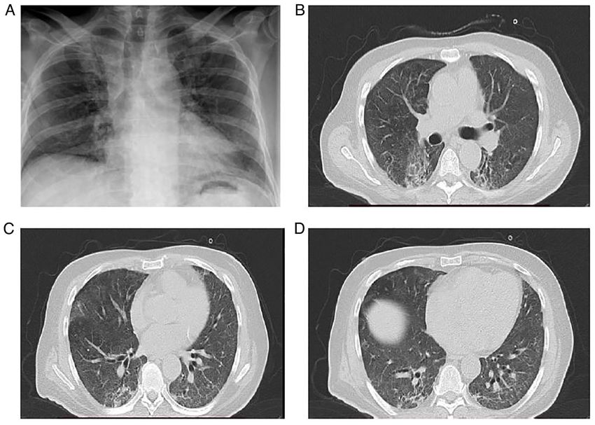
of COVID‑19‑associated pneumonia (9). Of note, the most Figure 1. Chest X‑ray illustrating diffuse infiltrates in all lung fields in the
common findings on chest computed tomography (CT) scans patient in case 1 upon admission.
in patients with COVID‑19 are peripheral GGOs, consolida‑
tion or both, mostly in bilateral and multifocal distributions
that highly resemble a CT pattern of OP (10). Moreover, post‑ respiratory deterioration and required oxygen therapy with a
mortem lung pathological examinations have demonstrated high‑flow nasal cannula [oxygen flow rate, 60 l/min; fraction
that the majority of patients with COVID‑19‑associated of inspired oxygen (FiO2), 90%]. He also received a single dose
pneumonia have secondary OP (11). of intravenous tocilizumab (400 mg).
Research concerning COVID‑19‑associated OP is still The patient's fever, cough, dyspnea and inflammatory
limited. The present study reports two cases of patients indices improved after the treatment was commenced, and on
with COVID‑19 with radiological evidence of OP following day 8 of hospitalization (day 15 of illness), his SpO2 was 97%
the initial infection, who responded well to treatment with while breathing through a high‑flow nasal cannula with lower
corticosteroids. settings (oxygen flow rate, 40 l/min; FiO2, 70%). However,
on day 14 of hospitalization (day 21 of illness), he developed
Case report rapidly progressive respiratory failure, and his SpO2 decreased
to 90%; He thus required oxygen therapy with a high‑flow
Case 1. A 51‑year‑old male patient with no notable previous nasal cannula at maximum settings (oxygen flow rate, 60 l/min;
medical history was admitted to the Emergency Department FiO2, 100%).
(ED) of Laiko General Hospital complaining of fever, cough The patient underwent a new chest X‑ray which revealed
and dyspnea over the last 7 days. The patient had been diag‑ worsening infiltrates in all lung fields (Fig. 2A). He also
nosed with COVID‑19 by reverse transcription polymerase underwent a chest CT scan and chest computed pulmonary
chain reaction (RT‑PCR) of a nasopharyngeal swab sample for angiogram (CTPA), which revealed bilateral peripheral GGO
SARS‑CoV‑2 4 days prior to his admission. The patient was infiltrates and consolidation in both lower lung lobes, with
unvaccinated against SARS‑CoV‑2. areas of reversed halo sign (Fig. 2B‑D). There were no findings
Upon admission, his body temperature was 37.4˚C, his suggesting pulmonary embolism. Simultaneously with this
blood pressure was 120/70 mmHg, his heart rate was 94 beats radiological and respiratory deterioration, the patient presented
per minute, his respiratory rate was 32 breaths per minute, and with recurrent fever and significantly elevated CRP levels
his oxygen saturation (SpO2) was 92% in room air. A chest (279.31 mg/l; reference range, 0‑5 mg/l). He received antimicro‑
examination revealed crackles on auscultation in all lung bial therapy with intravenous piperacillin‑tazobactam at 4.5 g
fields. Arterial blood gases analysis revealed a partial pres‑ four times daily and intravenous linezolid 600 mg twice daily.
sure of oxygen (pO2) of 56 mmHg, partial pressure of carbon Blood and sputum culture did not reveal any infectious micro‑
dioxide (pCO2) 31 mmHg, pH 7.51, HCO3 24.7 mmol/l on organisms. In addition, serum procalcitonin levels were within
room air. A chest X‑ray revealed diffuse infiltrates in all lung the normal range. The patient did not exhibit any improve‑
fields (Fig. 1). The laboratory findings included an increased ment with antibiotics. Based on clinical and radiological
white blood cell (WBC) count (11.22 k/µl; reference range, data, COVID‑19‑associated OP was suspected, and systemic
4.5‑11 k/µl) with neutrophilia (87.4%; reference range, 40‑74%) corticosteroid therapy (methylprednisolone 1 mg/kg/day) was
and lymphopenia (6.63%; reference range, 19‑48%), elevated initiated. On day 23 of hospitalization (day 30 of illness),
C‑reactive protein (CRP) levels (151.77 mg/l; reference range 3 days following the commencement of corticosteroid
0‑5 mg/l), elevated lactate dehydrogenase (LDH) levels therapy, his oxygenation level markedly improved. A chest
(331 U/l; reference range, 135‑225 U/l) and elevated ferritin X‑ray and CT imaging performed on day 14 following the
levels (1,200 ng/ml; reference range, 30‑400 ng/ml). commencement of corticosteroid therapy (day 34 of illness)
Based on these findings, treatment with intravenous remde‑ revealed an improvement of lung infiltrates (Fig. 3). The levels
sivir (200 mg on the first day, followed by 100 mg daily for the of CRP also returned to normal. The methylprednisolone
following 4 days) and dexamethasone (6 mg/day) for 10 days administration was decreased to 40 mg, and the patient was
was commenced for SARS‑CoV‑2 infection. The patient also discharged on day 39 following admission (day 46 of illness).
received oxygen therapy with a Venturi mask, delivering an Following discharge, the methylprednisolone administration
oxygen concentration of 60%. The patient exhibited further was decreased to 32 mg for 10 days, 16 mg for 10 days and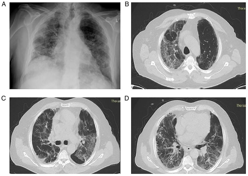
EXPERIMENTAL AND THERAPEUTIC MEDICINE 24: 453, 2022                                                                3

Figure 2. Chest X‑ray and computed tomography of the patient in case 1 on day 14 of hospitalization. (A) Chest X‑ray illustrating lung infiltrates in all lung
fields. (B) Chest computed tomography illustrating bilateral peripheral ground glass infiltrates in upper lung lobes. (C and D) Chest computed tomography
illustrating consolidation in both lower lung lobes, with areas of reverse halo sign.

Figure 3. Chest X‑ray and computed tomography of the patient in case 1 performed on day 14 following the commencement of corticosteroid therapy. (A) Chest
X‑ray illustrating an improvement in lung infiltrates in all lung fields. (B) Chest computed tomography reveals improvement in bilateral peripheral ground glass
infiltrates in upper lung lobes. (C and D) Chest computed tomography reveals improvement in lung lesions compared to Fig. 2C and D.
4                                     SIAFARIKAS et al: COVID-19-ASSOCIATED ORGANIZING PNEUMONIA

                                                                                     Based on these findings, treatment with intravenous
                                                                                 remdesivir (200 mg on the first day, followed by 100 mg daily
                                                                                 for the following 4 days) and dexamethasone (6 mg/day) was
                                                                                 commenced for SARS‑CoV‑2 infection for 10 days. The patient
                                                                                 also received oxygen therapy with a Venturi mask, delivering
                                                                                 an oxygen concentration of 60%. The patient exhibited further
                                                                                 respiratory deterioration with a further elevation of CRP levels
                                                                                 (174 mg/l; reference range, 0‑5 mg/l) on the second day of
                                                                                 hospitalization (day 15 of illness) and required oxygen therapy
                                                                                 with a high‑flow nasal cannula (oxygen flow rate, 60 l/min;
                                                                                 FiO2, 90%). He also received antimicrobial treatment with
                                                                                 intravenous ceftriaxone at 2 g once daily and linezolid 600 mg
                                                                                 twice daily for 7 days.
                                                                                     The patient's clinical symptoms and inflammatory indices
                                                                                 (CRP, 17 mg/l; reference range, 0‑5 mg/l) improved after
                                                                                 commencing the treatment, and on day 10 of hospitalization
                                                                                 (day 23 of illness), his SpO2 was 97%, while breathing through
                                                                                 a Venturi mask delivering an oxygen concentration of 60%.
                                                                                 The patient did not exhibit any further improvement for the
Figure 4. Chest X‑ray illustrating diffuse infiltrates in all lung fields with
                                                                                 following 2 days. On day 13 of hospitalization (day 26 of illness)
consolidations in the right upper and middle lung field and in the left middle
lung field of the patient in case 2 upon admission.                              the patient developed recurrent low‑grade fever and a concurrent
                                                                                 new increase in CRP levels (49 mg/l; reference range, 0‑5 mg/l).
                                                                                     The patient underwent a new chest X‑ray, which revealed
                                                                                 persistent infiltrates in all lung fields with consolidations
8 mg for 10 days, and discontinued thereafter. The patient did                   in the right upper and middle lung field, and in the left
not present with a relapse on a follow‑up at 3 months after                      middle lung field (Fig. 5A). He also underwent a chest
discharge.                                                                       CT scan and CTPA, which revealed bilateral consolida‑
                                                                                 tions with areas of reversed halo sign in all lung fields
Case 2. A 71‑year‑old male patient presented to the ED of                        and peripheral GGO infiltrates in both lower lung lobes
Laiko General Hospital complaining of fever, cough and                           (Fig. 5B‑D). There were no findings suggesting pulmonary
dyspnea over the last 24 h. The patient had been diagnosed                       embolism. No infectious microorganisms were isolated
with COVID‑19 by RT‑PCR testing of a nasopharyngeal swab                         from blood and sputum cultures. Based on the clinical and
sample for SARS‑CoV‑2 14 days prior to his admission. The                        radiological data, COVID‑19‑associated OP was suspected,
patient reported low‑grade fever then for only 2 days without                    and systemic corticosteroid therapy (methylprednisolone at
any other symptoms and had not received any specific therapy                     1 mg/kg/day) was initiated on day 15 (day 28 of illness).
for COVID‑19.                                                                    On day 18, at 3 days following the commencement of
    The patient had a medical history of asthma, arterial                        corticosteroid therapy (day 31 of illness), his oxygenation
hypertension, dyslipidemia, appendectomy and inguinal                            level improved considerably. A chest X‑ray and CT imaging
hernia surgery. His current medications included inhaled                         performed on day 13 following the commencement of
formoterol/budesonide (160/4.5 µg twice daily), olmesartan/                      corticosteroid therapy (day 28 of hospitalization, day 41 of
hydrochlothiazide (40/12 mg once daily) and atorvastatin                         illness) revealed a notable improvement in previously noted
(20 mg once daily). The patient was unvaccinated against                         lung infiltrates (Fig. 6). The levels of CRP also returned to
SARS‑CoV‑2.                                                                      normal. Methylprednisolone administration was decreased
    Upon admission, his body temperature was 38.5˚C, his                         to 40 mg, and the patient was discharged on day 33 after
blood pressure was 140/80 mmHg, his heart rate was 90 beats                      admission (day 46 of illness). Following discharge, meth‑
per minute, his respiratory rate was 28 breaths per minute,                      ylprednisolone administration was decreased to 32 mg for
and his SpO2 was 87% in room air. A chest examination                            7 days, 16 mg for 7 days and 8 mg for 7 days, and discon‑
revealed crackles on auscultation in all lung fields. Arterial                   tinued thereafter. The patient did not present with a relapse
blood gas analysis revealed a pO2 of 52 mmHg, a pCO2 of                          and he had improvement in chest X‑ray at a follow‑up
29 mmHg, pH 7.50 and HCO3 22.7 mmol/l in room air. A                             2 months after discharge (Fig. 7).
chest X‑ray revealed diffuse infiltrates in all lung fields with
consolidations in the right upper and middle lung field and                      Discussion
in the left middle lung field (Fig. 4). The laboratory findings
included an increased WBC count (13.56 k/µl; reference                           The present study describes the cases of two individuals who
range, 4.5‑11 k/µl) with neutrophilia (84.8%; reference range,                   had rapidly worsening respiratory symptoms after the initial
40‑74%) and lymphopenia (8.7%; reference range, 19‑48%),                         phase of COVID‑19 infection. In numerous aspects, the
elevated CRP levels (93 mg/l; reference range, 0‑5 mg/l),                        patients' symptoms are compatible with those of OP caused by
elevated LDH levels (500 U/l; reference range, 135‑225 U/l)                      SARS‑CoV‑2. Following early treatment for their COVID‑19
and elevated ferritin levels (816 ng/ml; reference range,                        infection, both patients had rapidly deteriorating respiratory
30‑400 ng/ml).                                                                   symptoms that were not responding to optimal therapy.
EXPERIMENTAL AND THERAPEUTIC MEDICINE 24: 453, 2022 5 Figure 5. Chest X‑ray and computed tomography of the patient in case 2 on day 13 of hospitalization. (A) Chest X‑ray illustrating persistent infiltrates in all lung fields with consolidations in the right upper and middle lung field and in the left middle lung field. (B and C) Chest computed tomography illustrating bilateral consolidations with areas of reverse halo sign in upper lung fields. (D) Chest computed tomography illustrating bilateral consolidations with areas of reverse halo sign and peripheral ground glass infiltrates in both lower lung lobes. Figure 6. Chest X‑ray and computed tomography of the patient in case 2 performed on day 13 following the commencement of corticosteroid therapy. (A) Chest X‑ray illustrating an improvement in lung infiltrates compared to Fig. 5A. (B and C) Chest computed tomography illustrating an improvement in lung infiltrates in upper lung fields. (D) Chest computed tomography reveals improvement in lung infiltrates in both lower lung lobes.
6 SIAFARIKAS et al: COVID-19-ASSOCIATED ORGANIZING PNEUMONIA
A chest CT scan may be beneficial for ruling out complica‑
tions, such as pulmonary embolism, secondary pneumonia, or
OP in patients with SARS‑CoV‑2 infection who have clinical
deterioration despite optimal treatment and ventilatory support,
or who have worsening symptoms following an initial recovery.
Both patients in the present study waited an average of 21 days
(20‑22 days) from symptom initiation to undergo a chest
CT scan, and their average duration of stay after commencing
treatment with steroids was 10 days (5‑15 days). In the case that
additional imaging analyses are required for such individuals,
this time frame may be an attractive approach.
From the beginning of the pandemic, some researchers
have expressed their concerns about potential widespread
failure to identify and treat COVID‑19‑associated OP (9).
Radiological evidence of OP has been documented during the
later course of SARS‑CoV‑2 infection. In a previous study,
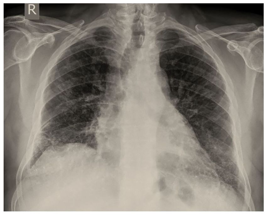
Figure 7. Chest X‑ray illustrating a significant improvement in lung infiltrates the observed prevalence of COVID‑19‑associated OP was
of the patient in case 2 at the follow‑up of 2 months. Please compare this to 12.5% (27). In another observational study, the authors studied
the image in Fig. 5A.
persistent lung changes following COVID‑19 infection and
described ongoing symptoms in 39% of participants. In that
study, OP was observed in 4.8% of these patients, with signifi‑
Of note, the patient in case 1 who was younger than the cant radiological and clinical improvement following steroid
patient in case 2 developed rapidly progressive respiratory administration (28). In addition, in a study on respiratory inter‑
failure on the day 14, while the patient in case 2 recovered more mediate care unit patients, 58% of patients that underwent a
easily under the same treatment regimen. Genetic factors may chest CT scan had a radiological pattern consistent with OP and
be a possible explanation for this event. Some patients have a significantly decreased need for intubation and in‑hospital
heritable, single‑gene mutations that influence their immune mortality compared to those with a GGO pattern (29).
systems. Such distinctive mutations have been detected in Of note, both patients described herein were unvaccinated
numerous cases of young individuals who are healthy, yet against SARS‑CoV‑2. A recent study demonstrated that
suddenly develop a life‑threatening infection (12). vaccination with at least two doses of COVID‑19 vaccine was
Furthermore, no substantial superimposed infection was associated with a significant decrease in reporting the most
proven in the patients described herein. Moreover, their condi‑ common post‑acute COVID‑19 symptoms, such as fatigue,
tion markedly improved following the initiation of high‑dose headache, weakness and persistent muscle pain (30). However,
corticosteroid treatment, suggesting that OP was the cause of whether vaccination against SARS‑CoV‑2 exerts a protective
their decompensated respiratory symptoms. effect against the development of OP following COVID‑19
Clinical and investigation data regarding COVID‑19- remains to be determined.
associated OP are limited. The diagnosis of OP was made in The role of corticosteroids in COVID‑19‑associated
some cases, by using only radiological data (13‑19), and in other pneumonia has been well‑established. Dexamethasone
cases, by performing transbronchial biopsy and a histological therapy is effective for COVID‑19‑associated pneumonia (31).
examination of the obtained tissue (14,20‑25). The cases of Methylprednisolone has been reported to increase the survival
COVID‑19‑associated organizing pneumonia reported in the of hospitalized patients who experience severe COVID‑19
literature are summarized in Table I. pneumonia (31). A previous meta‑analysis revealed a posi‑
A pathological investigation is essential for definitive tive effect of corticosteroids on short‑term mortality and a
diagnosis of COVID‑19‑associated OP, as radiological mani‑ decrease in the need for mechanical ventilation in patients
festations of bacterial co‑infection and OP may be similar. with COVID‑19 (32).
Therefore, it does not appear plausible to confirm the pres‑ The cornerstone of OP treatment is corticosteroids.
ence of OP in patients with SARS‑CoV‑2‑related pneumonia Prednisone at a dosage of 0.75‑1.5 mg/kg/day for 4 weeks,
based solely on radiological abnormalities (25). However, in tapered over a period of 3‑6 months, is the current recommen‑
both cases present herein, the conduction of lung biopsy was dation (33). In COVID‑19‑associated OP, case reports and case
considered extremely dangerous and was thus not performed, series, including ours, have documented favorable outcomes
due to high hypoxic conditions. with corticosteroid treatment, sometimes with high doses and
Notably, not every case of OP is confirmed by pathological prolonged therapy, often weeks to months (13‑19, 22‑25). A
data, as is well‑recognized. The primary goal of a biopsy in single‑center prospective observational study discovered that
individuals with suspected OP is to rule out other possible almost a quarter of post‑COVID‑19 pneumonia patients had
causes of comparable symptoms. As the histopathologic recurrent dyspnea with radiological signs of OP, and that
characteristics of OP are similar to those of other interstitial restarting corticosteroids resulted in clinical and radiological
lung diseases, relying on transbronchial biopsies may increase improvement in these patients (27). Of interest, the sponta‑
the risk of OP misdiagnosis or delay (13). Furthermore, the neous remission of OP without corticosteroid administration
radiographic characteristics of OP are distinct, and a chest CT has been described in two cases of histologically diagnosed
scan has a good diagnostic accuracy (79%) for OP (26). COVID‑19‑associated OP (20,21).EXPERIMENTAL AND THERAPEUTIC MEDICINE 24: 453, 2022 7
Table I. Cases of COVID‑19‑associated organizing pneumonia identified in the literature.
Case Age
no. Author/(Refs.) (years)/sex Diagnosis Management Outcome
1 Alsulami et al (13) 71/Μ Radiological Corticosteroids Recovery
2 Alsulami et al (13) 54/M Radiological Corticosteroids Recovery
3 Alsulami et al (13) 57/M Radiological Corticosteroids Recovery
4 Alsulami et al (13) 49/M Radiological Corticosteroids Recovery
5 Alsulami et al (13) 56/F Radiological Corticosteroids Recovery
6 Alsulami et al (13) 83/F Radiological Corticosteroids Recovery
7 Ng et al (14) 58/F Radiological Corticosteroids Recovery
8 de Oliveira Filho et al (15) 52/Μ Radiological Corticosteroids Recovery
9 de Oliveira Filho et al (15) 60/F Radiological Corticosteroids Recovery
10 de Oliveira Filho et al (15) 63/F Radiological Corticosteroids Recovery
11 Horii et al (16) 70/F Radiological Corticosteroids Recovery
12 Okamori et al (17) 60/M Radiological Corticosteroids Recovery
13 Okamori et al (17) 61/F Radiological Corticosteroids Recovery
14 Kim et al (18) 71/M Radiological Corticosteroids Recovery
15 Simões et al (19) 71/M Radiological Corticosteroids Recovery
16 Simões et al (19) 83/M Radiological Corticosteroids Recovery
17 Ng et al (14) 81/M TBLB, histopathological Corticosteroids Recovery
examination
18 Seo et al (20) 50/M TBLB, histopathological Spontaneous Recovery
examination remission
19 Funk et al (21) 49/M TBLB, histopathological Spontaneous Recovery
examination remission
20 Golbets et al (22) 36/M TBLB, histopathological Corticosteroids Recovery
examination
21 Kanaoka et al (23) 56/M TBLB, histopathological Corticosteroids Recovery
examination
22 Kanaoka et al (23) 84/F TBLB, histopathological Corticosteroids Recovery
examination
23 Cortés Colorado et al (24) 62/M TBLB, histopathological Corticosteroids Recovery
examination
24 Vadász et al (25) 57/M TBLB.histopathological Corticosteroids Recovery
examination
25 Vadász et al (25) 70/M TBLB,histopathological Corticosteroids Recovery
examination
26 Vadász et al (25) 76/M TBLB, histopathological Corticosteroids Recovery
examination
F, female; M, male; TBLB, transbronchial lung biopsy.
A previous study demonstrated that approximately one Large, multicenter randomized control trials are required
third of OP cases exhibited a complete resolution of lesions at to determine the optimal time to start, dosage, duration and
1‑2 months following disease onset, while those with greater benefit of corticosteroid therapy in COVID‑19‑associated OP.
areas of consolidation and more extensive lung involvement Clinicians should take the possible risks and advantages of
were more likely to develop fibrotic‑like changes (34). There corticosteroids into account and utilize CT as an evaluation
is currently no evidence to indicate that corticosteroid therapy tool for the distinct phases of CT patterns in patients with
can prevent relapses of OP or minimize the development of COVID‑19.
residual pulmonary fibrosis in COVID‑19‑associated OP (35). In conclusion, the importance of detecting OP arises from
There has been no research to date, at least to the best of our the fact that patients who receive treatment with corticosteroids
knowledge, on the impact of corticosteroids on the resolution have a better prognosis and outcome. Increasing awareness of
of COVID‑19‑related OP and long‑term pulmonary outcomes. this diagnosis may lead to more effective treatment approaches8 SIAFARIKAS et al: COVID-19-ASSOCIATED ORGANIZING PNEUMONIA
for COVID‑19 disease, further reduction of requirements 4. Oikonomou A and Hansell DM: Organizing pneumonia: The
many morphological faces. Eur Radiol 12: 1486‑1496, 2002.
for ventilatory support, and improved survival. Patients 5. Cottin V and Cordier JF: Cryptogenic organizing pneumonia.
with COVID‑19 that exhibit a minimal or no improvement Semin Respir Crit Care Med 33: 462‑475, 2012.
despite optimal therapy should be evaluated further with lung 6. Mandal RV, Mark EJ and Kradin RL: Organizing pneumonia and
pulmonary lymphatic architecture in diffuse alveolar damage.
imaging, as they may benefit from early diagnosis and targeted Hum Pathol 39: 1234‑1238, 2008.
treatment. The optimal time for corticosteroid administration, 7. Cornejo R, Llanos O, Fernández C, Carlos Díaz J, Cardemil G,
the dose and the duration of therapy need to be addressed. Salguero J, Luengo C, Tobar E, Romero C and Gálvez LR:
Organizing pneumonia in patients with severe respiratory
failure due to novel A (H1N1) influenza. BMJ Case Rep 2010:
Acknowledgements bcr0220102708, 2010.
8. Tse GM, To KF, Chan PK, Lo AW, Ng KC, Wu A, Lee N,
Wong HC, Mak SM, Chan KF, et al: Pulmonary pathological
Not applicable. features in coronavirus associated severe acute respiratory
syndrome (SARS). J Clin Pathol 57: 260‑265, 2004.
Funding 9. Kory P and Kanne JP: SARS‑CoV‑2 organising pneumonia:
‘Has there been a widespread failure to identify and treat this
prevalent condition in COVID‑19?’. BMJ Open Respir Res 7:
No funding was received. e000724, 2020.
10. Kanne JP, Little BP, Chung JH, Elicker BM and Ketai LH:
Essentials for radiologists on COVID‑19: An update‑radiology
Availability of data and materials scientific expert panel. Radiology 296: E113‑E114, 2020.
11. Copin MC, Parmentier E, Duburcq T, Poissy J, Mathieu D and
The datasets used and/or analyzed during the current study are Lille COVID‑19 ICU and Anatomopathology Group: Time
to consider histologic pattern of lung injury to treat critically
available from the corresponding author on reasonable request. ill patients with COVID‑19 infection. Intensive Care Med 46:
1124‑1126, 2020.
Authors' contributions 12. Andreakos E, Abel L, Vinh DC, Kaja E, Drolet BA, Zhang Q,
O'Farrelly C, Novelli G, Rodríguez‑Gallego C, Haerynck F, et al:
A global effort to dissect the human genetic basis of resistance to
SM, SS and IE conceptualized the study. PP, PK, NM, PS, NT SARS‑CoV‑2 infection. Nat Immunol 23: 159‑164, 2022.
and SC obtained medical images, and prepared the tables and 13. Alsulami F, Dhaliwal I, Mrkobrada M and Nicholson M: Post
Covid‑19 organizing pneumonia: Case series for 6 patients with
figures. VEG, CSi, CSt and AT advised on patient treatment post‑COVID interstitial lung disease. J Lung Pulm Respir Res 8:
and wrote and prepared the draft of the manuscript. DAS and 108‑111, 2021.
VEG analyzed patient data and provided critical revisions. 14. Ng BH, Ban AY, Nik Abeed NN and Faisal M: Organising pneu‑
monia manifesting as a late‑phase complication of COVID‑19.
CSi, CSt, AT, NM, PK, PP, SC, PS and NT made substantial BMJ Case Rep 14: e246119, 2021.
contributions to conception and design, and analysis and inter‑ 15. de Oliveira Filho CM, Vieceli T, de Fraga Bassotto C,
pretation of data. VEG and SM confirm the authenticity of all da Rosa Barbato JP, Garcia TS and Scheffel RS: Organizing
pneumonia: A late phase complication of COVID‑19 responding
the data. All authors contributed to manuscript revision and dramatically to corticosteroids. Braz J Infect Dis 25: 101541, 2021.
have read and approved the final manuscript. 16. Horii H, Kamada K, Nakakubo S, Yamashita Y, Nakamura J,
Nasuhara Y and Konno S: Rapidly progressive organizing pneu‑
monia associated with COVID‑19. Respir Med Case Rep 31:
Ethics approval and consent to participate 101295, 2020.
17. Okamori S, Lee H, Kondo Y, Akiyama Y, Kabata H, Kaneko Y,
Not applicable. Ishii M, Hasegawa N and Fukunaga K: Coronavirus disease
2019‑associated rapidly progressive organizing pneumonia with
fibrotic feature: Two case reports. Medicine (Baltimore) 99:
Patient consent for publication e21804, 2020.
18. Kim T, Son E, Jeon D, Lee SJ, Lim S and Cho WH: Effectiveness
of steroid treatment for SARS‑CoV‑2 pneumonia with cryp‑
Written informed was obtained from the patients for the publi‑ togenic organizing pneumonia‑like reaction: A case report.
cation of the data. A copy of the written consent is available for Disaster Med Public Health Prep 26: 1‑4, 2020.
review by the Editor‑in‑Chief of this journal on request. 19. Simões JP, Alves Ferreira AR, Almeida PM, Trigueiros F,
Braz A, Inácio JR, Medeiros FC, Braz S and Pais de Lacerda A:
Organizing pneumonia and COVID‑19: A report of two cases.
Competing interests Respir Med Case Rep 32: 101359, 2021.
20. Seo H, Jung J, Kim MJ, Jang SJ and Kim SH: Radiologically
suspected organizing pneumonia in a patient recovering from
DAS is the Editor‑in‑Chief for the journal, but had no personal COVID‑19: A case report. Infect Chemother 54: 208‑212, 2022.
involvement in the reviewing process, or any influence in 21. Funk GC, Nell C, Pokieser W, Thaler B, Rainer G and Valipour A:
terms of adjudicating on the final decision, for this article. The Organizing pneumonia following Covid19 pneumonia. Wien
Klin Wochenschr 133: 979‑982, 2021.
other authors declare that they have no competing interests. 22. Golbets E, Kaplan A, Shafat T, Yagel Y, Jotkowitz A, Awesat J
and Barski L: Secondary organizing pneumonia after recovery of
References mild COVID‑19 infection. J Med Virol 94: 417‑423, 2022.
23. Kanaoka K, Minami S, Ihara S, Tanaka T, Yasuoka H and
Komuta K: Secondary organizing pneumonia after coronavirus
1. Wang F, Kream RM and Stefano GB: Long‑term respiratory disease 2019: Two cases. Respir Med Case Rep 32: 101356, 2021.
and neurological sequelae of COVID‑19. Med Sci Monit 26: 24. Cortés Colorado JM, Cardona Ardila LF, Aguirre Vásquez N,
e928996, 2020. Gómez Calderón KC, Lozano Álvarez SL and Carrillo Bayona JA:
2. Vasarmidi E, Tsitoura E, Spandidos DA, Tzanakis N and Organizing pneumonia associated with SARS‑CoV‑2 infection.
Antoniou KM: Pulmonary fibrosis in the aftermath of the Radiol Case Rep 16: 2634‑2639, 2021.
COVID‑19 era (Review). Exp Ther Med 20: 2557‑2560, 2020. 25. Vadász I, Husain‑Syed F, Dorfmüller P, Roller FC, Tello K,
3. Roberton BJ and Hansell DM: Organizing pneumonia: A Hecker M, Morty RE, Gattenlöhner S, Walmrath HD,
kaleidoscope of concepts and morphologies. Eur Radiol 21: Grimminger F, et al: Severe organising pneumonia following
2244‑2254, 2011. COVID‑19. Thorax 76: 201‑204, 2021.EXPERIMENTAL AND THERAPEUTIC MEDICINE 24: 453, 2022 9
26. Johkoh T, Müller NL, Cartier Y, Kavanagh PV, Hartman TE, 31. RECOVERY Collaborative Group, Horby P, Lim WS,
Akira M, Ichikado K, Ando M and Nakamura H: Idiopathic Emberson JR, Mafham M, Bell JL, Linsell L, Staplin N,
interstitial pneumonias: diagnostic accuracy of thin‑section CT Brightling C, Ustianowski A, et al: Dexamethasone in hospital‑
in 129 patients. Radiology 211: 555‑560, 1999. ized patients with Covid‑19. N Engl J Med 384: 693‑704, 2021.
27. Shi H, Han X, Jiang N, Cao Y, Alwalid O, Gu J, Fan Y and 32. Edalatifard M, Akhtari M, Salehi M, Naderi Z, Jamshidi A,
Zheng C: Radiological findings from 81 patients with COVID‑19 Mostafaei S, Najafizadeh SR, Farhadi E, Jalili N, Esfahani M, et al:
pneumonia in Wuhan, China: A descriptive study. Lancet Infect Intravenous methylprednisolone pulse as a treatment for hospi‑
Dis 20: 425‑434, 2020. talised severe COVID‑19 patients: Results from a randomised
28. Myall KJ, Mukherjee B, Castanheira AM, Lam JL, Benedetti G, controlled clinical trial. Eur Respir J 56: 2002808, 2020.
Mak SM, Preston R, Thillai M, Dewar A, Molyneaux PL and 33. van Paassen J, Vos JS, Hoekstra EM, Neumann KMI, Boot PC
West AG: Persistent post‑COVID‑19 interstitial lung disease. An and Arbous SM: Corticosteroid use in COVID‑19 patients: A
observational study of corticosteroid treatment. Ann Am Thorac systematic review and meta‑analysis on clinical outcomes. Crit
Soc 18: 799‑806, 2021. Care 24: 696, 2020.
29. Rocha AS, Meireles M, Vilaça H, Guimarães TC, Martins MD, 34. Cordier JF: Cryptogenic organising pneumonia. Eur Respir J 28:
Santos LR, Castro A and Mesquita M: Outcomes of Covid‑19 422‑446, 2006.
organizing pneumonia in critically ill patients. J Infect 83: 35. Wang Y, Jin C, Wu CC, Zhao H, Liang T, Liu Z, Jian Z, Li R,
496‑522, 2021. Wang Z, Li F, et al: Organizing pneumonia of COVID‑19:
30. Kuodi P, Gorelik Y, Zayyad H, Wertheim O, Beiruti Wiegler K, Time‑dependent evolution and outcome in CT findings. PLoS
Abu Jabal K, Dror AA, Nazzal S, Glikman D and Edelstein M: One 15: e0240347, 2020.
Association between vaccination status and reported incidence
of post‑acute COVID‑19 symptoms in Israel: A cross‑sectional This work is licensed under a Creative Commons
study of patients tested between March 2020 and November Attribution-NonCommercial-NoDerivatives 4.0
2021. medRxiv: Jan 17, 2022 (Epub ahead of print). International (CC BY-NC-ND 4.0) License.You can also read