Mycoplasma pneumoniae pneumonia with pulmonary embolism

Page created by Roberto Richards
 
CONTINUE READING
Mycoplasma pneumoniae pneumonia with pulmonary embolism
EXPERIMENTAL AND THERAPEUTIC MEDICINE 21: 201, 2021

  Mycoplasma pneumoniae pneumonia with pulmonary embolism:
       A study on pediatric cases in Jilin province of China
                   CHU‑QIAO SHENG, CHUN‑FENG YANG, YU AO, ZHI‑YUE ZHAO and YU‑MEI LI

   Department of Pediatric Intensive Care Unit, The First Hospital of Jilin University, Changchun, Jilin 130021, P.R. China

                                     Received February 13, 2020; Accepted November 26, 2020

                                                      DOI: 10.3892/etm.2021.9634

Abstract. Mycoplasma is one of the most common patho‑                   During the operation, the local tissues were determined to
gens causing community‑acquired pneumonia in pediatric                  be infarcted and the pathological diagnosis was consistent
patients. In recent years, the number of refractory or severe           with pulmonary infarction. Among the 5 cases, 2 died of
cases with drug resistance has been gradually increasing                Acute Respiratory Distress Syndrome at 3‑8 days after the
and cases that developed embolism after Mycoplasma pneu‑                operation. The remaining patients underwent 6‑12 months
moniae (M. pneumoniae) infection have been reported. The                of follow‑up and respiratory rehabilitation and their quality
present study retrospectively analyzed the clinical features,           of life is now good. In conclusion, compared with healthy
diagnosis and treatment of M. pneumoniae pneumonia                      individuals, pediatric patients with critical MPP have an
(MPP) combined with pulmonary embolism (PE) in a series                 elevated risk of embolism. It is necessary to be vigilant
of 7 cases encountered between January 1st, 2016 to August              regarding whether MMP is combined with PE and perform
1st, 2019 at the Department of Pediatric Intensive Care                 timely CTPA examination. Early detection, early treatment
Unit of The First Hospital of Jilin University (Changchun,              and surgical intervention (if necessary) may significantly
China). Combined with relevant Chinese and international                reduce the risk of mortality and disability.
studies published during the last two decades, a comprehen‑
sive analysis was performed. All of the pediatric patients              Introduction
of the present study had fever, cough and dyspnea respira‑
tory symptoms at onset and the disease progressed rapidly.              Mycoplasma pneumoniae (M. pneumoniae) is the most
Thereafter, PE was confirmed by a series of examinations.               common pathogen in pediatric patients with commu‑
Pulmonary CT indicated patchy inflammations and signifi‑                nity‑acquired pneumonia (1,2) which breaks out every
cantly elevated D‑dimer levels, accompanied by positive                 3‑5 years (3). In recent years, the number of refractory
anticardiolipin antibodies. Furthermore, a filling defect in            or severe cases with drug resistance has been gradually
the pulmonary artery branch was observed on CT pulmonary                increasing (4‑6). In addition to pulmonary inflammation as
angiography (CTPA) examination. In 2 cases, the condi‑                  the most common manifestation, M. pneumoniae infection
tion was improved with anti‑infection and anticoagulation               may also cause damage to multiple systems and organs (7‑9).
treatment with low‑molecular‑weight heparin and warfarin,               In the last 20 years, ~60 cases of M. pneumoniae infection
respectively, and the pulmonary embolism disappeared                    with thrombotic disease in pediatric patients have been
after 3‑4 months. A total of 5 cases, who were not respon‑              reported worldwide (10‑15). The present study reported on
sive to the drug treatment, underwent surgical resection.               a series of 7 cases of M. pneumoniae pneumonia (MPP)
                                                                        accompanied by pulmonary embolism (PE) encountered
                                                                        January 1st, 2016 to August 1st, 2019 at the Department of
                                                                        Pediatric Intensive Care Unit of The First Hospital of Jilin
Correspondence to: Professor Yu‑Mei Li, Department of Pediatric         University (Changchun, China), and the clinical data of these
Intensive Care Unit, The First Hospital of Jilin University, 1 Xinmin   cases were reviewed. The present study aimed to improve the
Street, Changchun, Jilin 130021, P.R. China                             understanding of clinicians regarding the laboratory exami‑
E‑mail: liyumei201912@126.com                                           nations, diagnosis and treatments for pediatric patients with
                                                                        MPP‑associated PE.
Abbreviations:    ACA, anticardiolipin antibody; CTPA,
computed tomographic pulmonary angiography; M. pneumoniae,
Mycoplasma pneumoniae; MPP, M. pneumoniae pneumonia; PE,                Case report
pulmonary embolism
                                                                        Cases. MPP‑associated PE was confirmed in 7 cases by radio‑
Key words: mycoplasma pneumonia, pediatric patients, pulmonary          logical examination combined with serological tests and the
embolism, refractory mycoplasma            pneumonia,      computed     corresponding clinical data were collected and summarized.
tomographic pulmonary angiography                                       The present study had been approved by the Ethics Committee
                                                                        of the First Hospital of Jilin University (Changchun, China;
                                                                        approval no. 2019‑253).
Mycoplasma pneumoniae pneumonia with pulmonary embolism
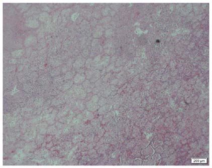
2                                              SHENG et al: PEDIATRIC MPP WITH PE

Baseline data. The seven cases were aged between 6 and             Table I. Baseline demographic and clinical characteristics of
11 years (median, 8.0 years) with a male/female ratio of 4:3,      the patients (n=7).
as presented in Table I. All patients were otherwise physically
healthy. Patients with a family history of thrombophilia and a     Characteristic                                       Value
history of allergy were excluded.
                                                                   Demographics
Clinical symptoms and physical signs. All of the patients had       Age (years)                                       8 (6, 11)
a cough and fever as the initial symptoms, typically irritable      Male sex                                          4 (57.14)
cough with viscous sputum, and remittent fever. Among              Anthropometry
them, 5 cases developed dyspnea within 2 to 6 days and were         Body weight (kg)                             24.2 (21.3, 30)
hospitalized on day 3‑12 from onset. These cases developed          Body height (cm)                            123.4 (119.5, 130)
PE on day 10‑14 and their condition soon deteriorated. Older        BMI z‑score                                   0.5 (‑0.5, 1)
pediatric patients complained of chest pain, chest tightness
or sudden dyspnea; younger patients were unable to describe        Clinical symptoms
their symptoms, but physical examinations revealed spiritless‑      Cough                                             7 (100)
ness, aggravating dyspnea, flapping of nasal wings, reduced         Fever                                             7 (100)
respiratory movement amplitude on the affected side and weak        Dyspnea                                           5 (71.43)
breath sounds. One case was combined with swelling in the           Swelling in limb                                  1 (14.29)
right lower limb.                                                  Radiological examination
                                                                    Pulmonary CT
Results of auxiliary examinations                                    Extensive diffuse inflammatory                   6 (85.71)
Laboratory tests. The serum biochemistry results of the cases
                                                                     Subcutaneous emphysema                           1 (14.29)
are presented in Table II. The serum M. pneumoniae antibody
                                                                     Pleural effusion                                 4 (57.14)
titers (Particle Agglutination assay; SERODIA‑MYCOII;
Fujirebio) were increased by varying degrees [from negative          Pericardial effusion                             1 (14.29)
titer at presentation to positive titer at the 2nd examination      Pulmonary arterial embolism
(n=1) or >4‑fold increased antibody titers during admission          Bilateral multiple branches                      2 (28.57)
(n=6)]. All cases had a significant increase in the platelet         Upper lobe of the right lung                     2 (28.57)
count and D‑dimer level. The levels of protein C and protein         Lower lobe of the right lung                     2 (28.57)
S first transiently decreased and were then restored to normal.      Upper lobe of the left lung                      1 (14.29)
Furthermore, two cases were weakly positive for anticardio‑
lipin antibody (ACA). In addition, four cases were positive        Values are expressed as the median (interquartile range) or n (%).
for ACA as detected by ELISA (16) (QUANTA Lite ACA                 BMI, body mass index.
IgG III; Inova). A total of four cases received bronchoscopy,
through which endobronchitis and necrotizing pneumonia
were revealed. The bronchoalveolar lavage fluid was tested
positive for M. pneumoniae (DNA sequence copy number,              Treatment. After admission, all of the cases were given
7,880‑16,343) and positivity for the macrolide resistance gene     the standard anti‑infection therapy of macrolides and
was detected in 2 cases.                                           certain patients received concurrent antibiotic therapy with
                                                                   third‑generation cephalosporins or carbapenems. Over the
Radiological examination. All 7 cases received dynamic             same period, moxifloxacin was selected for the 2 cases with
monitoring by chest X‑ray, pulmonary CT scan and computed          positivity for the drug resistance gene. Those patients with
tomographic pulmonary angiography (CTPA) during hospital‑          dyspnea were treated by tracheal intubation and mechanical
ization. Furthermore, 6 cases were indicated to have extensive     ventilation. In the meantime, other systemic treatments,
diffuse inflammatory changes in the two lungs upon chest           such as organ protection and nutritional support, were given.
X‑ray or pulmonary CT scan (Figs. 1 and 2) and 1 case had          Risk stratification was performed based on the guidelines
subcutaneous emphysema (Fig. 3). Furthermore, 4 cases were         of the American College of Chest Physicians (17), along
combined with a moderate amount of pleural effusion and            with the thrombolysis and anti‑coagulation therapies.
1 case was combined with mild pericardial effusion (Fig. 4).       Low‑molecular‑weight heparin calcium was injected subcu‑
CTPA indicated that 2 cases had a pulmonary arterial embo‑         taneously at the dose of 50 IU/kg per time, twice daily.
lism in multiple branches bilaterally (Fig. 5, arrows); 2 cases    During the treatment, the pediatric patients were properly
had a pulmonary arterial embolism in the upper lobe of the         immobilized to avoid violent cough and movement. Since
right lung and one of the two lesions was located at the distal    these pediatric patients did not have a basic history of
end of the right upper lung; 1 case had filling defects in the     congenital heart disease and presented with no pulmo‑
pulmonary artery branches in the upper lobe of the left lung;      nary thrombosis, no shock due to PE and no deep vein
2 cases had distal pulmonary artery embolism in the lower          thrombosis, thrombophilia was excluded and thrombolytic
lobe of the right lung. Furthermore, 1 case with swelling in the   therapy was therefore not selected. However, anticoagulant
lower limbs received local vascular ultrasound examination,        therapy had no curative effect in 5 cases and the disease
through which thrombosis in the common femoral vein was            progressed to pulmonary infarction; thus, surgical resec‑
detected.                                                          tions were conducted.
Mycoplasma pneumoniae pneumonia with pulmonary embolism
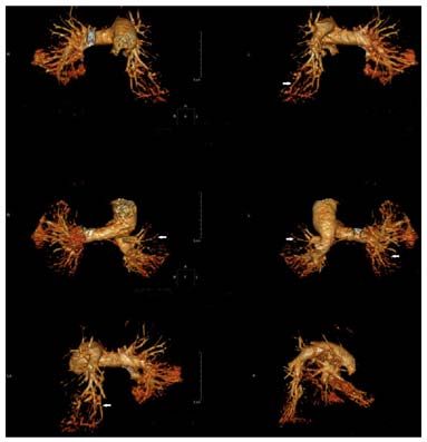
Table II. Results of auxiliary examinations of the patients.

                                                                                                                    Serum                                                              Macrolides                                                                                                                                                     Images included
Patient                                                                             WBC       PLT       CRP     M. pneumoniae		                                                        resistance                               Other		                                                                                        D‑dimer     ‑‑‑‑‑‑‑‑‑‑‑‑‑‑‑‑‑‑‑‑‑‑‑‑‑‑‑‑‑‑‑‑‑‑‑‑‑‑‑‑‑‑‑‑‑‑‑‑‑‑‑‑‑
no.                                                                                (x109/l) (x109/l)   (mg/l)      antibodya BAL‑DNA                                                  gene (BALF)                              pathogen ACA                                                                                     (µg/l)        CT            CTPA Pathology Treatment                                                                         Outcomes

1                                                                                      34.62   755      265        1:320      16343                                                                                 Positive   Cpn         Negative                                                                                 6883   Fig. 2       ‑             ‑                                                                               OP      Died
2                                                                                      27.55   810      210        1:160      14555                                                                                 Positive   S. aureus   Positive                                                                                 6500   Fig. 3       ‑             ‑                                                                               OPs,    Alive
3                                                                                       5.93   480       80        1:160        NA                                                                                  NA         NA          Positive                                                                                 1800     ‑        Fig. 5          ‑                                                                                ‑      Alive
4                                                                                      12.33   550      110        1:160      13880                                                                                 NA         Cpn         Positive                                                                                 1200     ‑          ‑           Fig. 7                                                                            OPs,    Died
5                                                                                      20.12   610      164        1:160        NA                                                                                  NA         Cpn         Weakly positive                                                                          5868   Fig. 1       ‑             ‑                                                                               OP,     Alive
6                                                                                       9.84   490     71.4        1:40        7880                                                                                 NA         Cpn         Positive                                                                                 1024     ‑          ‑           Fig. 6                                                                             ‑      Alive
7                                                                                      18.13   680      123        1:160        NA                                                                                  NA         S. aureus   Weakly positive                                                                          2109   Fig. 4       ‑             ‑                                                                               OPs,    Alive
a
 Determined during the 2nd examination, which was performed 6.7±1.4 days after the 1st examination. ACA, anti‑cardiolipin antibody; BAL‑DNA, M. pneumoniae DNA sequence copy number in BALF;
BALF, bronchoalveolar lavage fluid; M. pneumoniae, Mycoplasma pneumoniae; Cpn, Chlamydia pneumoniae; CRP, C‑reactive protein; CTPA, computed tomographic pulmonary angiography; OP,
operation; PLT, platelets; S. aureus, Staphylococcus aureus; WBC, white blood cells; NA, not available.
                                                                                                                                                                                                                                                                                                                                                                                                                                                                        EXPERIMENTAL AND THERAPEUTIC MEDICINE 21: 201, 2021

                                                                                                                                                                                                                                                       dation in the right lung of an 8 year old male.

                                                                                                                                      subcutaneous emphysema of an 11 year old male.
                                                                                                                                                                                                                                                                                                                                                                       atelectasis of the dorsal lobes of a 7 year old male.

    of the left lung (upper panel) and mild pericardial effusion (lower panel).
                                                                                                                                      Figure 3. Severe inflammatory changes in the bilateral lungs with extensive

    the left lung of a 6 year old female, with partial atelectasis in the lower lobe
                                                                                                                                                                                                                                                                                                                                                                       Figure 1. Bilateral pulmonary extensive diffuse inflammation, with bilateral

    Figure 4. Inflammation in the middle lobe of the right lung and all lobes of
                                                                                                                                                                                                                                                       Figure 2. Bilateral pulmonary extensive diffuse inflammation with consoli‑
                                                                                                                                                                                                                                                                                                                                                                                                                                                                        3
4                                                       SHENG et al: PEDIATRIC MPP WITH PE

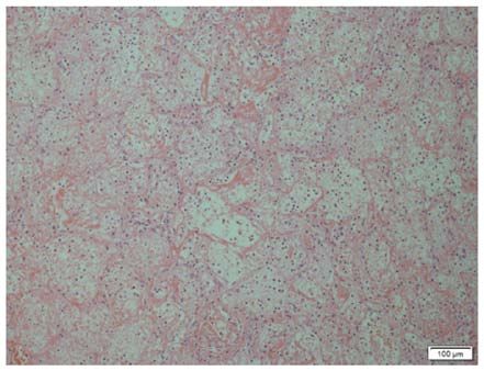
                                                                               Figure 6. Histological image displaying pulmonary infarction and necrosis
                                                                               with abscess formation in an 8 year old female (H&E staining; scale bar,
                                                                               200 µm).
Figure 5. Bilateral pulmonary arterial filling defect of a 6 year old male;
arterial embolism is visible in multiple branches, with mild dilation of the
pulmonary trunk (white arrows; scale bar, 5 cm).

Outcomes and follow‑up. Anticoagulant therapy is the first
choice for all pediatric patients with PE if there is no contra‑
indication (18). A total of 2 patients achieved significant
improvement after 15‑21 days of treatment as detected by
chest radiological examination and D‑dimer test. They were
discharged after the symptoms improved, and the PE disap‑
peared 3‑5 months later as detected during the follow‑up. The
5 remaining cases exhibited no improvement in the local PE,
which was confirmed by pulmonary CT scan on day 14‑21
during the anticoagulation therapy. Pulmonary infarction
was considered in certain patients who had local nodular
                                                                               Figure 7. Extensive infarction and necrosis of the resected pulmonary tissues
solid lesions in the lungs with cavitation and the condition                   with infiltration of a large amount of inflammatory cells in an 11 year old
deteriorated to high dependence on oxygen. Among them,                         female (H&E staining; scale bar, 100 µm).
2 patients had considerable pleural effusion (pus) and required
surgical resection. Intraoperative findings included dark‑red
resected pulmonary tissues or yellowish‑white infarct‑like
changes. These tissues had no contraction and dilation func‑                   treatment process; and ii) Studies published within the last
tions, with high tension and pus coating on their surface. The                 20 years. Exclusion criteria was incomplete clinical infor‑
pathological diagnosis of the resected pulmonary tissues was                   mation. Ultimately, the clinical data of 10 pediatric cases
pulmonary infarction (Figs. 6 and 7). Among them, 2 cases                      with MPP and PE were reported and their details are listed
were combined with Acinetobacter baumannii infection                           in Table III (19‑26).
after surgery and Acute Respiratory Distress Syndrome                              These cases were aged between 6 and 13 years (median
occurred 3‑8 days later. Although the patients were treated                    age, 9.0 years) with a male/female ratio of 5:1. The levels of
with extracorporeal membrane oxygenation, their condition                      M. pneumoniae antibody were significantly increased, along
did not improve and they eventually died. The remaining                        with a transient decrease in protein S and protein C. The
surviving pediatric patients underwent 6‑12 months of                          lesions were located at the lower lobe close to the hilus of the
follow‑up and respiratory rehabilitation and they recovered                    lung. After receiving anti‑infective and anti‑coagulant treat‑
to a normal state.                                                             ments, 8 cases improved but 1 patient died.

Literature search and review. Using ‘Pulmonary Embolism’                       Discussion
and ‘Mycoplasma pneumoniae pneumonia’ with ‘pediatric’
as the keywords, relevant articles were searched in the                        Pediatric patients with critical MPP complicated with PE was
PubMed, MEDLINE, Update, Web of Science and Embase                             rarely reported. Cases with mild PE may be asymptomatic,
databases. The Chinese subject heading terms used in the                       while severe cases may suffer from pulmonary arterial hyper‑
Wanfang, Chinese National Knowledge Infrastructure and                         tension, unstable hemodynamics or even sudden death (27,28).
Chongqing VIP databases were the same as those above.                          The common symptoms include shortness of breath, chest
The inclusion criteria were as follows: i) Pediatric patients                  pain and even dyspnea (19‑26). Missed diagnosis may occur
with a confirmed diagnosis of MPP and PE during the                            if young pediatric patients are not able to properly describe
Table III. Details of previous studies.

Author          Case		 Age Intervala       Antibody to
(year)           no. Sex (years) (days)   M. pneumoniae   Agglutination test     D reg           Treatment               Outcome             (Refs.)

Graw‑Panzer 1 M 13  5 ELISA IgM (1:128) Increased D‑dimer, protein Left popliteal         Heparin + warfarin     Radiographic chest           (19)
(2009)						                             S deficiency and positive   vein embolism		                             findings returned to
						                                   ACA.                        and PE.		                                   normal after 3 months
									                                                                                                        and the anemia
									                                                                                                        resolved gradually
									                                                                                                        over 5 months.
Chen (2013) 2 F 12 12 PA (1:160)         Increased D‑dimer,          Thrombosis in        Low‑molecular‑         The chest X‑ray was          (20)
						                                   positive ACA                right lower limb     weight heparin +       almost normal at
							                                                              and PE (left         warfarin               follow‑up after
							                                                              lower lobe).		                              6 months.
Brown       3 M  6 16 Complement binding Positive ACA and            Femoral vein         Not mentioned          Alive.                       (21)
(2008)					           (1:640)            acquired activated protein  embolism and
						                                   C resistance.               PE (left lower lobe)
Su (2012)   4 M  6 17 ELISA (1:128) CA   Increased D‑dimer, positive PE (left lower       Heparin + warfarin     At the 3‑month               (22)
					                 (1:1,024)          ACA and decreased activity lobe)		                                      follow‑up, Aca was
						                                   of plasma protein C.			                                                 negative, plasma
									                                                                                                        protein C activity
									                                                                                                        recovered and lung
									                                                                                                        lesions were absorbed.
Wei (2015)  5 M  9 23 Not mentioned      Increased D‑dimer and       PE (right lower      Heparin + warfarin     Chest radiographic           (23)
					                 (1:1,280)          positive ACA.               lobe).		                                    findings returned to
									                                                                                                        normal after 3 months.
Zhuo (2015) 6 M  9 10 PA (1:160)         Increased D‑dimer.          PE (mainly on        Low‑molecular‑weight   Died on the eighth           (24)
							                                                              the right side).     heparin + warfarin     day after admission.
                                                                                                                                                       EXPERIMENTAL AND THERAPEUTIC MEDICINE 21: 201, 2021

Qin (2019)  7 F 10 14 Not mentioned      Increased D‑dimer,          PE (bilateral        Low‑molecular‑         At the 8‑month               (25)
					                 (1:320)            anticardiolipin IgM         lung).               weight heparin         follow‑up, chest CT
						                                   antibody was positive, 		                        calcium + warfarin     indicated old lung
						                                   plasma protein C/S 			                                                  lesions in both
						                                   activity was not 			                                                    lungs, segmental
						                                   mentioned.			                                                           atelectasis of the right
									                                                                                                        upper lung accompanied
									                                                                                                        by bilateral lower lung
									                                                                                                        filaments and a small
									                                                                                                        amount of pleural
                                                                                                                                                       5

									                                                                                                        lesions in the left lung.
6                                                                                                           SHENG et al: PEDIATRIC MPP WITH PE

                                                                                                                                                                                                                                                                                                                                    their symptoms. Therefore, if PE is not discovered in a timely

                                                                                                                            Interval refers to the duration from fever to PE. PE, pulmonary embolism; M. pneumoniae, Mycoplasma pneumoniae; ACA, anticardiolipin antibody; CA, cold agglutinin; PA, particle agglutination assay;
                                 (Refs.)

                                                      (26)

                                                                                  (26)
                                                                                                                                                                                                                                                                                                                                    manner, the anti‑coagulation treatment is delayed and the
                                                                                                                                                                                                                                                                                                                                    disease may progress into acute pulmonary infarction or
                                                                                                                                                                                                                                                                                                                                    even death. When encountering pediatric cases with MPP,

                                                      5.5 months. The lesions

                                                                                  4.5 months. The lesions
                                                                                                                                                                                                                                                                                                                                    the patient or the parents should be asked whether there is a
                                                                                                                                                                                                                                                                                                                                    family history of protein C/protein S deficiency, recent history
                                                      The total course of

                                                                                  The total course of
                                 Outcome

                                                                                                                                                                                                                                                                                                                                    of surgeries or presence of congenital vascular malformation,
                                                      were absorbed.

                                                                                  were absorbed.
                                                                                                                                                                                                                                                                                                                                    so as to preliminarily assess the risk of PE. In the present
                                                      treatment was

                                                                                  treatment was
                                                                                                                                                                                                                                                                                                                                    study, evaluation at the early stage of admission indicated a
                                                                                                                                                                                                                                                                                                                                    low risk of thrombosis in all cases. However, their symptoms
                                                                                                                                                                                                                                                                                                                                    kept on deteriorating during the treatment and chest radio‑
                                                                                                                                                                                                                                                                                                                                    logical examination indicated poor recovery. In combination
                                                                                                                                                                                                                                                                                                                                    with laboratory tests and chest radiological examination, the
                                                                            Methylprednisolone +

                                                                            Methylprednisolone +

                                                                                                                                                                                                                                                                                                                                    diagnosis of PE was confirmed and the standard treatment was
                                                      posterior tibial vein Nadroparin calcium

                                                                            Nadroparin calcium

                                                                                                                                                                                                                                                                                                                                    provided.
                                 Treatment

                                                                                                                                                                                                                                                                                                                                        For such pediatric cases, physicians should begin early
                                                                                                                                                                                                                                                                                                                                    dynamic monitoring and examination, which may be able to
                                                                                                                                                                                                                                                                                                                                    effectively control disease progression, reduce surgical rates
                                                                                                                                                                                                                                                                                                                                    and mortality. However, with the current technological stan‑
                                                                                                                                                                                                                                                                                                                                    dards available, it is still limited to perform the interventional
                                                      in both lower limbs
                                                      and PE (bilateral

                                                      saphenous vein in
                                                      right lower limb and

                                                                                                                                                                                                                                                                                                                                    treatment and implement thrombolysis therapy for young pedi‑
                                                                                                                                                                                                                                                                                                                                    atric patients with PE. More work, such as a more appropriate
                                                      the bilateral lung).
                                                      Thrombophlebitis

                                                      PE (lower lobe in

                                                                                                                                                                                                                                                                                                                                    design using more sophisticated instruments, still needs to be
                                                      Thrombosis of
                                 D reg

                                                                                                                                                                                                                                                                                                                                    done to overcome this deficiency in the future.
                                                      of the great

                                                                                                                                                                                                                                                                                                                                        The pathogenesis of M. pneumoniae infection with
                                                                                                                                                                                                                                                                                                                                    thrombosis remains to be fully elucidated, but it may be asso‑
                                                      lung).

                                                                                                                                                                                                                                                                                                                                    ciated with immune damage mediated by infection (7,29‑31).
                                                                                                                                                                                                                                                                                                                                    Since the membrane proteins and glycolipids of M. pneu‑
                                                                                            III, protein C/S were normal.

                                                                                                                                                                                                                                                                                                                                    moniae have certain common antigens in the heart, liver,
                                                                                            III, protein C were normal.

                                                                                                                                                                                                                                                                                                                                    lung, brain, kidney and smooth muscle tissues of the human
                                                                                            activities of antithrombin

                                                                                            activities of antithrombin

                                                                                            Protein S was decreased.
                                 Agglutination test

                                                                                            Increased D‑dimer, the

                                                                                            Increased D‑dimer, the

                                                                                                                                                                                                                                                                                                                                    body, upon infection of the host with M. pneumoniae, the
                                                                                                                                                                                                                                                                                                                                    corresponding antibodies are produced and the immune
                                                                                                                                                                                                                                                                                                                                    complex is formed to activate complement, which produces
                                                                                                                                                                                                                                                                                                                                    neutrophils. Previous studies reported that embolism may
                                                                                                                                                                                                                                                                                                                                    affect multiple sites of the body after M. pneumoniae infec‑
                                                                                                                                                                                                                                                                                                                                    tion, including the brain, lower extremity veins, spleen and

                                                                                                                                                                                                                                                                                                                                    pulmonary arteries (10‑15). Chemokines, which attract
                                                                                                                                                                                                                                                                                                                                    a large number of white blood cells to invade the lesion,

                                                                                                                                                                                                                                                                                                                                    release a large number of inflammatory mediators and
                                                                                                                                                                                                                                                                                                                                    lysosomal enzymes, causing inflammatory damage to target
                        M. pneumoniae

                                                                            ELISA (1:1,280)

                                                                            ELISA (1:1,280)
                         Antibody to

                                                                                                                                                                                                                                                                                                                                    organs. It was reported that patients with MPP and thrombus
                                                                                                                                                                                                                                                                                                                                    were positive for ACA (32,33). ACA is an autoantibody that
                                                                                                                                                                                                                                                                                                                                    targets antigens in platelets and cardiolipin on endothelial
                                                                                                                                                                                                                                                                                                                                    membranes and is associated with thrombogenesis. The
                                                                                                                                                                                                                                                                                                                                    present study concluded that M. pneumoniae infection may

                                                                                                                                                                                                                                                                                                                                    cause vascular endothelial cell injury and ACA positivity,
                        Case		 Age Intervala

                                                                                                                                                                                                                                                                                                                                    leading to a temporary hypercoagulability state that induces
                         no. Sex (years) (days)

                                                                         20

                                                                          6

                                                                                                                                                                                                                                                                                                                                    thrombosis. Under severe conditions, M. pneumoniae infec‑
                                                                                                                            D reg, damaged region; M, male; F, female.

                                                                                                                                                                                                                                                                                                                                    tion further affects the synthesis of coagulation factors and
                                                                                                                                                                                                                                                                                                                                    thrombin (e.g., protein C, protein S and antithrombin III),
                                                                                                                                                                                                                                                                                                                                    resulting in embolism. Certain patients may acquire protein
                                                                       8

                                                                       5

                                                                                                                                                                                                                                                                                                                                    C or protein S deficiency/resistance (21,34). The D‑dimer test
                                                                                                                                                                                                                                                                                                                                    is an important screening method for PE and a negative result
                                                                     F

                                                                     F

                                                                                                                                                                                                                                                                                                                                    may exclude PE with 100% certainty (35,36). While CTPA is
Table III. Continued.

                                                                                                                                                                                                                                                                                                                                    considered as the gold standard for PE diagnosis (37). Based
                                                      Zhang (2019) 8

                                                      Zhang (2019) 9

                                                                                                                                                                                                                                                                                                                                    on the above points, in a child with severe MPP, the D‑dimer
                                                                                                                                                                                                                                                                                                                                    test, ACA test, Protein C test, Protein S test and CTPA should
                                                                                                                                                                                                                                                                                                                                    be considered to prevent the occurrence of PE.
                                                                                                                                                                                                                                                                                                                                        At present, the major treatment for pediatric patients
                        Author
                        (year)

                                                                                                                                                                                                                                                                                                                                    with acute PE is anticoagulant therapy, the purpose of which
                                                                                                                                      a
EXPERIMENTAL AND THERAPEUTIC MEDICINE 21: 201, 2021                                                   7

is to prevent acute thrombosis and expansion. If available,        References
local thrombus therapy or interventional thrombus therapy
may be performed. During the anticoagulation therapy with           1. Jain S, Williams DJ, Arnold SR, Ampofo K, Bramley AM,
                                                                       Reed C, Stockmann C, Anderson EJ, Grijalva CG, Self WH, et al:
low‑molecular‑weight heparin, dynamic monitoring of the                Community‑acquired pneumonia requiring hospitalization
coagulation status and treatment outcome is required to avoid          among U.S. children. N Engl J Med 372: 835‑845, 2015.
bleeding, whose risk is considerable. Furthermore, coopera‑         2. Tashiro M, Fushimi K, Kawano K, Takazono T, Saijo T,
                                                                       Yamamoto K, Kuriha ra S, Imamura Y, Miyaza ki T,
tion between the pediatric thoracic surgery department and the         Yanagihara K, et al: Comparison of efficacy of antimicrobial
vascular surgery department is preferred and surgical inter‑           agents among hospitalized patients with Mycoplasma pneu‑
vention may be provided if necessary.                                  moniae pneumonia in Japan during large epidemics of
                                                                       macrolide‑resistant M. pneumoniae infections: A nationwide
    Critical M. pneumoniae infection in pediatric patients is          observational study. Clin Infect Dis 65: 1837‑1842, 2017.
associated with a high risk of PE. During clinical treatment,       3. Omori R, Nakata Y, Tessmer HL, Suzuki S and Shibayama K:
such cases should be screened for high‑risk factors and                The determinant of periodicity in Mycoplasma pneumoniae
                                                                       incidence: An insight from mathematical modelling. Sci Rep 5:
patients could be closely monitored for any manifestations of          14473, 2015.
PE. Necessary examinations, particularly CTPA, should be            4. Tanaka T, Oishi T, Miyata I, Wakabayashi S, Kono M, Ono S,
performed to confirm the diagnosis and to initiate standard            Kato A, Fukuda Y, Saito A, Kondo E, et al: Macrolide‑resistant
                                                                       Mycoplasma pneumoniae infection, Japan, 2008‑2015. Emerg
treatment as early as possible. Surgical intervention is another       Infect Dis 23: 1703‑1706, 2017.
important salvage to reduce poor prognosis, if the disease          5. Huang L, Chen H and Peng S: Spontaneous pneumomediastinum,
progresses to pulmonary infarction, and therefore results in           emphysema, and pulmonary bullae associated with refrac‑
                                                                       tory Mycoplasma pneumoniae pneumonia in a child. Pediatr
serious and life threatening complications.                            Pulmonol 52: E77‑E80, 2017.
                                                                    6. Pereyre S, Goret J and Bébéar C: Mycoplasma pneumoniae:
Acknowledgements                                                       Current knowledge on macrolide resistance and treatment. Front
                                                                       Microbiol 7: 974, 2016.
                                                                    7. Narita M: Classification of extrapulmonary manifestations due
Not applicable.                                                        to Mycoplasma pneumoniae infection on the basis of possible
                                                                       pathogenesis. Front Microbiol 7: 23, 2016.
                                                                    8. Olson D, Watkins LK, Demirjian A, Lin X, Robinson CC, Pretty K,
Funding                                                                Benitez AJ, Winchell JM, Diaz MH, Miller LA, et al: Outbreak
                                                                       of Mycoplasma pneumoniae‑associated Stevens‑Johnson
No funding was received.                                               syndrome. Pediatrics 136: e386‑e394, 2015.
                                                                    9. Dhaliwal K and Enright K: Rare extrapulmonary complications
                                                                       of Mycoplasma pneumoniae infection. BMJ Case Rep 2016:
Availability of data                                                   bcr2015214044, 2016.
                                                                   10. Witmer CM, Steenhoff AP, Shah SS and Raffini LJ:
                                                                       Mycoplasma pneumoniae, splenic infarct, and transient
The datasets used and/or analysed during the current study             antiphospholipid antibodies: A new association? Pediatrics 119:
are available from the corresponding author on reasonable              e292‑e295, 2007.
request.                                                           11. Bao Y, Li X, Wang K, Zhao C, Ji X and Jiang M: Central
                                                                       retinal artery occlusion and cerebral infarction associated with
                                                                       Mycoplasma pneumonia infection in children. BMC Pediatr 16:
Authors' contributions                                                 210, 2016.
                                                                   12. Kang B, Kim DH, Hong YJ, Son BK, Lim MK, Choe YH and
                                                                       Kwon YS: Complete occlusion of the right middle cerebral artery
CQS conceived the current study and drafted and revised the            associated with Mycoplasma pneumoniae pneumonia. Korean J
manuscript. CFY collected the literature and reviewed and              Pediatr 59: 149‑152, 2016.
revised the manuscript. YA and ZYZ collected the data and          13. Oeser C, Andreas M, Rath C, Habertheuer A and Kocher A:
                                                                       Left ventricular thrombus in a patient with cutaneous T‑cell
performed initial analyses. YML coordinated and supervised             lymphoma, hypereosinophilia and Mycoplasma pneumoniae
data collection, and critically reviewed the manuscript. CQS           infection‑a challenging diagnosis: A case report. J Cardiothorac
and YML confirm the authenticity of all the raw data. All              Surg 10: 21, 2015.
                                                                   14. Bakshi M, Khemani C, Vishwanathan V, Anand RK and
authors read and approved the final manuscript.                        Khubchandani RP: Mycoplasma pneumonia with antiphospho‑
                                                                       lipid antibodies and a cardiac thrombus. Lupus 15: 105‑106, 2006.
Ethics approval and consent to participate                         15. Jin X, Zou Y, Zhai J, Liu J and Huang B: Refractory
                                                                       Mycoplasma pneumoniae pneumonia with concomitant acute
                                                                       cerebral infarction in a child: A case report and literature review.
The research followed international and national regulations           Medicine (Baltimore) 97: e0103, 2018.
in accordance with the Declaration of Helsinki. The study was      16. Tincani A, Balestrieri G, Allegri F, Cinquini M, Vianelli M,
                                                                       Taglietti M, Sanmarco M, Ichikawa K, Koike T, Meroni P and
approved by the Ethics Committee of the First Hospital of Jilin        Boffa MC: Overview on anticardiolipin ELISA standardization.
University. (Changchun, China; approval no. 2019‑253).                 J Autoimmun 15: 195‑197, 2000.
                                                                   17. Kearon C, Akl EA, Ornelas J, Blaivas A, Jimenez D,
                                                                       Bounameaux H, Huisman M, King CS, Morris TA, Sood N, et al:
Patient consent for publication                                        Antithrombotic therapy for VTE disease: CHEST guideline and
                                                                       expert panel report. Chest 149: 315‑352, 2016.
Written informed consents were obtained from the patients'         18. Monagle P, Cuello CA, Augustine C, Bonduel M, Brandão LR,
legal guardians for the publication of any accompanying                Capman T, Chan AKC, Hanson S, Male C, Meerpohl J, et al:
                                                                       American society of hematology 2018 Guidelines for manage‑
images prior to submission.                                            ment of venous thromboembolism: Treatment of pediatric venous
                                                                       thromboembolism. Blood Adv 2: 3292‑3316, 2018.
Competing interests                                                19. Graw‑Panzer KD, Verma S, Rao S, Miller ST and Lee H: Venous
                                                                       thrombosis and pulmonary embolism in a child with pneumonia
                                                                       due to Mycoplasma pneumoniae. J Natl Med Assoc 101: 956‑958,
The authors declare that they have no competing interests.             2009.
8                                                 SHENG et al: PEDIATRIC MPP WITH PE

20. Chen Y, Huang P, Chen Q, Lin Z and Tian W: Two separated           30. Narita M: Pathogenesis of neurologic manifestations of
    thrombi in deep veins associated with pulmonary embolism after         Mycoplasma pneumoniae infection. Pediatr Neurol 41: 159‑166,
    Mycoplasma pneumoniae infection: A case in adolescent female.          2009.
    Transl Pediatr 2: 198‑201, 2013.                                   31. Sotgiu S, Pugliatti M, Rosati G, Deiana GA and Sechi GP:
21. Brown S, Padley S, Bush A, Cummins D, Davidson S and                   Neurological disorders associated with Mycoplasma pneu‑
    Buchdahl R: Mycoplasma pneumonia and pulmonary embo‑                   moniae infection. Eur J Neurol 10: 165‑168, 2003.
    lism in a child due to acquired prothrombotic factors. Pediatr     32. Nagashima M, Higaki T, Satoh H and Nakano T: Cardiac
    Pulmonol 43: 200‑202, 2008.                                            thrombus associated with Mycoplasma pneumoniae infection.
22. Su HY, Jin WJ, Zhang HL and Li CC: Clinical analysis of                Interact Cardiovasc Thorac Surg 11: 849‑851, 2010.
    pulmonary embolism in a child with Mycoplasma pneu‑                33. Senda J, Ito M, Atsuta N, Watanabe H, Hattori N, Kawai H
    moniae pneumonia. Zhonghua Er Ke Za Zhi 50: 151‑154, 2012              and Sobue G: Paradoxical brain embolism induced by
    (In Chinese).                                                          Mycoplasma pneumoniae infection with deep venous thrombus.
23. Wei H, Chang Y and Lu S: A case report of pulmonary embolism           Intern Med 49: 2003‑2005, 2010.
    associated with Mycoplasma pneumoniae pneumonia. Zhonghua          34. Ascer E, Marques M and Gidlund M: M pneumoniae infection,
    Er Ke Za Zhi 53: 143‑144, 2015 (In Chinese).                           pulmonary thromboembolism and antiphospholipid antibodies.
24. Zhuo Z, Li F, Chen X, Jin P, Guo Q and Wang H: Mycoplasma              BMJ Case Rep 2011: bcr1220103561, 2011.
    pneumonia combined with pulmonary infarction in a child. Int J     35. van Es N, van der Hulle T, van Es J, den Exter PL, Douma RA,
    Clin Exp Med 8: 1482‑1486, 2015.                                       Goekoop RJ, Mos IC, Galipienzo J, Kamphuisen PW,
25. Qin Y, Wang H, Wang Y, Zhang W, He L and Liu C:                        Huisman MV, et al: Wells rule and d‑Dimer testing to rule out
    Mycoplasma pneumoniae pneumonia complicated with pulmo‑                pulmonary embolism: A systematic review and individual‑patient
    nary embolism in children: A case report. J Clin Pediatr 37:           data meta‑analysis. Ann intern Med 165: 253‑261, 2016.
    765‑768, 2019 (In Chinese).                                        36. Konstantinides SV, Barco S, Lankeit M and Meyer G:
26. Zhang J, Liu F, Guo C, et al: A report of 2 cases of pediatric         Management of pulmonary embolism: An update. J Am Coll
    refractory Mycoplasma pneumonia complicated with pulmonary             Cardiol 67: 976‑990, 2016.
    embolism. Chin J Pract Pediatr 34: 1043‑1045, 2019 (In Chinese).   37. Moore AJE, Wachsmann J, Chamarthy MR, Panjikaran L,
    doi: 10.19538/j.ek2019120617.                                          Tanabe Y and Rajiah P: Imaging of acute pulmonary embolism:
27. Dijl FN, Curtin J, Lord D and Fitzgerald DA: Pulmonary embo‑           An update. Cardiovasc Diagn Ther 8: 225‑243, 2018.
    lism in children. Paediatr Respir Rev 13: 112‑122, 2012.
28. Simmons BP and Aber RC: Mycoplasma pneumoniae pneu‑                                 This work is licensed under a Creative Commons
    monia. Symptoms mimicking pulmonary embolism with                                   Attribution-NonCommercial-NoDerivatives 4.0
    infarction. JAMA 241: 1268‑1269, 1979.                                              International (CC BY-NC-ND 4.0) License.
29. Li T, Yu H, Hou W, Li Z, Han C and Wang L: Evaluation of
    variation in coagulation among children with Mycoplasma pneu‑
    moniae pneumonia: A case‑control study. J Int Med Res 45:
    2110‑2118, 2017.
You can also read