Recurrent variation in the active NOR sites in the monkey frogs of the genus Pithecopus Cope, 1866 (Phyllomedusidae, Anura)
←
→
Page content transcription
If your browser does not render page correctly, please read the page content below
CompCytogen 13(4): 325–338 (2019)                                                                         COMPARATIVE                 A peer-reviewed open-access journal

                                                    Cytogenetics of Pithecupus
doi: 10.3897/CompCytogen.v13i4.37687                     RESEARCH ARTICLE
                                                                                                                    325
                                                                                                          Cytogenetics
http://compcytogen.pensoft.net                                                                                  International Journal of Plant & Animal Cytogenetics,
                                                                                                                       Karyosystematics, and Molecular Systematics

       Recurrent variation in the active NOR sites in the
       monkey frogs of the genus Pithecopus Cope, 1866
                  (Phyllomedusidae, Anura)

       Joana Moura Gama1, Camilla Borges Gazolla1, Deborah Yasmin de Souza1,
               Shirlei Maria Recco-Pimentel2, Daniel Pacheco Bruschi1

1 Programa de Pós-Graduação em Genética (PPG-GEN). Departamento de Genética, Universidade Federal
do Paraná (UFPR), Setor de Biológicas, Av. Coronel Francisco H. Dos Santos, 100, 81531-980, Curitiba, PR,
Brazil 2 Departamento de Biologia Estrutural and Funcional, Universidade Estadual de Campinas (UNI-
CAMP), Cidade Universitária, 13083-863, Campinas, SP, Brazil

Corresponding author: Daniel Pacheco Bruschi (danielpachecobruschi@gmail.com)

Academic editor: L. Kupriyanova | Received 25 June 2019 | Accepted 20 September 2019 | Published 21 October 2019

                                 http://zoobank.org/4A76D35B-97A9-4824-B793-6A5B0553E77D

Citation: Gama JM, Gazolla CB, de Souza DY, Recco-Pimentel SM, Bruschi DP (2019) Recurrent variation in the
active NOR sites in the monkey frogs of the genus Pithecopus Cope, 1866 (Phyllomedusidae, Anura). Comparative
Cytogenetics 13(4): 325–338. https://doi.org/10.3897/CompCytogen.v13i4.37687

Abstract
Treefrogs of the genus Pithecopus Cope, 1866 exhibit expressive chromosomal homogeneity which con-
trasts with a high variation frequency of the nucleolus organizer region (NOR) related to the group.
Currently, the genus contains eleven species and no chromosomal data are available on P. palliatus Peters,
1873, P. ayeaye Lutz, 1966 and P. megacephalus Miranda-Ribeiro, 1926. Here, we describe the karyotypes
of these three species based on Giemsa staining, C-banding, silver impregnation and in situ hybridization
(FISH). We were also analyze the evolutionary dynamic of the NOR-bearing chromosome in species of
genus under a phylogenetic view. The results indicate that P. palliatus, P. ayeaye, and P. megacephalus have
similar karyotypes, which are typical of the genus Pithecopus. In P. palliatus the NOR was detected in the
pericentromeric region of pair 9p whereas in P. ayeaye and P. megacephalus we report cases of the multiple
NOR sites in karyotypes. In P. ayeaye the NOR was detected in the pericentromeric region of pair 9p in
both homologues and additional sites was detected in pairs 3q, 4p, and 8q, all confirmed by FISH experi-
ments. Already in P. megacephalus the NOR sites were detected in pericentromeric region homologues of
pair 8q and additionally in one chromosome of pair 13q. A comparative overview of all the Pithecopus
karyotypes analyzed up to now indicates the recurrence of the NOR-bearing chromosome pairs and the
position of the NORs sites on these chromosomes. We hypothesized that this feature is a result of a poly-
morphic condition present in the common ancestor of Pithecopus. In such case, the lineages derived from

Copyright Joana Moura Gama et al. This is an open access article distributed under the terms of the Creative Commons Attribution License (CC
BY 4.0), which permits unrestricted use, distribution, and reproduction in any medium, provided the original author and source are credited.
326 Joana Moura Gama et al. / Comparative Cytogenetics 13(4): 325–338 (2019)
polymorphic ancestor have reached fixation independently after divergence of lineages, resulting in a high
level of homoplasy observed in this marker. Our findings help to fill the gaps in the understanding of the
karyotype of the genus Pithecopus and reinforce the role of the evolutionary dynamics of the rDNA genes
in karyotype diversification in this group.
Keywords
chromosomal evolution, Pithecopus
Introduction
Duellman et al. (2016) recognized the genus Pithecopus Cope, 1986 (the monkey
frogs) as a distinct taxon from the genus Phyllomedusa Wagler, 1930, with which it had
previously been synonymized, and Frost (2019) concluded that the genus contains 11
valid species. The genus is distributed throughout Central America from east of the
Andes and northern Argentina (Frost 2019). Molecular inferences (Faivovich et al.
2010; Bruschi et al. 2014; Duellman et al. 2016; Haga et al. 2017) have recovered
two well-supported clades in Pithecopus with a strong biogeographic component. One
clade includes primarily lowland species (Pithecopus azureus Cope, 1862, Pithecopus
araguaius Haga, Andrade, Bruschi, Recco-Pimentel & Giaretta, 2017, Pithecopus hypo-
chondrialis Daudin, 1800, Pithecopus palliatus Peters, 1873 and Pithecopus nordestinus
Caramaschi, 2006), while the second clade encompasses species that inhabit highland
regions and plateaus (Pithecopus ayeaye Lutz, 1966, Pithecopus centralis Bokermann,
1965, Pithecopus megacephalus Miranda-Ribeiro, 1926, Pithecopus oreades Brandão,
2002, and Pithecopus rusticus Bruschi, Lucas, Garcia & Recco-Pimentel, 2014), with
the exception of Pithecopus rohdei Mertens, 1926, which is distributed throughout the
altitudinal gradient of the Brazilian Atlantic Forest. Interestingly, high levels of end-
emism (Magalhães et al. 2018) and cryptic diversity (Faivovich et al. 2010, Ramos et
al. 2019) have been reported in the “highland” clade. Cytogenetic data have already
indicated interpopulational variability in P. rohdei (Barth et al. 2009, Paiva et al. 2009,
Bruschi et al. 2012), which could be the first step to speciation. Population genetic
divergence was recently confirmed by a molecular analysis using nuclear and mito-
chondrial markers (Ramos et al. 2019), which emphasizes the potential contribution
of karyotype data as complementary evidence for the identification of cryptic diversity.
No published chromosomal data are available on P. palliatus, P. ayeaye, and P. mega-
cephalus. Pithecopus palliatus is a member of the lowland clade (Faivovich et al. 2010,
Duellman et al. 2016), and inhabits temporary pools in the tropical rainforests of the
upper Amazon basin in Ecuador, Peru, northern Bolivia and western Brazil (Frost 2019).
By contrast, P. ayeaye and P. megacephalus have more restricted geographic ranges in
southeastern Brazil, where they form small, highly structured and isolated populations
with a discontinuous distribution in mountaintop isolates (“sky islands”) in highland
Rockfield (“campo rupestre”) ecosystems (Magalhães et al. 2018, Ramos et al. 2018).
Pithecopus ayeaye is endemic to high altitudes in southeastern Brazil. This species
is listed as critically endangered (CR) by the International Union for Conservation ofCytogenetics of Pithecupus 327
Nature, IUCN (Caramaschi et al. 2016), although reports of new occurrence localities
(Araújo et al. 2007, Baêta et al. 2009) led to the removal of the species from the Brazilian
List of Endangered Species (ICMBio 2014). Magalhães et al. (2018) recently identified
three different evolutionary significant units (ESUs) of P. ayeaye in distinct campo rupestre
ecosystems using multilocus DNA sequences and emphasized the need for the inclusion
of the genetic profile of this species in the definition of regional conservation policies.
Pithecopus megacephalus occurs at high elevations (above 800 m a.s.l.) in the campo
rupestre systems of the Southern Espinhaço Mountain Range (Oliveira et al. 2012).
Using multilocus analyses, Ramos et al. (2018) found considerable genetic structuring
among three P. megacephalus populations from different “sky islands” in the Espinhaço
Range, and evidence of low gene flow among these populations.
Here, we advance our understanding of the cytogenetics of the genus Pithecopus
and compile the karyotype data available on the genus to discuss its chromosomal fea-
tures from a phylogenetic perspective.
Material and methods
Biological samples
We analyzed populations of P. ayeaye, P. megacephalus and P. palliatus sampled in Brazil-
ian localities (Table 1). Specimen collection was authorized by the Biodiversity Infor-
mation System (SISBIO) of the Chico Mendes Institute for Biodiversity Conservation
(ICMBio), through license 45183-3. Voucher specimens were deposited in the “Prof.
Dr. Adão José Cardoso” Museum of Zoology (ZUEC) at University of Campinas
(UNICAMP), in São Paulo state, Brazil.
Cytogenetic analyses
Metaphase cells were obtained from the intestines and testes of animals previously
treated with 2% colchicine (Sigma – Aldrich; 0.02 ml per 1 g of body weight), follow-
ing procedures modified from King and Rofe (1976) and Schmid (1978). Prior to the
removal of the organs, the animals were anesthetized profoundly with 5% Lidocaine,
Table 1. Details of the Pithecopus species and specimens sampled for the cytogenetic analyses presented
in this study.
Species Number of specimens Locality/State1 Geographic coordinates ZUEC2 number
P. ayeaye 03 ♂ Brumadinho/MG 20°29'S, 44°19'W 16403–16405
P. megacephalus 03 ♂ Santana do Riacho/MG 19°10'S, 43°42'W In the accept
P. palliatus 12 ♂ + 3 ♀ Boca do Acre/AM 8°44'S, 67°23'W 17037–17051
AM = Amazonas; MG = Minas Gerais; 2ZUEC = “Prof. Dr. Adão Cardoso” Museum of Zoology at University of
1
Campinas (UNICAMP).328 Joana Moura Gama et al. / Comparative Cytogenetics 13(4): 325–338 (2019)
applied to the skin, to minimize suffering, as recommended by the Herpetological
Animal Care and Use Committee (HACC) of the American Society of Ichthyologists
and Herpetologists (available at http//www.asih.org/publications). The chromosome
preparations were stained with 10% Giemsa and C-banded (Sumner 1972). The C-
banded chromosomes of P. ayeaye were stained with fluorochrome AT-specific DAPI
and GC-specific Mytramycin (MM).
The nucleolus organizer regions (NOR) were revealed by the silver nitrate im-
pregnation technique (Ag-NOR) following Howell and Black (1980). Fluorescent
in situ hybridization (FISH) was used to confirm the presence of multiple NORs in
the P. ayeaye karyotype. The FISH assays followed the protocol of Viegas-Péquignot
(1992). The 28S rDNA probe were isolated from Pithecopus hypochondriasis, cloned
and sequenced by Bruschi et al. (2012) and sequence is available in GenBank database
under accession number HM639985. The probe was labeled with digoxigenin 11-
dUTP (Roche Applied Science). The hybridized signals were detected using an anti-
digoxigenin antibody conjugated with rhodamine (600 ng/mL) and counterstained
with 0.5 mg/ml of DAPI.
We analyzed twenty metaphase plates per individual for each of the applied meth-
ods. The metaphases were photographed under an Olympus microscope and analyzed
using the Image Pro-Plus software, version 4 (Media Cybernetics, Bethesda, MD,
USA). The chromosomes were ranked and classified according to the scheme of Green
and Sessions (1991).
Results
All three species analyzed here had a diploid number of 26 chromosomes. The chro-
mosomal complement of all three species (Figs 1A, 2A, and 3A) consisted of the four
metacentric pairs (1, 4, 8 and 11), six submetacentric pairs (2, 3, 5, 6, 12 and 13),
and three subtelocentric pairs (7, 9 and 10). A secondary constriction was detected in
the pericentromeric region of the short arm of the homologs of pair 9 in P. ayeaye and
P. palliatus, although in the P. megacephalus karyotype, the secondary constriction was
observed in the pericentromeric region of the long arm of the homologs of pair 8. Ad-
ditional secondary constrictions were observed heterozygously in the interstitial region
of the long arms of chromosomes 3 and 8 in all the individuals analyzed, as well as in
the pericentromeric region of the short arm of chromosome 4 (Fig. 1A).
The heterochromatin revealed by the C-banding was arranged in centromeric blocks
in the karyotypes of all three species studied here (Figs 1B, 2B and 3B). In P. ayeaye, we
detected C-positive bands in the pericentromeric region of the long arm of pairs 6 and 8,
and in the short arm of pair 11 (Fig. 1B). In P. ayeaye karyotype C-banded chromosomes
were sequentially stained with DAPI and MM fluorochromes to reveal the A:T and C:G
richness and resulted in brilliant signals in regions coincident with heterochromatic blocks
detected by C-banding (Fig. 1C). We also detected MM-positive fluorescence signals that
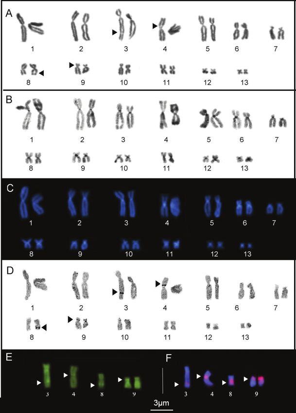
coincided with the secondary constrictions observed by conventional staining (Fig. 1E).Cytogenetics of Pithecupus 329 Figure 1. Karyotype of P. ayeaye prepared by conventional Giemsa staining (A) C- banding (B) Ag-NOR (D) DAPI staining after C-banding (C). Chromosomes submitted to Mytramicim (MM) (E) and FISH experiments with a nucleolar 28S rDNA probe (F). The arrow indicates indicates secondary constrictions; the arrowheads indicate multiple NOR site.
330          Joana Moura Gama et al. / Comparative Cytogenetics 13(4): 325–338 (2019)

     In all the karyotypes, the secondary constrictions revealed by conventional Giemsa
staining coincided with the NOR sites detected by the Ag-NOR method. In P. ayeaye the
NORs were detected in the pericentromeric region of the short arm of the both homologs
of pair 9 (Fig. 1D), besides of the additional sites in the interstitial region of the long arm
of chromosomes 3 and 8 and in pericentromeric region of the short arm of chromosome
4 (Fig. 1D). The additional sites (pairs 3, 4 and 8) were found in all the individuals ana-
lyzed, invariably in the heterozygous condition. The FISH assays realized in P. ayeaye con-
firmed additional NOR sites in the pair 9 (Fig. 1F), which are MM-positive, as is typical
of the anuran chromosome. In the P. palliatus the NOR sites also were detected in the
pericentromeric region of the short arm of the homologs of pair 9 (Fig. 2C). Already in
P. megacephalus the NORs were located in the pericentromeric region of the long arm of
the homologs of pair 8 (Fig. 3C) and additionally in one homologue of pair 13 (Fig. 3C).

Figure 2. Karyotype of P. palliatus prepared by conventional Giemsa staining (A) C-banding (B) and
Ag-NOR method (C). Secondary constrictions observed coincided with the Ag-NOR sites (C).
Cytogenetics of Pithecupus 331 Figure 3. Karyotype of P. megacephalus prepared by conventional Giemsa staining (A) C-banding (B) and Ag-NOR method (C). The arrow indicates secondary constrictions in the pair 8 correspond to NOR sites. Note the additional NOR in one homologue of pair 13. Discussion Karyotype conservation in the subfamily Phyllomedusinae The analysis of the chromosomes of the three Pithecopus species, presented here, rein- forces the conclusion that the macrostructure of the karyotypes of the members of this genus (diploid number and chromosome morphology) is highly conserved (Barth et
332         Joana Moura Gama et al. / Comparative Cytogenetics 13(4): 325–338 (2019)

al. 2009; Bruschi et al. 2013, Bruschi et al. 2014). The extreme homogeneity of these
karyotypes allows for the proposal of a number of different hypotheses on the interspe-
cific chromosomal homologies found in the genus. To begin with, the presence of 26
chromosomes in Pithecopus represents the plesiomorphic condition in the subfamily
Phyllomedusinae (Schmid et al. 1995, Morand and Hernando 1997, Gruber et al.
2013, Bruschi et al. 2014b, Barth et al. 2014, Schmid et al. 2018). Currently, this sub-
family assemble 65 species distributed in eight genus (Agalychnis Cope, 1864, Callime-
dusa Duellman, Marion & Hedges, 2016, Cruziohyla Faivovich, Haddad, Garcia, Frost,
Campbell & Wheeler, 2005, Hylomantis Peters, 1873 “1872”, Phasmahyla Cruz, 1991,
Phrynomedusa Miranda-Ribeiro, 1923, Phyllomedusa Wagler, 1830, Pithecopus Cope,
1866) and only 22 species have been karyotyped (Perkins et al. 2019). The karyotype of
the phyllomedusines is highly conserved (Barth et al. 2013; Gruber et al. 2013; Bruschi
et al. 2014; Schmid et al. 2018). The unique variation in chromosome morphology
found in the species of the genus Phyllomedusa karyotype, in particular in the P. tarsius
group (P. camba De la Riva, 1999, P. tarsius Cope, 1868, P. neildi Barrio-Amorós, 2006,
and P. trinitatis Mertens, 1926), with three telocentric chromosome pairs (pairs 7, 10,
and 12), may represent a possible synapomorphy in this group (Bruschi et al. 2014b).
     Like the other species of the genus Pithecopus (Bruschi et al. 2012, 2013, 2014),
the heterochromatin in P. palliatus and P. ayeaye is found essentially in the centro-
meric regions of the all chromosomes, with no distinct band or other marking that
permits the differentiation of the karyotypes. The only Pithecopus species that can be
distinguished based on its C-banding pattern is P. nordestinus, which is characterized
by a considerable accumulation of heterochromatin, primarily in centromeric regions,
extending to the pericentromeric portions of both arms of the chromosome 9 (Bruschi
et al. 2012), which is a characteristic of this species.

Multiple rDNA sites in the karyotype of Pithecopus
The extreme chromosomal conservation observed in the Pithecopus species contrasts
with its considerable inter- and intrapopulation variation in the chromosomal pairs
that carry the 28S rDNA gene clusters. In the present study, two new cases of multiple
NOR sites were recorded in the genus Pithecopus, in the karyotypes of P. ayeaye and P.
megacephalus. However, a comparative overview of all the Pithecopus karyotypes ana-
lyzed up to now indicates the recurrence of the NOR-bearing chromosome pairs, and
the position of the NORs on these chromosomes, in particular in pairs 3, 4, 8, 9, 11,
and 13. Multiple NORs are common in this genus, and have been recorded in practi-
cally all the species (Morand and Hernando 1997, Barth et al. 2009, 2013, Paiva et
al. 2009, Bruschi et al. 2012, 2013 and present study). In most cases, the karyotypes
shown a NOR-bearing pair (homozygosis), detected in all specimens of population
whereas the additional NOR-sites occurred in heterozygous and polymorphic condi-
tion (Morand and Hernando 1997; Barth et al. 2009, 2013; Paiva et al. 2009; pre-
sent study). Although intrapopulation variation in the number of NORs is a frequent
Cytogenetics of Pithecupus 333
condition in anuran species, the configuration found in Pithecopus reflects the unique
evolutionary dynamics of this chromosomal marker.
The interesting feature of the genus Pithecopus is that when the polymorphic con-
dition is recorded in the different species, it to be located in the same chromosomes
and NOR positions. Thus, it is difficult to recognize the phylogenetic signal of this
marker for the application of a parsimonious evolutionary analysis. Here, we suggest
two possible scenarios to explain this variation: (i) the NOR in pair 9q represents the
plesiomorphic condition in Pithecopus, with subsequent rearrangements resulting in
the repositioning of the NOR to pair 8 in P. azureus and in the ancestor of P. hypochon-
drialis + P. araguaius, with the NOR in pair 8q also representing an autapomorphy in
P. megacephalus. Subsequent independent events of the loss or gain of rDNA would
have resulted in the appearance of the rDNA sites in chromosomes 3, 4, 7, 11, and
13 in the species with the polymorphic condition. In this context, the NOR in pair
9q should be present in the most recent common ancestor (TMRC) of the Pithecopus
genus (see Figure 4). Alternatively (ii) an ancestral polymorphism would be the source
of the extreme variation in the NOR found in this genus.
While the first of these explanations depends on high rates of loss/gain of copies
of the rDNA in the genomes of the species, the second hypothesis would depend on
the recurrence of the same pairs as the NOR-bearing chromosomes in the different
species in the genus Pithecopus (see Fig. 4), which would be consistent with the idea of
an ancestral polymorphism as the source of the complex scenario observed in the pre-
sent day. If this hypothesis is accepted, any attempt to trace an evolutionary pathway
from this chromosomal marker will inevitably generate a high degree of homoplasy in
the phylogenetic inferences, which is typical of the multiple paralogous copies of this
marker in the genome (Robinson et al. 2008).
Assuming the ancestral polymorphism hypothesis, the total reproductive isolation
of each evolutionary lineage would have resulted in the fixation of the principal active
NOR sites in at least one pair of homologous chromosomes (the homozygous condi-
tion), which would permit the degeneration of the other sites, or at least the reduction
or silencing of their expression. In P. nordestinus and P. ayaye, respectively, the position
of the active NOR detected by Ag-NOR was confirmed by the FISH using 18S/28S
rDNA probes (Barth et al. 2013 and present study), which is consistent with the ob-
servation of a homozygous principal pair, together with additional, heterozygous sites,
that bear the rDNA gene. While a cell requires at least one cluster of active rDNA
to satisfy its demand for ribosomal RNAs, there does not appear to be any restric-
tion on the maximum number of copies in a genome (Cazaux et al. 2011). The case
of the species of the genus Mus is an example of this, in which 1–21 clusters of the
rDNA are found in a given karyotype (Cazaux et al. 2011). Given this, not all rDNA
sites are being expressed in the cells, and some may be silenced or even lost during
the diversification of the different lineages (e.g., Derjusheva et al. 1998; Santos et al.
2002). The number, chromosomal distribution and inheritance of NOR are an impor-
tant character to genome comparison in Anuran genomes, as observed in water frogs
Pelophylax lessonae Camerano, 1882, Pelophylax ridibundus Pallas, 1771 and in their334 Joana Moura Gama et al. / Comparative Cytogenetics 13(4): 325–338 (2019)
Figure 4. The active NOR-bearing chromosomes found in the karyotypes of the Pithecopus species and
the broader phylogenetic context of the genus. Two possible scenarios to explain NOR variation are
shown in inset (see details in discussion). The phylogenetic arrangement was reconstructed from Du-
ellman et al. (2016) and Haga et al. (2017). Chromosomes within brackets present additional NOR sites
in the polymorphic condition within the population. The NOR site of the underlined pairs (black lines)
was confirmed by FISH using the rDNA probe. Species with unknown karyotypes are indicated by the
“?” symbol. Species suspected to contain cryptic diversity are represented by triangles. The letters within
brackets indicate the following references: [a] Present study; [b] Bruschi et al. (2012); [c] Barth et al.
(2009); [d] Paiva et al. (2009); [e] Bruschi et al. (2013); [f ] Morand and Hernando (1997); [g] Barth et
al. (2013); [h] Bruschi et al. (2014a); [i] karyotype described by Bruschi et al. (2013) and recognized as a
new species by Haga et al. (2017). The asterisks (*) represent the heteromorphic condition resulting from
the paracentric inversion found in the Alta Floresta population by Bruschi et al. (2013).
natural hybrids (Pelophylax esculentus Fitzinger, 1843) (Zalesna et al. 2017). In this
case, active NOR variability are relationships with ploidy level in hybrids and denote
the intragenomic behavior of this chromosomal marker.
One particularly illustrative example of this scenario is the variation in P. hypochon-
drialis found by Bruschi et al. (2013), who detected a pronounced population struc-
ture based on the analysis of fragments of mitochondrial and nuclear genes. This study
found clear differences among populations, and geographical coherence between the
clades recuperated by phylogenetic analysis and the NOR-bearing chromosome, which
indicates the possible fixation of distinct chromosomes that bear the transcriptionally-
active rDNA genes in populations connected by little gene flow. The principal NOR-
bearing chromosomes in this species were pairs 4, 7, and 8, in addition to a polymor-
phic population with extra sites in pairs 3 and 4. This regional chromosomal variationCytogenetics of Pithecupus 335
may reflect the role of population dynamics in the fixation of the active NOR from
the pool of rDNA sites present in the ancestral genome. Once fixed one chromosome
pair with NOR site at a population level, the additional copies of rDNA may either
(i) become free of selective pressure and degenerate through stochastic events which
would account for the absence of hybridization signals in the FISH experiment or (ii)
remain silenced in genome and for consequence undetectable by Ag-NOR method. It
is important to note here that Bruschi et al. (2013) did not design the experiment to
evaluate these specific questions.
The results of the present study also indicate clearly a predominance of rDNA
sites located in the pericentromeric and/or subterminal regions of the chromosomes
(Fig. 4). Similar results have been obtained for many examples in Anuran karyo-
types, as observed in species of the hylid tribe Cophomantini (see Ferro et al. 2018)
or in species of the Agalychnis Cope, 1864 and Scinax Wagner, 1830 genus (Schmid
et al. 2018), for example. A number of studies indicate that the NOR-bearing sites
in the chromosomes act as hotspots of chromosomal rearrangement (Cazaux et al.
2011). The mechanisms recognized traditionally include the occurrence of unequal
crossovers, ectopic recombination, and invasion by mobile genetic elements, all of
which have been invoked to account for the observed variation and dispersal of the
copies of the NOR in the genome (Poletto et al. 2010; Cazaux et al. 2011; Silva et
al. 2013). The evidence points to the possible occurrence of intrachromosomal rear-
rangements (peri- and paracentric inversions) as the source of the variation in the
position of the NOR, such as the distinct positions (8p and 8q) that the rDNA site
occupies in the homologs of pair 8 in the different populations of P. hypochondrialis
(see Fig. 4), for example.
Conclusions
Our findings help to fill the gaps in the knowledge of the karyotype variability
of the genus Pithecopus and constitute a good example of the complex role of the
rDNA genes in karyotype evolution. Ours results reveals that evolutionary dynam-
ics of the NOR sites in genus and its potential as hotspot of chromosomal rear-
rangements, which implies that it may be a fundamental feature of chromosomal
evolution in the genome of Pithecopus.
Acknowledgements
We thank the Fundação de Amparo a Pesquisa do Estado de São Paulo (FAPESP
2016/07717-6), Coordenação de Aperfeiçoamento de Pessoal de Nível Superior
(CAPES/PROAP – Finance Code 001) for the scholarships provided to JGM, CBG
and DYS. We thank the Multi-User Confocal Microscopy Center of the Federal Uni-
versity of Paraná for the capture of the images included in this study.336 Joana Moura Gama et al. / Comparative Cytogenetics 13(4): 325–338 (2019)
Reference
Araújo CO, Condez TH, Haddad CFB (2007) Amphibia, Anura, Phyllomedusa ayeaye (B.
Lutz, 1966): Distribution extension, new state record, and geographic distribution map.
Check List 3(2): 156–158. https://doi.org/10.15560/3.2.156
Barth A, Solé M, Costa MA (2009) Chromosome Polymorphism in Phyllomedusa roh-
dei Populations (Anura: Hylidae). Journal of Herpetology 43(4): 676–679. https://doi.
org/10.1670/08-210.1
Barth A, Souza VA, Solé M, Costa MA (2013) Molecular cytogenetics of nucleolar organizer
regions in Phyllomedusa and Phasmahyla species (Hylidae, Phyllomedusinae): a cytotaxo-
nomic contribution. Genetics and Molecular Research 12(3): 2400–2408. https://doi.
org/10.4238/2013.July.15.3
Barth A, Vences M, Solé M, Costa MA (2014) Molecular cytogenetics and phylogenetic
analysis of Brazilian leaf frog species of the genera Phyllomedusa and Phasmahyla species
( Hylidae, Phyllomedusinae). Canadian Journal of Zoology 92(9): 795–802. https://doi.
org/10.1139/cjz-2013-0301
Baêta D, Caramaschi U, Cruz CAG, Pombal Jr JP (2009) Phyllomedusa itacolomi Caramaschi,
Cruz & Feio, 2006, a junior synonym of Phyllomedusa ayeaye (B. lutz, 1966) (Hylidae,
Phyllomedusinae). Zootaxa 2226: 58–65. https://doi.org/10.11646/zootaxa.2226.1.5
Bruschi DP, Lucas EM, Garcia PCA, Recco-Pimentel S (2014a) Molecular and morphological
evidence reveals a new species in the Phyllomedusa hypochondrialis group (Hylidae, Phyl-
lomedusinae) from the Atlantic forest of the highlands of southern Brazil. PlosOne 9(8):
e105608. https://doi.org/10.1371/journal.pone.0105608
Bruschi DP, Busin CS, Siqueira S, Recco-Pimentel SM (2012) Cytogenetic analysis of two spe-
cies in the Phyllomedusa hypochondrialis group (Anura, Hylidae). Hereditas 149(1):34–30.
https://doi.org/10.1111/j.1601-5223.2010.02236.x
Bruschi DP, Rivera M, Lima AP, Zúñiga AB, Recco-Pimentel SM (2014b) Interstitial Telomeric
Sequences (ITS) and major rDNA mapping reveal insights into the karyotypical evolution
of Neotropical leaf frogs species (Phyllomedusa, Hylidae, Anura). Molecular Cytogenetics
7(22): 1–12. https://doi.org/10.1186/1755-8166-7-22
Bruschi DP, Busin CS, Toledo LF, Vasconcellos GA, Strussmann C, Weber LN, Lima AP, Lima
JD, Recco-Pimentel SM (2013) Evaluation of the taxonomic status of populations as-
signed to Phyllomedusa hypochondrialis (Anura, Hylidae, Phyllomedusinae) based on mo-
lecular, chromosomal and morphological approach. BMC Genetics 14(70): 1–14. https://
doi.org/10.1186/1471-2156-14-70
Caramaschi U, Cruz CAG, Lima R, Brandão R (2016) Pithecopus ayeaye. The IUCN
Red List of Threatened Species. http://doi.org/10.2305/IUCN.UK.2016-3.RLTS.
T55839A107295713.en [Accessed 21 June 2019]
Cazaux B, Catalan J, Veyrunes F, Douzery EJP, Britton-Davidian J (2011) Are ribosomal DNA
clusters rearrangement hotspots? A case study in the genus Mus (Rodentia, Muridae). BMC
Evolutionary Biology 11(124): 1–14. https://doi.org/10.1186/1471-2148-11-124
Derjusheva SE, Loginova JA, Parada R, Chiryaeva OG, Smirnov AF, Jaszczak K (1998) The com-
parative analysis of NOR polymorphism detected by FISH and Ag-staining on horse chro-
mosomes. Caryologia 51(1): 1–11. https://doi.org/10.1080/00087114.1998.10589115Cytogenetics of Pithecupus 337
Duellman WE, Marion AB, Hedges SB (2016) Phylogenetics, classification, and biogeogra-
phy of the treefrogs (Amphibia: Anura: Arboranae). Zootaxa 4101(1): 1–109. https://doi.
org/10.11646/zootaxa.4104.1.1
Faivovich J, Haddad CFB, Baêta D, Jungfer KH, Álvares FR, Brandão RA, Sheil C, Barrientos
LS, Barrio-Amorós CL, Cruz CAG, Wheeler WC (2010) The phylogenetic relationships of
the charismatic poster frogs Phyllomedusinae (Anura, Hylidae). Cladistics 26(3): 227–261.
https://doi.org/10.1111/j.1096-0031.2009.00287.x
Ferro JM, Cardozo DE, Suárez P, Boeris JM, Blasco-Zúñiga A, Barbero G, Gomes A, Gazoni T,
Costa W, Nagamachi CY, Rivera M, Parise-Maltempi PP, Wiley JE, Pieczarka JC, Haddad
CFB, Faivovich J, Baldo D (2018) Chromosome evolution in Cophomantini (Amphibia, An-
ura, Hylidae). PlosOne 13(2): eO192861. https://doi.org/10.1371/journal.pone.0192861
Frost DR (2019) Amphibian species of the world: an online reference. Version 6.0 American
Museum of Natural History, New York, USA. http://research.amnh.org/herpetology/am-
phibia/index.html [Accessed 18 June, 2019]
Green DM, Sessions SK (1991) Nomenclature for chromosomes. In: Green DM, Sessions
SK (Eds) Academic Press. Amphibian cytogenetics and evolution. San Diego, 431–432.
https://doi.org/10.1016/B978-0-12-297880-7.50021-4
Gruber SL, Silva ANZ, Haddad CFB, Kasahara S (2013) Cytogenetic analysis of Phyllomedusa
distincta Lutz, 1950 (2n=2×=26), P. tetraploidea Pombal and Haddad, 1992 (2n=4×=52),
and their natural triploid hybrids (2n=3×=39) (Anura, Hylidae, Phyllomedusinae). BMC
Genetics 14(75): 1–13. https://doi.org/10.1186/1471-2156-14-75
Haga IA, Andrade FS, Bruschi DP, Recco-Pimentel SM, Giaretta AA (2017) Unrevealing
the leaf frogs Cerrado diversity: A new species of Pithecopus (Anura, Arboranae, Phyl-
lomedusinae) from the Mato Grosso state, Brazil. PlosOne 12(9): e0184631. https://doi.
org/10.1371/journal.pone.0184631
Howell WM, Black DA (1980) Controlled silver staining of nucleolar organizer regions with
a protective colloidal developer a 1-step method. Experientia 36: 1014–1015. https://doi.
org/10.1007/BF01953855
King M, Rofe R (1976) Karyotypic variation in the Australian gekko Phyllodactylus marmoratus
(Gray) (Gekkonidae: Reptilia). Chromosoma 54: 75–87. https://doi.org/10.1007/BF00331835
Magalhães RF, Rocha PC, Santos FR, Strüssmann C, Giaretta AA (2018) Integrative taxonomy
helps to assess the extinction risk of anuran species. Journal for Nature Conservation 45:
1–10. https://doi.org/10.1016/j.jnc.2018.07.001
Morand M, Hernando A (1997) Localización cromossômica de genes ribosomales activos em
Phyllomedusa hypochondrialis y P. sauvagii (Anura: hylidae). Cuadernos de Herpetologia
11(1–2): 31–36. http://sedici.unlp.edu.ar/handle/10915/6276
Oliveira FFR, Nogueira PAG, Eterovick PC (2012) Natural history of Phyllomedusa mega-
cephala (Miranda-Ribeiro, 1926) (Anura:Hylidae) in southeastern Brazil, with descriptions
of its breeding biology and male territorial behavior. Journal of Natural History 46(1–2):
117–129. https://doi.org/10.1080/00222933.2011.626127
Paiva CR, Nascimento J, Silva APZ, Bernarde OS, Ananias F (2009) Karyotypes and Ag-NORs
in Phyllomedusa camba De La Riva, and P. rhodei Mertens, 1926 (Anura, Hylidae, Phyl-
lomedusinae): cytotaxonomic considerations. Italian Journal of Zoology 77(1): 116–121.
https://doi.org/10.1080/11250000903187585338 Joana Moura Gama et al. / Comparative Cytogenetics 13(4): 325–338 (2019)
Perkins RD, Gamboa JR, Jonika MM, Lo J, Shum A, Adams RH, Blackmon H (2019) The
Amphibian Karyotype Database. Chromosome Research. https://doi.org/10.1007/
s10577-019-09613-1
Poletto AB, Ferreira IA, Martins C (2010) The B chromosomes of the African cichlid fish Hap-
lochromis obliquidens harbout 18S rRNA gene copies. BMC Genetics 11(1): 1–8. https://
doi.org/10.1186/1471-2156-11-1
Ramos EKS, Magalhães RF, Marques NCS, Baêta D, Garcia PCA, Santos FR (2019) Cryptic
diversity in Brazilian endemic monkey frogs (Hylidae, Phyllomedusinae, Pithecopus) re-
vealed by multispecies coalescents and integrative approaches. Molecular Phylogenetics
and Evolution 132: 105–116. https://doi.org/10.1016/j.ympev.2018.11.022
Ramos EKS, Magalhães RF, Sari HER, Rosa AHB, Garcia PCA, Santos FR (2018) Population
genetics and distribution data reveal conservation concerns to the sky island. Endemic
Pithecopus megacephalus (Anura, Phyllomedusidae). Conservation Genetics 19(1): 99–110.
https://doi.org/10.1007/s10592-017-1013-z
Robinson TJ, Ruiz-Herrera A, Avise JC (2008) Hemiplasy and homoplasy in the karyotypic
phylogenies of mammals. Proceedings of the National Academy of Sciences of the United
States of America 105(38): 14477–14481. https://doi.org/10.1073/pnas.0807433105
Santos N, Fagundes V, Yonenaga-Yassuda Y, Souza MJ (2002) Localization of rDNA genes in
Phyllostomidae bates reveals silent NORs in Artibeus cinereus. Hereditas 136: 137–143.
https://doi.org/10.1034/j.1601-5223.2002.1360208.x
Schmid M (1978) Chromosome banding in Amphibia. I. Constitutive heterochromatin and
nucleolus organizer regions in Bufo and Hyla. Chromosoma 66(1): 361–388. https://doi.
org/10.1007/BF00328536
Schmid M, Borgat JP, Hedges SB (2018) The Arboranan frogs: evolution, Biology and Cy-
togenetics. Cytogenetics Genome Research 155: 1–5. https://doi.org/10.1159/isbn.978-
3-318-06405-6
Schmid M, Feichtinger W, Weimer R, Mais C, Bolaños F, Leon P (1995) Chromosome band-
ing in Amphibia. XXI. Inversion polymorphism and multiple nucleolus organizer re-
gions in Agalychnis callidryas (Anura, Hylidae). Cytogenetic 69(1–2): 18–26. https://doi.
org/10.1159/000133929
Silva DMZA, Pansonato-Alves JC, Utsunomia R, Daniel SN, Hashimoto DT, Oliveira C,
Porto-Foresti F, Foresti F (2013) Chromosomal organization of repetitive DNA sequences
in Astyanax bockmanni (Teleostei, Characiformes): dispersive location, association and
co-localization in the genome. Genetica 141(7–9): 329–336. https://doi.org/10.1007/
s10709-013-9732-7
Sumner AT (1972) A simple technique for demonstrating centromeric heterochromatin. Ex-
perimental Cell Research 75: 304–306. https://doi.org/10.1016/0014-4827(72)90558-7
Viegas-Péquignot E (1992) In situ hybridization to chromosomes with biotinylated probes. In:
Willernson D (Ed.) In situ hybridization: a practical approach. Oxford University Press-
IRL Press, Oxford, 137–158.
Zalesna A, Florek M, Rybacki M, Ofielska M (2017) Variability of NOR patterns in European
water frogs of different genome composition and ploidy level. Comparative Cytogenetic
11(2): 249–266. https://doi.org/10.3897/CompCytogen.v11i2.10804You can also read