Renal Mechanisms of Diuretic Resistance in Congestive Heart Failure
←
→
Page content transcription
If your browser does not render page correctly, please read the page content below
Review
Renal Mechanisms of Diuretic Resistance in Congestive
Heart Failure
Norbert Lameire

                                         Department of Nephrology, Ghent University Hospital, 9000 Ghent, Belgium; lameire.norbert@outlook.com

                                         Abstract: This study reviews the renal aspects of diuretic resistance occurring in diuretic treatment,
                                         mostly with loop diuretics of congestive heart failure. A short discussion on the different classes
                                         of diuretics, including the recently introduced sodium-glucose transporter 2 inhibitors, and their
                                         mechanism of action in the nephron is provided, followed by a summary of recent data discussing
                                         the different causes and pathophysiological mechanisms of diuretic resistance. The major cause
                                         of diuretic resistance appears to be localized within the distal tubule. Traditionally, the concept of
                                         compensatory post-diuretic sodium reabsorption (CPDSR) was considered the major cause of diuretic
                                         resistance; however, recent studies have disputed this traditional concept and demonstrated that
                                         patients with congestive heart failure are in constant sodium-avid state. Finally, the different options
                                         of therapeutic strategies, combining different classes of diuretics are summarized.

                                         Keywords: congestive heart failure; mechanisms of diuretic resistance; combined diuretic strategies

                                         1. Introduction
                                               Diuretic resistance (DR) can be defined as a failure to increase fluid and sodium output
                                         sufficiently to relieve generalized edema, volume overload or congestion despite treatment
                                         with a full dose of a loop diuretic [1–3].
                                               Diuretic resistance is a frequent problem in severe congestive heart failure (CHF), and
                                         extensive literature is published in the cardiology literature. This contribution summarizes
                                         selected and recent insights in diuretic resistance for a nephrology readership.
                                               Treatment of generalized edema primarily relies on the administration of natriuretic
Citation: Lameire, N. Renal              agents which diminish sodium reabsorption at different sites in the nephron, thereby
Mechanisms of Diuretic Resistance in     increasing urinary sodium and water losses. The terms “diuretics” and the more exact term
Congestive Heart Failure. Kidney Dial.   of “natriuretics” are often used interchangeably.
2023, 3, 56–72. https://doi.org/               “Aquaretic” agents are vasopressin 2 anta-agonists (VRAs), acting at the collecting
10.3390/kidneydial3010005                duct, which increase free water excretion, and are, strictly speaking, not “diuretics”.
Academic Editor: Vladimir Tesar                The molecular targets of diuretic drugs are predominantly sodium (Na+ ) transport
                                         pathways at the apical (luminal) surface of the kidney tubule cells. When coupled with the
Received: 18 November 2022               basolateral cell membrane Na+ /K+ -ATPase, these pathways permit the vectorial transport
Revised: 13 December 2022
                                         of sodium.
Accepted: 14 December 2022
                                               Traditionally, diuretics are classified according to their predominant sites of action
Published: 5 January 2023
                                         in the tubular segments of the nephron and by the mechanism by which they inhibit
                                         transport [4].
                                               Figure 1 modified from [1] illustrates the sites of action in the nephron of different
Copyright:   © 2023 by the author.
                                         diuretics in relation to the percentage of filtrate reabsorption at the different nephron
Licensee MDPI, Basel, Switzerland.       segments.
This article is an open access article         Table 1 adapted from [5] summarizes the classification of commonly used diuretics
distributed under the terms and          and aquaretics based on the site and mechanism of action.
conditions of the Creative Commons             The discussion on osmotic diuretics (i.e., mannitol) is beyond the scope of this paper.
Attribution (CC BY) license (https://          Diuretics block the function of sodium transport proteins in the apical plasma mem-
creativecommons.org/licenses/by/         brane of kidney epithelial cells and, except for mineralocorticoid receptor blockers, secretion
4.0/).                                   into the tubular fluid is required for all “traditional” diuretics [4,6].

Kidney Dial. 2023, 3, 56–72. https://doi.org/10.3390/kidneydial3010005                                 https://www.mdpi.com/journal/kidneydial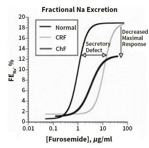
KidneyDial.
Kidney Dial.2023,
2023,33, FOR PEER REVIEW 572
Figure 1.1. Schematic
Figure Schematic of aa nephron
nephron shows
shows sites
sites of
of action
action of
of diuretics
diuretics along
along the
the various
various segments.
segments.
Adapted with permission from Ref. [1]. 2022, Elsevier. (DCT: distal convoluted tubule;
Adapted with permission from Ref. [1]. 2022, Elsevier. (DCT: distal convoluted tubule; CNT: CNT: con-
necting tubule).
connecting tubule).
Table Table 1 adapted
1. Diuretics from
classified [5] summarizes
according the classification
to their localization and theirofmechanisms
commonlyof used diuretics
action on the
and aquaretics
sodium based on
and vasopressin the site and
transporters mechanism
in the of action.
nephron. Adapted with permission from Ref. [5] 2022,
Oxford Publishing Limited.
Table 1. Diuretics classified according to their localization and their mechanisms of action on the
Natriuretics sodium and vasopressin transporters in the nephron. Adapted with permissionAquaretics from Ref. [5] 2022,
Proximal diuretics Oxford Publishing
Loop diuretics Limited.
Distal convoluted tubule Cortical collecting tubule Collecting duct
Carbonic anhydrase
Natriuretics
inhibitors Na channel blockers (ENAC Aquaretics
Proximal diuretics Loop diuretics
Na-K-CI (NKCC2) Distal convoluted tubule inhibitors)
Na-CI (NCC) Cortical collecting tubule Vasopressor
Collecting duct
receptor
Sodium-glucose
Carbonic anhydrase transport inhibitors cotransporter inhibitors blockers
cotransporter 2 Aldosterone antagonists
inhibitors Na channel blockers (ENAC
inhibitors
Acetazolamide Na-K-CI (NKCC2) Na-CI (NCC) cotransporter inhibitors) Vasopressor receptor
Sodium-glucose transport inhibitors Hydrochlorothiazide
inhibitors Amiloride blockers
Furosemide
Metolazone Triameterene
Empagliflozin 2
cotransporter Bumetanide Aldosterone Conivaptan
antagonists
Chlorthalidone
Dapagliflozin
inhibitors Torasemide Tolvaptan
Indapamide b Spironolactone
Canagliflozin Ethacrynic acid a
Acetazolamide Others Eplerenone
Others... Hydrochlorothiazide Amiloride
Furosemide
a mechanism of ethacrynic acid differs from that of other loop diuretics; b indapamide and metolazone may have
Metolazone Triameterene
Empagliflozin Bumetanide
other actions, as well. Conivaptan
Chlorthalidone
Dapagliflozin Torasemide Tolvaptan
The SGLT2 Indapamide
inhibitors are b
believed to beSpironolactone
freely filtered, where they bind to SGLT2s in
Canagliflozin Ethacrynic acid a
the luminal membraneOthers of the epithelial cellsEplerenone
in the proximal tubule [7].
Others...
The mineralocorticoid blocker, spironolactone, and the new nonsteroidal mineralo-
a mechanism of ethacrynic acid differs from that of other loop diuretics; b indapamide and metola-
corticoid blocker, finerenone, act within cells and do not require secretion into the tubule
zone may have other actions, as well.
lumen [4].
Detailed pharmacokinetic
The discussion on osmoticand pharmacodynamic
diuretics (i.e., mannitol) aspects of thethe
is beyond different
scope ofclasses of
this pa-
diuretics
per. are available in [1,4,5,8,9].
Diuretics block the function of sodium transport proteins in the apical plasma
2. Diuretics Acting on the Proximal Tubule (PT)
membrane of kidney epithelial cells and, except for mineralocorticoid receptor blockers,
The proximal tubules reabsorb around 70% of the filtrate. Despite this high absorptive
secretion into the tubular fluid is required for all “traditional” diuretics [4,6].
capacity, large losses of sodium and water by diuretics acting in the PT, including aceta-
The SGLT2 inhibitors are believed to be freely filtered, where they bind to SGLT2s
zolamide and SGLT2 inhibitors, cannot be expected because almost all of the excess fluid
in the luminal membrane of the epithelial cells in the proximal tubule [7].
delivered out of the PT is reabsorbed in the loop of Henle and the distal tubule.
The mineralocorticoid blocker, spironolactone, and the new nonsteroidal mineralo-
corticoid blocker, finerenone, act within cells and do not require secretion into the tubule
2.1. Acetazolamide
lumen [4].
Acetazolamide inhibits the activity of carbonic anhydrases, a ubiquitous superfamily
of enzymes that catalyze the hydration of CO2 to bicarbonate and protons which plays an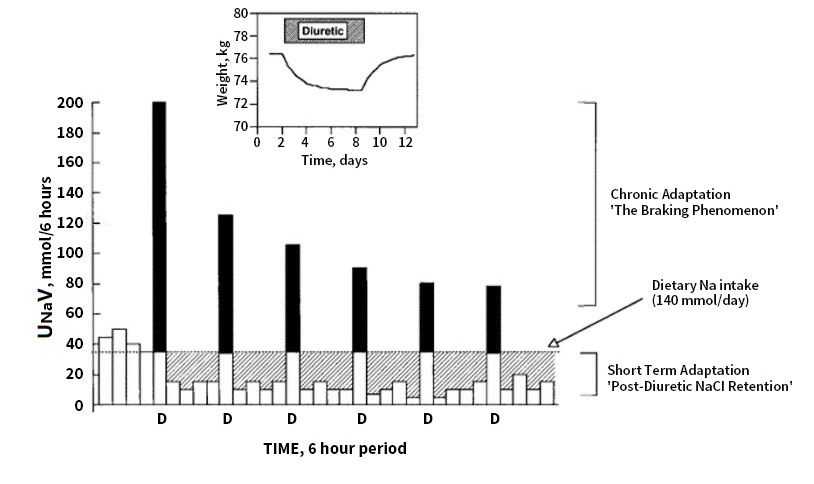
Kidney Dial. 2023, 3                                                                                                58

                       important role in proximal bicarbonate, sodium, and chloride reabsorption. As a result,
                       this agent results in the loss of both NaCl and NaHCO3 [5,8].

                       2.2. SGLT2 Inhibitors (SGLT2i)
                             SGLT2 inhibitors decrease proximal tubular sodium and chloride reabsorption and
                       increase macula densa sodium and chloride delivery. This occurs via the tubulo-glomerular
                       feedback (TGF) mechanism adenosine-induced vasoconstriction of the afferent arteriole
                       and increased prostaglandin secretion with dilatation of the efferent arteriole, resulting in a
                       decline in intraglomerular filtration pressure and in glomerular filtration rate (GFR) [10].
                       SGLT2 inhibitors further decrease proximal tubular sodium reabsorption via inhibition of
                       the sodium–hydrogen transporter (NHE3) [11].
                             Recent reviews have summarized the cardiovascular and renal benefits of SGLT2
                       inhibitors in diabetic as well as nondiabetic patients with heart failure with reduced and
                       preserved ejection fraction and patients with renal dysfunction [12–16].
                             One of the potential mechanisms of the benefits in cardiovascular protection of the
                       SGLT2 inhibitors may be related to their diuretic effects.
                             However, the diuretic effects of SGLT2 inhibitors are limited and the natriuresis
                       disappears after 4 days of treatment [17].
                             Several mechanisms besides the increased diuresis and natriuresis have been suggested
                       to mediate the cardiovascular benefits with SGLT2 inhibitors (for review see [18]).
                             Despite the high capacity for sodium reabsorption in the PT, relatively large losses
                       of sodium and water by diuretics acting in the PT, including acetazolamide and SGLT2
                       inhibitors cannot be expected because almost all of the excess fluid delivered out of the PT
                       is reabsorbed more distally, in both the loop of Henle and the distal tubule.
                             The role of acetazolamide and SGLT2 inhibitors in the management of DR will be
                       discussed below.

                       3. Diuretics Acting in the Loop of Henle
                             Filtered sodium chloride enters the cells in the thick ascending limb of the loop of
                       Henle via cotransporter Na-K-2Cl carrier isoform 2 (NKCC2) at the apical membrane of the
                       tubular cells and the macula densa [8,19]. The macula densa cells are affected by luminal
                       chloride concentration.
                             Loop diuretics (bumetanide, ethacrynic acid, furosemide, and torsemide) act princi-
                       pally by blocking the luminal NKCC2 transporter. This inhibition leads to increased renin
                       secretion and increase in intraglomerular pressures by preventing feedback (TGF) [5,8]. The
                       blockade of the sodium chloride reabsorption at the macula densa disrupts TGF [20] and
                       maintains the GFR, despite ongoing diuresis, and causes the release of volume-independent
                       renin from the juxtaglomerular apparatus and subsequent renin-angiotensin-aldosterone
                       system (RAS) activation.
                             By inhibiting the NaCl reabsorption in the water-impermeable parts of Henle’s loop,
                       loop diuretics decrease the tonicity in the renal interstitium resulting in increased free water
                       excretion. Because the thick ascending limb reabsorbs 20–25% of the filtered sodium load,
                       these drugs are among the strongest diuretics available [8,19,21].
                             Though loop diuretics are effective in natriuresis in the acute setting, chronic use can
                       lead to a compensatory increase in sodium reabsorption, which distally mitigates their
                       natriuretic effects [22] (see below).
                             Loop diuretics circulate bound to proteins (>90%), limiting their volumes of distribution.
                             The oral absorption of furosemide is highly variable, with bioavailability ranging
                       between 10% and 100% (on average, 50%). In congested patients, bowel edema can reduce
                       its absorption, causing lower concentrations of peak plasma.
                             Bumetanide and torsemide have higher and more consistent oral bioavailability than
                       furosemide (>90%), and do not exhibit absorption-limited kinetics, making oral and intra-
                       venous doses similar [19].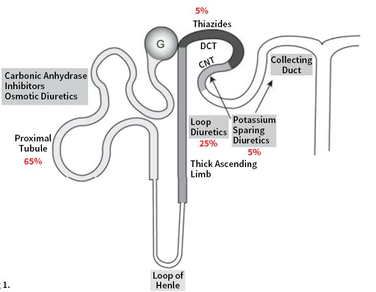
Kidney Dial. 2023, 3                                                                                              59

                            Doses of different loop diuretics can be converted to furosemide equivalents with 1 mg
                       bumetanide = 20 mg torsemide = 80 mg furosemide for oral, and 1 mg bumetanide = 20 mg
                       torsemide = 40 mg furosemide for intravenous diuretics [23–25].

                       4. The Distal Nephron Acting Diuretics
                            The sodium chloride co-transporter (NCC), also known as the “thiazide-sensitive”
                       transporter, is mainly located at the distal convoluted tubule (DCT). Thiazides and thiazide-
                       likes (indapamide (INDA) and chlorthalidone (CLTD) achieve their diuretic action via
                       inhibition of the NCC, which then prevents the co-transport of sodium.
                            The majority of thiazides may also interact with multiple sodium transport pathways
                       such as carbonic anhydrase, pendrin, and the sodium-dependent chloride/bicarbonate
                       exchanger (NDCBE). The distal convoluted tubule is part of the diluting nephron segment,
                       and blocking salt reabsorption with thiazides impairs urinary dilution, potentially resulting
                       in hyponatremia.
                            Because the distal convoluted tubule is responsible for only around 5% of sodium
                       reabsorption, thiazide diuretics only have a limited capacity to change sodium balance.

                       5. Diuretics Mainly Acting on the Collecting Duct
                           The collecting duct contributes to up to 2% of absorption of the filtered salt load.
                           The potassium-sparing diuretics include two drug classes, namely, epithelial sodium
                       channel (ENaC) blockers and mineralocorticoid receptor antagonists (MRAs).

                       5.1. The Potassium-Sparing Diuretics
                            ENaC is the primary channel for sodium reabsorption in the principal cells of the
                       connecting tubules and collecting ducts [26,27]. Amiloride and triamterene directly but
                       reversibly block luminal ENaCs without affecting the mineralocorticoid receptor.
                            The inhibition of ENaC disrupts the normal lumen-negative potential in the collect-
                       ing duct, inhibiting potassium and hydrogen ion secretion and promoting calcium and
                       magnesium reabsorption.
                            Pendrin is a Cl− /HCO3− exchanger located in type B intercalated cells and non-type
                       A/non-type B intercalated cells, as well as in thyroid, inner ear, and the adrenal medulla. In
                       people, suffering from Pendred syndrome, and in mice, pendrin gene ablation can lead to
                       hypothyroidism, deafness, volume contraction, and reduced blood pressure (for review [28]).
                            Angiotensin II and aldosterone stimulate pendrin, which increases renal chloride
                       absorption, thereby contributing to the pressor response seen in response to these hormones.
                       In kidney, pendrin increases renal ENaC activity and abundance, thereby stimulating the
                       renal absorption of both Na+ and Cl− . Metabolic alkalosis stimulates pendrin, thereby
                       increasing the secretion of HCO3− into the pro-urine, which mitigates the alkalosis.
                            Of physiological and clinical significance is the role of these pendrin-positive inter-
                       calated cells in blood pressure regulation, which occurs, at least in part, through pendrin-
                       mediated renal Cl− absorption, as well as their effect, on the epithelial Na+ channel, ENaC.
                       Aldosterone stimulates ENaC directly through principal cell mineralocorticoid hormone
                       receptor (ligand) binding and also indirectly through its effect on pendrin expression and
                       function. Thus, pendrin contributes to the aldosterone pressor response.
                            Pendrin may also modulate blood pressure in part through its action in the adrenal
                       medulla, where it modulates the release of catecholamines, or through an indirect effect on
                       vascular contractile force [28].
                            Recent findings suggest pendrin inhibition as a future novel approach to potentiate
                       the action of loop diuretics for treatment of hypertension and edema, including diuretic-
                       resistant edema [29].

                       5.2. Aldosterone Antagonists
                            Aldosterone is a mineralocorticoid hormone produced in the zona glomerulosa of
                       the adrenal cortex and acts on principal-like cells in the late DCT, the connecting tubule
Kidney Dial. 2023, 3 60
(CNT), and the cortical collecting duct (CCD), favoring sodium and water reabsorption and
potassium excretion while also contributing to the acid–base balance. As with all steroid
hormones, aldosterone passes through cell membranes to bind to cytoplasmic receptors
which translocate to the nucleus to influence mRNA transcription and subsequently protein
synthesis. Aldosterone stimulates ENaC directly through principal cell mineralocorti-
coid hormone receptor (ligand) binding and also indirectly through its effect on pendrin
expression and function [30].
The mineralocorticoid receptor antagonists (MRAS) include, in order of increasing
mineralocorticoid receptor selectivity, spironolactone, eplerenone, and finerenone.
In the kidney, spironolactone and its metabolites bind to cytosolic mineralocorticoid
receptors, and act as competitive inhibitors of the endogenous hormone. Spironolactone
induces a mild increase in sodium excretion (1–2%) and a decrease in potassium and
hydrogen ion excretion [5]. Eplerenone is more selective for mineralocorticoid receptors,
and therefore less likely to cause side effects [31,32].
Finerenone, is the first of a new class of nonsteroidal MRAs with stronger mineralo-
corticoid receptor binding compared with eplerenone and spironolactone [33,34]. The
FDA recently approved the use of finenerone to decrease the risk of CKD progression and
cardiovascular events in patients with diabetic kidney disease [9,35,36].
6. Diuretic Resistance (DR) in Congestive Heart Failure
Congestion is the major cause for hospital admission in acute decompensated heart
failure (ADHF) and DR leading to refractory congestion is associated with worse outcomes,
increased length of stay, and other intermediate complications [22,24,37,38].
The small increases in serum creatinine (SCr) or Cystatin C, often observed in ADHF
patients during decongestion therapy with either diuretics or ultrafiltration, are frequently
labelled as worsening renal function (WRF). These fluctuations in GFR are intrinsic to the
intrarenal hemodynamics of decongestion and do not reflect tubular injury as observed
in acute kidney injury (AKI). Increasing levels of SCr (up to 0.5 mg/dL) and cystatin
C reflecting adequate decongestion) are associated with better chances of 6-month sur-
vival [39]. However, if WRF occurs in the setting of DR and persistent congestion, prognosis
is exceedingly poor [40,41]. Therefore, WRF by itself should not be a reason to withdraw
decongestive therapies and should not limit achieving euvolemia [39,42].
Quantitative definitions of DR include failure of oral furosemide (160 mg twice daily
or equivalent) to increase sodium excretion by at least 90 mmol over 3 days. Alternatively,
a spot urine sample with a sodium outputKidney Dial. 2023, 3 61
The well-known loop DR observed in acute kidney disease (AKD), including acute
kidney injury (AKI), and chronic kidney disease (CKD) is probably the combined effect of
lower functioning nephron mass, associated metabolic acidosis and the interference at the
proximal tubular secretory sites by drugs and/or uremic toxic anions. Defects in glomerular
filtration are less important mediators of diuretic resistance than tubular mechanisms [47].
Recent epidemiological studies identified hypochloremia, rather than hyponatremia,
as the primary ion driving the poor survival in heart failure [48–51]. Chloride, and
not sodium, has a predominant role in renal salt-sensing mechanisms, and TGF and
renin release are primarily driven by chloride [52–54]. Chloride is a critical regulator of
sodium transporter pathways through phosphorylation of a family of serine–threonine
kinases [55,56].
Hanberg et al. [57] found that hypochloremia is associated with neurohormonal
activation and diuretic resistance; sodium-free chloride supplementation was associated
with increases in serum chloride and beneficial changes in several cardiorenal parameters.
For a recent review on hypochloremia in heart failure, see [58].
Recent opinions consider that high dietary sodium intake does not directly reduce
diuretic response and, if anything, increases it [47].
8. Contribution of Proximal versus Distal Tubular Mechanisms of Diuretic
Resistance (DR)
A recent study [59] revealed that distal tubular mechanisms explained >70% of the
variability in diuretic response, indicating that natriuresis is determined primarily by distal
tubular mechanisms.
The contributions of proximal and distal nephron’s sodium reabsorption after IV
furosemide were explored by comparing the differences in the fractional urinary excretion
of Lithium (FELi) and fractional urinary excretion of sodium (FENa) between patients in
CHF and controls [60]. The FELi reflects an in vivo assessment of proximal tubular and loop
of Henle sodium handling and assesses sodium loop exit after loop diuretic administration.
FENa assesses the net sodium excreted into the urine. Lithium is not reabsorbed in the
distal nephron; thus, a disproportionate increase in FELi after furosemide administration
suggests that distal sodium reabsorption is at play. The prediuretic FELi was 16.2%, similar
to that in a control cohort without HF not receiving diuretics. IV furosemide increased
FELi by 12.6%, but increased FENa by only 4.8%, meaning that only 34% of the estimated
diuretic-induced sodium release did not undergo distal reabsorption. These data suggest
that a high-dose loop diuretic in patients with CHF yields meaningful increases in sodium
exit from the proximal tubule/loop of Henle, indicating adequate drug delivery; however,
little of this sodium seems to reach the urine, indicating that the nephron distal to the loop
reabsorbs most of that sodium.
The majority of diuretic resistance in CHF is thus not from impaired drug delivery,
but from a change in the nephron’s response [61].
9. Distal Tubular Mechanisms in DR
One rather exceptional mechanism of diuretic resistance at the level of the distal/
collecting tubule may play a role in diuretic-resistant edema in nephrotic syndrome. This
is attributed to a direct activation of the sodium transporter ENaC by plasmin, present
in nephrotic urine [1,62]. In agreement, nephrotic urine contains soluble plasmin that
proteolytically activates ENaC in vitro [63]. Blocking ENaC by amiloride [61] or tri-
amterene [1,64,65] showed a therapeutic effect in cases with resistant nephrotic edema.
As reviewed and explained in several recent papers, compensatory post-diuretic
sodium reabsorption (CPDSR) was, until recently, considered to be the main cause of
diuretic resistance in ADHF [3,9,22,37,38,44,60,66].
Two mechanisms with a major effect on the diuretic response in the acute and chronic
post diuretic period can play an important role in the pathogenesis of DR and are illustrated
in Figure 2 (taken from [67]).nephrotic edema.
As reviewed and explained in several recent papers, compensatory post-diuretic
sodium reabsorption (CPDSR) was, until recently, considered to be the main cause of
diuretic resistance in ADHF [3,9,22,37,38,44,60,66].
Two mechanisms with a major effect on the diuretic response in the acute and
Kidney Dial. 2023, 3 chronic post diuretic period can play an important role in the pathogenesis of DR and 62
are illustrated in Figure 2 (taken from [67]).
Figure 2. Effects of diuretics on urinary Na excretion and extracellular fluid (ECF) volume. Inset:
Figure 2. Effects of diuretics on urinary Na excretion and extracellular fluid (ECF) volume. Inset:
effect of a diuretic on body weight, taken as an index of ECF volume. Bars represent 6-hour periods
effect of a diuretic on body weight, taken as an index of ECF volume. Bars represent 6-hour
beforebefore
periods (in Na balance) and afterand
(in Na balance) doses of loop
after diuretic
doses (D).diuretic
of loop The dotted
(D).line
Theindicates
dotted dietary Na intake.
line indicates
The solid portion of the bars indicates the amount by which Na excretion
dietary Na intake. The solid portion of the bars indicates the amount by which Na excretion exceeds intake during
natriuresis.
exceeds intake The hatched
during areas indicate
natriuresis. the amount
The hatched of positive
areas indicate theNa balance
amount of after the Na
positive diuretic effect
balance
hasthe
after worn off. Net
diuretic Na has
effect balance
wornduring 24 h
off. Net is balance
Na the difference
duringbetween
24 h is the
the hatched area
difference (postdiuretic
between the
hatched area (postdiuretic
NaCl retention) NaClarea
and the solid retention) and the solid
(diuretic-induced area (diuretic-induced
natriuresis). After the firstnatriuresis).
dose of the After
diuretic
thethe
first dosenatriuresis
excess of the diuretic thea excess
triggers natriuresis
compensatory triggers asodium
post-diuretic compensatory post-diuretic
reabsorption sodium
of equal magnitude,
reabsorption
resulting inofa equal
net evenmagnitude, resulting
sodium balance in a net evenpost
(compensatory sodium balance
diuretic sodium (compensatory
reabsorption).postWith
diuretic sodium reabsorption). With chronic administration of the same dose of diuretic, chronic
chronic administration of the same dose of diuretic, chronic adaptation is indicated by progressively
adaptation is indicated by progressively smaller peak natriuretic effects (the braking
smaller peak natriuretic effects (the braking phenomenon) and is mirrored by a return to neutral
phenomenon) and is mirrored by a return to neutral balance, as indicated in the inset, where the
balance,
solid as indicated
and hatched areasin are
the inset,
equal.where
Notethe solid
that and hatched
steady state is areas are within
reached equal. Note that steady
6–8 days, state
despite
is reached within 6–8 days, despite continued diuretic administration.
continued diuretic administration. Reprinted with permission from Ref. [67]. 2022, Karger.Reprinted with permission
from Ref. [67]. 2022, Karger.
The post diuretic effect is observed within hours, and sodium loss is thought to play a
role in the upregulation of proximal and distal sodium transporters and distal tubular hyper-
trophy in the long term. High doses of loop diuretics, when used chronically, induce distal
nephron remodeling, with hypertrophy and hyperplasia of the distal convoluted tubule,
connecting tubule, and collecting duct, which increase the number of apical NaCl sym-
porters as well as the combined physiologic effects of pendrin and the sodium-dependent
chloride/bicarbonate exchanger available for sodium reabsorption [68,69]. CPDSR is a
mechanism by which, despite an adequate acute diuretic response, patients fail to achieve
a negative sodium/fluid balance secondary to an equal amount of compensatory/rebound
sodium reabsorption in the post-diuretic period [19,22,70].
The braking phenomenon occurring after repetitive administration of the diuretic
acts as a protective mechanism triggered by acute sodium and water loss to avoid over-
diuresis. The initial fluid loss leads to activation of the renin–angiotensin–aldosterone
(RAAS), norepinephrine and sympathetic nervous systems can all promote tubular sodium
reabsorption (for review [8]).
Figure 3, taken from [67], shows that in CHF, the diuretic concentration–Na+ excretion
curve is displaced downward and to the right, so that the threshold concentration of drug
required to achieve any diuretic effect rises, and the maximal diuresis that can be achieved
declines.
The “ceiling dose” refers to the single dose amount that is required to reach the ceiling
of diuretic efficiency; doses above this level do not meaningfully increase diuresis. While
80 mg of oral furosemide is a typical ceiling dose for “diuretic-naïve” individuals, increased
doses of oral diuretics can be required in the setting of impaired kidney drug delivery.
For example, in CKD, ceiling doses can be as high as 400 mg furosemide per dose. Once
the diuretic ceiling dose has been identified for an individual, this dose can be given uppost-diuretic period [19,22,70].
The braking phenomenon occurring after repetitive administration of the diuretic
acts as a protective mechanism triggered by acute sodium and water loss to avoid over-
diuresis. The initial fluid loss leads to activation of the renin–angiotensin–aldosterone
(RAAS), norepinephrine and sympathetic nervous systems can all promote tubular
Kidney Dial. 2023, 3 63
sodium reabsorption (for review [8]).
Figure 3, taken from [67], shows that in CHF, the diuretic concentration–Na+
excretion curve is displaced downward and to the right, so that the threshold
to every 6 h of
concentration to maximize the total
drug required daily urine
to achieve output. effect
any diuretic The ceiling
rises, dose can maximal
and the change in
an individual over time, pending the
diuresis that can be achieved declines. factors that impact loop diuretic drug delivery and
efficacy discussed in the previous section.
Figure
Figure 3. 3. Dose–response
Dose–response curves
curves forfor loop
loop diuretics
diuretics illustrate
illustrate thethe fractional
fractional NaNa excretion
excretion (FENa)
(FENa) asasaa
function of loop diuretic urine concentration. Compared with normal patients,
function of loop diuretic urine concentration. Compared with normal patients, patients with patients with chronic
renal failure
chronic (CRF) show
renal failure (CRF)a rightward shift in the
show a rightward curve,
shift owing
in the to impaired
curve, owing to diuretic secretion.
impaired The
diuretic
secretion.
maximalThe maximal
response response iswhen
is preserved preserved whenasexpressed
expressed FENa, but asnot
FENa,
whenbutexpressed
not when as expressed
absoluteas Na
absolute NaPatients
excretion. excretion.
withPatients withheart
congestive congestive
failure heart
(CHF)failure (CHF) ademonstrate
demonstrate rightward and a rightward
downwardand shift,
downward
even whenshift, even when
the response the response
is expressed is expressed
as FENa, and thus asare
FENa, and thus
relatively are resistant.
diuretic relatively Reprinted
diuretic
resistant. Reprinted with permission from
with permission from Ref. [67]. 2022, Karger. Ref. [67]. 2022, Karger.
The
The“ceiling
idea thatdose” refers to the
compensatory single dosesodium
post-diuretic amount that is required
reabsorption (CPDSR)to reach the
is a major
ceiling of diuretic
cause of diureticresistance
efficiency; doses
in HF above
patients has this
been level
deriveddofrom
not observations
meaningfully increase
in euvolemic,
diuresis.
healthy While 80 mg
volunteers, andofhas
oral furosemide
“silently” beenisextrapolated
a typical ceiling dosewith
to patients for “diuretic-naïve”
acute HF. A recent
individuals, increased doses
landmark mechanistic paperof[71]
oralestablished
diuretics can
thatbeonrequired in the
a population setting
level, CPDSRof impaired
following
kidney drug delivery. For
loop-diuretic-induced example,
natriuresis, in CKD, in
as observed ceiling doses
healthy can be was
volunteers, as high
not anas important
400 mg
furosemide
driver of diuretic resistance in hypervolemic ACHF patients. On the contrary,forin an
per dose. Once the diuretic ceiling dose has been identified that
individual,
study the this dose results
opposite can beweregivenobserved;
up to every 6 h to diuretic-induced
increasing maximize the total daily urine
natriuresis with
output. The ceiling
loop diuretics wasdose can change
followed in an
by greater individual sodium
spontaneous over time, pending
excretion in the
the factors that
post-diuretic
periods.
impact loopImportantly,
diuretic drug thedelivery
intensification of diuretic
and efficacy therapies
discussed in theinprevious
poor diuretic responders
section.
was shown to increase natriuresis; however, the levels of post-diuretic sodium excretion
remained unchanged, indicating that patients with hypervolemic acute heart failure do
not have CPDSR. It thus appears that the strongest driver of both diuretic-induced and
post-diuretic sodium excretion was basal kidney sodium avidity. Patients with advanced
HF and DR seem to be in a continuous sodium-avid state [47,71].
10. Diagnosing Diuretic Resistance
Diagnosing DR and identifying the driving cause is difficult in practice. A spot 2 h
urine sodium concentration after administering a loop diuretic can help predict whether or
not natriuresis will be adequate and can facilitate early recognition of an impaired diuretic
response. In patients with congestion, an hourly urine output ofKidney Dial. 2023, 3 64
clinical parameters such as net fluid output and weight loss. Using a nurse-driven auto-
mated diuretic titration protocol, including the NRPE, resulted in a rapid and well-tolerated
decongestion [74]. Additionally, a metric of diuretic efficiency, calculated as the urine output
in ml per 40 mg of furosemide equivalents administered was developed [24].
A low diuretic efficiency during decongestive therapy portends poorer long-term
outcomes above and beyond traditional prognostic factors in patients hospitalized with
decompensated HF.
The most relevant causes of insufficient diuretic delivery, i.e., the pre-distal tubular
causes of DR, as described in Ref [43], should be excluded in individual patients.
11. Diagnosis of Congestion/Decongestion
The cardiologic assessment of hypervolemia is outside the scope of this review and
has been extensively reviewed [72,75].
12. Role of Diuretics in the Treatments of Diuretic Resistance
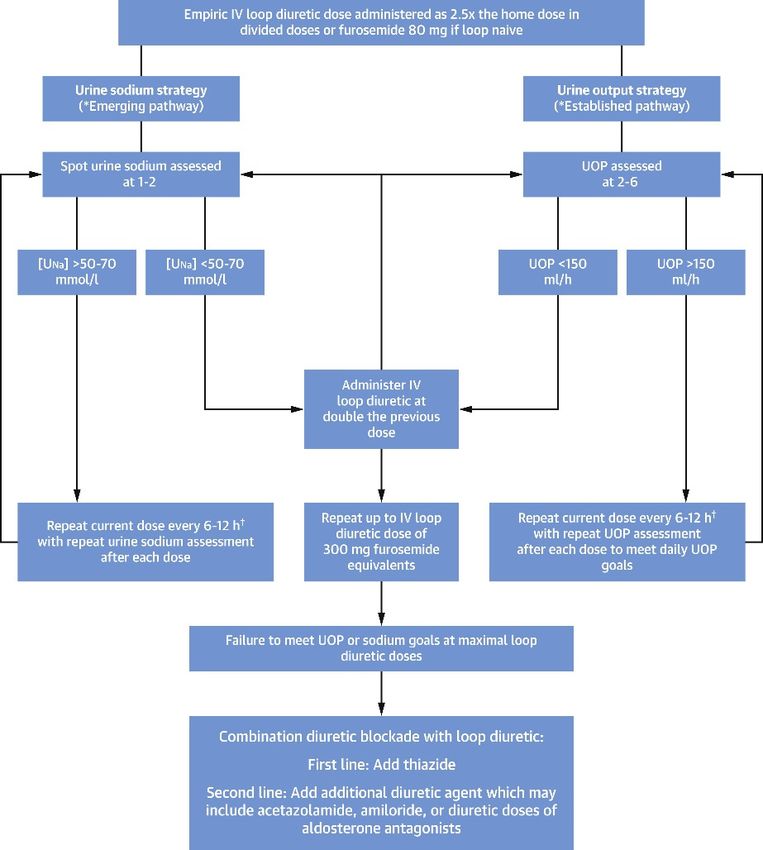
Figure 4, taken from ref [22] is an algorithm for titrating diuretic strategies in patients
Kidney Dial. 2023, 3, FOR PEER REVIEW 10
with persistent congestion and DR, on the basis of urine sodium measurement and urine
output.
Figure4.4.A proposed
Figure A proposed algorithm
algorithm for titrating
for titrating diureticdiuretic
strategiesstrategies
in patientsinwith
patients withcongestion
persistent persistent
congestion and diuretic resistance, on the basis of urine sodium measurement and urine output, is
and diuretic resistance, on the basis of urine sodium measurement and urine output, is shown. For
shown. For more details, see [22]. Reprinted with permission from Ref. [22] 2022, Elsevier. †: loop
more details, see [22]. Reprinted with permission from Ref. [22] 2022, Elsevier. †: loop diuretic doses
diuretic doses exceeding >1000 mg of furosemide equivalents/day have limited safety data and
exceeding
should be >1000 mg of furosemide
used cautiously. equivalents/day
Bumetanide have
may confer less limited
risk safety
at higher databecause
doses and should
it hasbe used
demon-
cautiously. Bumetanide may confer less risk at
strated reduced ototoxicity risk in animal models. higher doses because it has demonstrated reduced
ototoxicity risk in animal models.
Please note that in current clinical practice as the last step where combination diu-
retic blockades are recommended, SGLT2 inhibitors should also be included.
13. Co-Administration of Diuretics with AlbuminKidney Dial. 2023, 3 65
Please note that in current clinical practice as the last step where combination diuretic
blockades are recommended, SGLT2 inhibitors should also be included.
13. Co-Administration of Diuretics with Albumin
Severe hypoalbuminemia might impair diuretic effectiveness, owing to impaired
delivery to the kidney, and albumin administration might enhance natriuresis. However,
studies have produced mixed results. A meta-analysis concluded that this policy suggests
transient effects of modest clinical significance [76]. Some guidelines continue to suggest
that albumin infusion should be used as an adjunct to diuretics when nephrotic patients
appear to have vascular volume depletion (or appear to be “underfilled”) [77].
14. Continuous Infusion of Loop Diuretics
Compared with bolus therapy, continuous infusion of diuretics provides more sus-
tained and uniform drug delivery and prevents post-diuretic sodium retention.
The Diuretic Optimization Strategies Evaluation (DOSE) trial compared the efficacy
and safety of continuous vs. bolus furosemide therapy in 308 patients with acute HF [78].
There was no difference in symptom control or net fluid loss at 72 h in either group. Other
studies have shown more diuresis with continuous infusion than with a similarly dosed
bolus regimen [79]. However, at this point, definitive clinical evidence to support routine
use of continuous loop diuretic therapy is lacking.
15. Hypertonic Saline Infusion (HSS)
HSS along with diuretics induces fluid flow from interstitial spaces to the intravascular
space which leads to congestion relief by decreasing interstitial volume [80].
A meta-analysis demonstrated a significant decrease in all-cause mortality rate, reduced
length of hospital stay, increased weight loss, and fewer re-hospitalizations [81]. Additionally,
no electrolyte abnormalities requiring a change in dose or HSS discontinuation were observed.
A recent study reported that hypertonic saline administration was associated with
increased diuretic efficiency, as well as fluid and weight loss, without adverse respiratory
or neurological signals [82].
16. Combination Diuretic Therapy
Sequential nephron blockade with combination diuretic therapy is an important
therapeutic strategy against diuretic resistance and is the next step when the desired
diuretic response is not obtained with high doses of loop diuretic monotherapy.
16.1. Thiazides
Thiazide diuretics as monotherapy is not successful in DHF [83]; however, the addition
of 25–100 mg thiazide to a high dose of i.v. furosemide in 20 NYHA (The New York Heart
Association) class 3 and 4 HF patients with diuretic resistance increased body weight
reduction and urine output [84].
The most popular combination is a loop diuretic plus a thiazide, although no large-
scale placebo-controlled trials have been performed [85]. Metolazone (a thiazide-like
diuretic) is typically used due to its low cost and availability [86]. Metolazone also blocks
sodium reabsorption at the proximal tubule, which may contribute to its synergistic effect.
Chlorothiazide is available in an intravenous formulation and has a faster onset of action
than metolazone. However, studies have failed to detect any benefit of one over the other [87].
A recent propensity analysis, however, found that in DR HF patients, up-titration of
loop diuretics may be a preferred strategy over routine early addition of a thiazide type
diuretics when diuresis is inadequate [88].
16.2. Aldosterone Antagonists
A number of studies explored the potential advantages and safety of adding spirono-
lactone, mostly to loop diuretics.Kidney Dial. 2023, 3 66
The first major double-blind RALES study where spironolactone was added to an ACE
inhibitor, a loop diuretic, and in most cases digoxin, was discontinued early, because an
interim analysis showed lower mortality, a lower frequency of hospitalization for worsening
HF and a significant improvement in HF symptoms with spironolactone. Importantly, the
incidence of serious hyperkalemia was minimal in both groups of patients [89].
After reporting the RALES results, early single center [90] and later, a large epidemio-
logical survey [91] reported a clear rise in hospitalization for hyperkalemia associated with
a dramatic increase in mortality in the aldosterone-treated group [91].
In the ATHENA-HF trial, spironolactone in high doses (100 mg/d) was added to usual
therapy [83]. The primary end point was the change in NT-proBNP levels from baseline
to 96 h. Secondary end points included the clinical congestion score, dyspnea assessment,
net urine output, and net weight change. The result showed that adding spironolactone to
usual care was well tolerated, but did not improve the primary or secondary efficacy end
points.
16.3. Acetazolamide
A meta-analysis including nine studies up to mid-2017 in 229 patients with HF con-
cluded that, when compared with the placebo, acetazolamide significantly increased na-
triuresis and decreased the apnea-hypopnea index (and central apnea index) among HF
patients [92].
Results from an observational study [93] and a small, prospective, randomized
trial [94] suggest that the addition of acetazolamide (at a dose of 500 mg administered
intravenously once daily) to IV loop-diuretic therapy increased urinary sodium excretion
in patients with ADHF.
Mullens et al. [95] recently published the Acetazolamide in Decompensated Heart
Failure With Volume OveRload (ADVOR) study, a multicenter, placebo-controlled Belgian
trial where 519 patients with HF received standardized loop diuretics and were randomized
towards once daily IV acetazolamide (500 mg) versus placebo. The acetazolamide-loop
diuretic combination resulted in a more complete clinical decongestion than placebo at
3 days (42% vs. 31%), as well as the benefits associated with a shorter hospital stay. The
incidence of worsening kidney function, hypokalemia, hypotension, and adverse events
was similar in the two groups.
As underlined in an accompanying editorial [96], a limitation of this trial was the
exclusion in the trial of patients receiving SGLT2 inhibitors, which in contemporary clinical
care would probably be added in Acute Decompensated Heart Disease Failure (ADHDF)
therapy.
16.4. Multi-Nephron Segment Diuretic Therapy (MSDT)
MSDT is defined as the simultaneous use of 4 diuretic classes with actions along the
whole nephron is a potential method to overcome severe diuretic resistance in Acute Heart
Failure (AHF).
A retrospective analysis [97] of a study in 167 patients hospitalized with AHF and
diuretic resistance showed that MSDT was associated with increased median 24 h urine
output in the first day of therapy compared with the previous day (2.16 L to 3.08 L) in
the total cohort and in the severe diuretic resistance cohort (0.91 L to 2.08 L). The median
cumulative weight loss at day 7 or discharge was −7.4 kg (−15.3 to −3.4 kg). There were
no changes in serum chemistries or kidney function. More prospective studies of MSDT in
AHF and diuretic resistance are clearly warranted.
16.4.1. SGLT2 Inhibitors
As outlined above, the major mechanism of action of SGLT2 inhibitors occurs in
the proximal renal tubule (the same location as acetazolamide), and SGLT2 inhibitors
themselves have (weak) diuretic effects.Kidney Dial. 2023, 3 67
According to a pilot study, randomized, double-blind, placebo-controlled, multi-
center study on the effects of empagliflozin on clinical outcomes in patients with ADHF
(EMPARESPONSE-AHF), the use of empagliflozin did not affect the response to diuretics.
However, it was safe and reduced both mortality and the frequency of rehospitalization
60 days post discharge [98].
Wilcox et al. [99] investigated the interaction between a loop diuretic (bumetanide)
and dapagliflozin in normal subjects. There was no first-dose synergy between the two
diuretics; however, dapagliflozin administration for 1 week enhanced the sodium excretion
with bumetanide, and vice versa. Thus, there was significant two-way adaptive natriuretic
synergy. Griffin et al. [100] retrospectively analyzed patients who received adjuvant SGLT-
2i in 31 patients, 58% of whom had type 2 diabetes. Compared with the measurements
taken 24 h prior to SGLT-2i initiation, SGLT-2i’s improved weight loss, urine output, and
diuretic efficiency without worsening of creatinine, potassium, or blood pressure. The role
of SGLT2 inhibitors as adjuvant therapy in the diuretic treatment of resistance is still not
yet established; however, the mechanism of their diuretic action and the preliminary results
as described above make further clinical investigations necessary. When diuretic resistance
limits the efficient treatment of the congestion in SGLT2 inhibitor naïve patients, adding
these drugs to loop diuretics is recommended.
In patients treated with background SGLT2 inhibitors, additional dosages of acetazo-
lamide could further increase the efficacy of decongestion management.
16.4.2. Aquaretics
In hyponatremic patients with CHF, tolvaptan acutely increases serum sodium and
decreases body weight but does not improve long-term CHF morbidity or mortality [101].
In the TACTICS-HF trial, the addition of tolvaptan to a standardized furosemide regimen
did not improve the number of responders at 24 h despite greater weight loss [102]. Simi-
larly, the Short-Term Effects of Tolvaptan (SECRET) of CHF trial did not show significant
improvement in dyspnea in patients with AHF who were selected for greater potential
benefit from tolvaptan [103].
16.4.3. Ultrafiltration
This topic is beyond the topic of this contribution. It is sufficient to state that ultrafil-
tration has not demonstrated decongestive superiority over diuretic therapy in randomized
AHF trials and was associated with more complications [104,105].
17. Discussion
This review focuses on selected renal aspects of diuretic treatment in congestive heart
failure. The different classes of diuretics and their mechanism of action in the nephron
are discussed. Several mechanisms of loop diuretic resistance have traditionally been
proposed; however, recent evidence indicates that a constant sodium-avid state is the
main driver of diuretic resistance patients in congestive heart failure. Different therapeutic
options including combinations of diuretics within so-called nephron blockage strategies
are discussed.
18. Conclusions
Despite an impressive knowledge on the fascinating topic of congestive heart fail-
ure exists, ongoing research to optimally evaluate full decongestion (eu-volemia) and to
determine the ideal diuretic strategy is necessary.
Parenteral loop diuretic agents are the mainstay in the treatment of congestive heart
failure. Diuretic resistance in loop diuretic monotherapy may however develop, attenuating
the maximal diuretic effect and thereby preventing full decongestion. The mechanisms be-
hind diuretic resistance are diverse; however, contrary to traditional opinion, basal sodium
avidity, rather than diuretic-induced compensatory post-diuretic sodium reabsorption
(CPDSR), appears to be the predominant mechanism of diuretic resistance. A mechanism-Kidney Dial. 2023, 3 68
based classification can guide medical strategies to restore diuretic efficacy. Optimization
of loop diuretic regimens based upon diuretic response should be the primary strategy,
followed by combination nephron blockage with thiazides and an eventual low dose of al-
dosterone blockers. Recent studies show an emerging role for sodium- glucose transporter
2 inhibitors, possibly in combination with acetazolamide for the future management of
diuretic resistance.
Future research into the specific kidney transporters causing diuretic resistance in
heart failure is crucial to guiding future therapeutic treatment strategies.
Funding: This research received no external funding.
Institutional Review Board Statement: Not applicable.
Informed Consent Statement: Not applicable.
Data Availability Statement: Not applicable.
Conflicts of Interest: The author declares no conflict of interest.
References
1. Hoorn, E.J.; Ellison, D.H. Diuretic Resistance. Am. J. Kidney Dis. 2017, 69, 136–142. [CrossRef] [PubMed]
2. Mullens, W.; Martens, P.; Testani, J.M.; Tang, W.H.W.; Skouri, H.; Verbrugge, F.H.; Fudim, M.; Iacoviello, M.; Franke, J.; Flammer,
A.J.; et al. Renal effects of guideline-directed medical therapies in heart failure: A consensus document from the Heart Failure
Association of the European Society of Cardiology. Eur. J. Heart Fail. 2022, 24, 603–619. [CrossRef] [PubMed]
3. Wilcox, C.S.; Testani, J.M.; Pitt, B. Pathophysiology of Diuretic Resistance and Its Implications for the Management of Chronic
Heart Failure. Hypertension 2020, 76, 1045–1054. [CrossRef]
4. Ellison, D.H. Clinical Pharmacology in Diuretic Use. Clin. J. Am. Soc. Nephrol. 2019, 14, 1248–1257. [CrossRef]
5. Ellison, D.H.; Subramanya, A.R. Clinical Use of Diuretics. In Oxford Textbook of Clinical Nephrology, 4th ed.; Turner, N., Lameire, N.,
Goldsmith, D.J., Winearls, C.G., Himmelfarb, J., Remuzzi, G., Eds.; Oxford University Press: Oxford, UK, 2016; pp. 299–322.
6. Wu, W.; Bush, K.T.; Nigam, S.K. Key Role for the Organic Anion Transporters, OAT1 and OAT3, in the in vivo Handling of
Uremic Toxins and Solutes. Sci. Rep. 2017, 7, 4939. [CrossRef] [PubMed]
7. Wright, E.M. SGLT2 Inhibitors: Physiology and Pharmacology. Kidney360 2021, 2, 2027–2037. [CrossRef]
8. Brater, D.C.; Ellison, D.H. Mechanism of Action of Diuretics; Post, T.W., Ed.; UpToDate: Waltham, MA, USA, 2020; Volume 2020.
9. Novak, J.E.; Ellison, D.H. Diuretics in States of Volume Overload: Core Curriculum 2022. Am. J. Kidney Dis. 2022, 80, 264–276.
[CrossRef]
10. Vallon, V.; Thomson, S.C. The tubular hypothesis of nephron filtration and diabetic kidney disease. Nat. Rev. Nephrol. 2020,
16, 317–336. [CrossRef]
11. Onishi, A.; Fu, Y.; Patel, R.; Darshi, M.; Crespo-Masip, M.; Huang, W.; Song, P.; Freeman, B.; Kim, Y.C.; Soleimani, M.; et al. A
role for tubular Na(+)/H(+) exchanger NHE3 in the natriuretic effect of the SGLT2 inhibitor empagliflozin. Am. J. Physiol. Renal.
Physiol. 2020, 319, F712–F728. [CrossRef]
12. Aguilar-Gallardo, J.S.; Correa, A.; Contreras, J.P. Cardio-renal benefits of sodium-glucose co-transporter 2 inhibitors in heart
failure with reduced ejection fraction: Mechanisms and clinical evidence. Eur. Heart J. Cardiovasc. Pharmacother. 2022, 8, 311–321.
[CrossRef]
13. Bhatt, D.L.; Szarek, M.; Steg, P.G.; Cannon, C.P.; Leiter, L.A.; McGuire, D.K.; Lewis, J.B.; Riddle, M.C.; Voors, A.A.; Metra, M.;
et al. Sotagliflozin in Patients with Diabetes and Recent Worsening Heart Failure. N. Engl. J. Med. 2021, 384, 117–128. [CrossRef]
[PubMed]
14. Maddox, T.M.; Januzzi, J.L., Jr.; Allen, L.A.; Breathett, K.; Butler, J.; Davis, L.L.; Fonarow, G.C.; Ibrahim, N.E.; Lindenfeld, J.;
Masoudi, F.A.; et al. 2021 Update to the 2017 ACC Expert Consensus Decision Pathway for Optimization of Heart Failure
Treatment: Answers to 10 Pivotal Issues About Heart Failure with Reduced Ejection Fraction: A Report of the American College
of Cardiology Solution Set Oversight Committee. J. Am. Coll. Cardiol. 2021, 77, 772–810. [CrossRef] [PubMed]
15. McDonagh, T.A.; Metra, M.; Adamo, M.; Gardner, R.S.; Baumbach, A.; Böhm, M.; Burri, H.; Butler, J.; Čelutkienė, J.; Chioncel, O.;
et al. 2021 ESC Guidelines for the diagnosis and treatment of acute and chronic heart failure. Eur. Heart J. 2021, 42, 3599–3726.
[CrossRef]
16. McMurray, J.J.V.; Solomon, S.D.; Inzucchi, S.E.; Køber, L.; Kosiborod, M.N.; Martinez, F.A.; Ponikowski, P.; Sabatine, M.S.; Anand,
I.S.; Bělohlávek, J.; et al. Dapagliflozin in Patients with Heart Failure and Reduced Ejection Fraction. N. Engl. J. Med. 2019, 381,
1995–2008. [CrossRef] [PubMed]
17. Mordi, N.A.; Mordi, I.R.; Singh, J.S.; McCrimmon, R.J.; Struthers, A.D.; Lang, C.C. Renal and Cardiovascular Effects of SGLT2
Inhibition in Combination with Loop Diuretics in Patients with Type 2 Diabetes and Chronic Heart Failure: The RECEDE-CHF
Trial. Circulation 2020, 142, 1713–1724. [CrossRef] [PubMed]Kidney Dial. 2023, 3 69
18. Vallon, V.; Verma, S. Effects of SGLT2 Inhibitors on Kidney and Cardiovascular Function. Annu. Rev. Physiol. 2021, 83, 503–528.
[CrossRef] [PubMed]
19. Ellison, D.H.; Felker, G.M. Diuretic Treatment in Heart Failure. N. Engl. J. Med. 2017, 377, 1964–1975. [CrossRef]
20. Schnermann, J.; Briggs, J.P. Synthesis and secretion of renin in mice with induced genetic mutations. Kidney Int. 2012, 81, 529–538.
[CrossRef]
21. Ellison, D.H. Mechanistic Insights into Loop Diuretic Responsiveness in Heart Failure. Clin. J. Am. Soc. Nephrol. 2019, 14, 650–652.
[CrossRef]
22. Felker, G.M.; Ellison, D.H.; Mullens, W.; Cox, Z.L.; Testani, J.M. Diuretic Therapy for Patients with Heart Failure. JACC State Art
Rev. 2019, 75, 1178–1195. [CrossRef]
23. Brater, D.C.; Day, B.; Burdette, A.; Anderson, S. Bumetanide and furosemide in heart failure. Kidney Int. 1984, 26, 183–189.
[CrossRef] [PubMed]
24. Testani, J.M.; Brisco, M.A.; Turner, J.M.; Spatz, E.S.; Bellumkonda, L.; Parikh, C.R.; Tang, W.H. Loop diuretic efficiency: A metric
of diuretic responsiveness with prognostic importance in acute decompensated heart failure. Circ. Heart Fail. 2014, 7, 261–270.
[CrossRef] [PubMed]
25. Vargo, D.L.; Kramer, W.G.; Black, P.K.; Smith, W.B.; Serpas, T.; Brater, D.C. Bioavailability, pharmacokinetics, and pharmacody-
namics of torsemide and furosemide in patients with congestive heart failure. Clin. Pharmacol. Ther. 1995, 57, 601–609. [CrossRef]
[PubMed]
26. Pearce, D.; Manis, A.D.; Nesterov, V.; Korbmacher, C. Regulation of distal tubule sodium transport: Mechanisms and roles in
homeostasis and pathophysiology. Pflugers Arch. 2022, 474, 869–884. [CrossRef] [PubMed]
27. Pearce, D.; Soundararajan, R.; Trimpert, C.; Kashlan, O.B.; Deen, P.M.; Kohan, D.E. Collecting duct principal cell transport
processes and their regulation. Clin. J. Am. Soc. Nephrol. 2015, 10, 135–146. [CrossRef] [PubMed]
28. Wall, S.M.; Verlander, J.W.; Romero, C.A. The Renal Physiology of Pendrin-Positive Intercalated Cells. Physiol. Rev. 2020, 100,
1119–1147. [CrossRef]
29. Cil, O.; Haggie, P.M.; Phuan, P.W.; Tan, J.A.; Verkman, A.S. Small-Molecule Inhibitors of Pendrin Potentiate the Diuretic Action of
Furosemide. J. Am. Soc. Nephrol. JASN 2016, 27, 3706–3714. [CrossRef]
30. Soleimani, M. The multiple roles of pendrin in the kidney. Nephrol. Dial. Transplant. 2015, 30, 1257–1266. [CrossRef]
31. Brown, R.; Quirk, J.; Kirkpatrick, P. Eplerenone. Nat. Rev. Drug Discov. 2003, 2, 177–178. [CrossRef]
32. Weinberger, M.H.; Roniker, B.; Krause, S.L.; Weiss, R.J. Eplerenone, a selective aldosterone blocker, in mild-to-moderate
hypertension. Am. J. Hypertens. 2002, 15, 709–716. [CrossRef]
33. Agarwal, R.; Kolkhof, P.; Bakris, G.; Bauersachs, J.; Haller, H.; Wada, T.; Zannad, F. Steroidal and non-steroidal mineralocorticoid
receptor antagonists in cardiorenal medicine. Eur. Heart J. 2021, 42, 152–161. [CrossRef] [PubMed]
34. Rico-Mesa, J.S.; White, A.; Ahmadian-Tehrani, A.; Anderson, A.S. Mineralocorticoid Receptor Antagonists: A Comprehensive
Review of Finerenone. Curr. Cardiol. Rep. 2020, 22, 140. [CrossRef] [PubMed]
35. Bakris, G.L.; Agarwal, R.; Anker, S.D.; Pitt, B.; Ruilope, L.M.; Rossing, P.; Kolkhof, P.; Nowack, C.; Schloemer, P.; Joseph, A.; et al.
Effect of Finerenone on Chronic Kidney Disease Outcomes in Type 2 Diabetes. N. Engl. J. Med. 2020, 383, 2219–2229. [CrossRef]
[PubMed]
36. Pitt, B.; Filippatos, G.; Agarwal, R.; Anker, S.D.; Bakris, G.L.; Rossing, P.; Joseph, A.; Kolkhof, P.; Nowack, C.; Schloemer, P.; et al.
Cardiovascular Events with Finerenone in Kidney Disease and Type 2 Diabetes. N. Engl. J. Med. 2021, 385, 2252–2263. [CrossRef]
[PubMed]
37. Cox, Z.L.; Testani, J.M. Loop diuretic resistance complicating acute heart failure. Heart Fail. Rev. 2020, 25, 133–145. [CrossRef]
38. Ellison, D.H. Diuretic resistance: Physiology and therapeutics. Semin. Nephrol. 1999, 19, 581–597.
39. Ahmad, T.; Jackson, K.; Rao, V.S.; Tang, W.H.W.; Brisco-Bacik, M.A.; Chen, H.H.; Felker, G.M.; Hernandez, A.F.; O’Connor, C.M.;
Sabbisetti, V.S.; et al. Worsening Renal Function in Patients with Acute Heart Failure Undergoing Aggressive Diuresis Is Not
Associated with Tubular Injury. Circulation 2018, 137, 2016–2028. [CrossRef]
40. Fudim, M.; Loungani, R.; Doerfler, S.M.; Coles, A.; Greene, S.J.; Cooper, L.B.; Fiuzat, M.; O’Connor, C.M.; Rogers, J.G.; Mentz, R.J.
Worsening renal function during decongestion among patients hospitalized for heart failure: Findings from the Evaluation Study
of Congestive Heart Failure and Pulmonary Artery Catheterization Effectiveness (ESCAPE) trial. Am. Heart J. 2018, 204, 163–173.
[CrossRef]
41. Metra, M.; Davison, B.; Bettari, L.; Sun, H.; Edwards, C.; Lazzarini, V.; Piovanelli, B.; Carubelli, V.; Bugatti, S.; Lombardi, C.;
et al. Is worsening renal function an ominous prognostic sign in patients with acute heart failure? The role of congestion and its
interaction with renal function. Circ. Heart Fail. 2012, 5, 54–62. [CrossRef]
42. Rao, V.S.; Ahmad, T.; Brisco-Bacik, M.A.; Bonventre, J.V.; Wilson, F.P.; Siew, E.D.; Felker, G.M.; Anstrom, K.K.; Mahoney, D.D.;
Bart, B.A.; et al. Renal Effects of Intensive Volume Removal in Heart Failure Patients with Preexisting Worsening Renal Function.
Circ. Heart Fail. 2019, 12, e005552. [CrossRef]
43. Lo, K.B.; Rangaswami, J. Mechanistic Insights in Cardiorenal Syndrome. NEJM Evid. 2022, 1, EVIDra2200053. [CrossRef]
44. Verbrugge, F.H. Editor’s Choice-Diuretic resistance in acute heart failure. Eur. Heart J. Acute Cardiovasc. Care 2018, 7, 379–389.
[CrossRef] [PubMed]You can also read


