Renal stone disease: medical management - Jasmine Tan 21 April 2021 Green Lane Medical Specialists
←
→
Page content transcription
If your browser does not render page correctly, please read the page content below
Renal stone disease:
medical management
             Jasmine Tan
            21 April 2021
    Green Lane Medical Specialists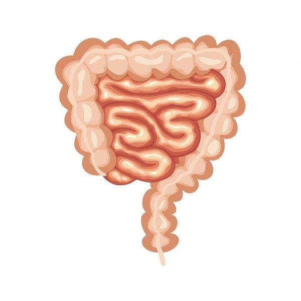
Increased incidence of renal stone disease
 • Radiological detection
 • Obesity (females > males)
        • HR 2.6 for stone recurrence in first time stone formers
        • 2-fold risk in those with ≥ 4 traits present (abdominal obesity, increased TG, decreased HDL,
          hypertension, or diabetes/IGT)

 • Diabetes and renal stones
        • Incident risk of nephrolithiasis in older women with DM 1.29 (95% CI: 1.05–1.58); younger
          women 1.60 (95% CI: 1.16–2.21) and in men 0.81 (95% CI:0.59–1.09)1
        • Similarly high rates of diabetes within 5 years of diagnosis of renal stone disease

1. Renal Failure, 2012; 34(10): 1348–1354
Am J Kidney Dis (2008) 51: 741–747
Eur J Epidemiol (2018) 33: 1033–1047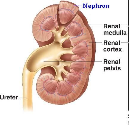
Renal Failure, 2012; 34(10): 1348–1354
Renal stone disease and CKD risk
• Absolute risk for ESRD is small
    • ESRD                HR 2.16
    • CKD stage 3b-5      HR 1.74
    • At risk: stone formers with cystinuria, uric acid or struvite stones, RTA, chronic bowel
      disorders

 J Nephrol (2016) 29:715 – 734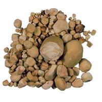
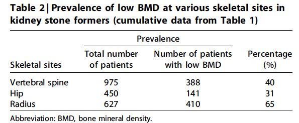
Renal stone disease and osteoporosis risk
• 4-fold cumulative risk of vertebral fractures compared to general non-stone-
  forming population
    • Higher associations for fractures in men> women
    • Low BMD present in both hypercalciuric and normocalciuric stone-forming subjects
    • Greater reduction in BMD in patients with hypercalciuria

 Kidney Int (1998) 53: 450 – 465
 Kidney Int (2011) 79, 393–403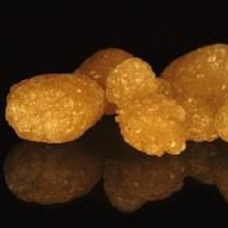
Approach to renal stone disease
                                                      Stones > 4mm
                                                      Obstructive uropathy
                                                      Associated urosepsis
                                                      Cystine, staghorn calculi
                                     Surgical

   Patient education         Dietary
                                                Medical
   Accurate and focused   intervention                        Metabolic evaluation
   dietary advice                                             Assessment for secondary causes
   Weight loss                                                Pharmacological therapy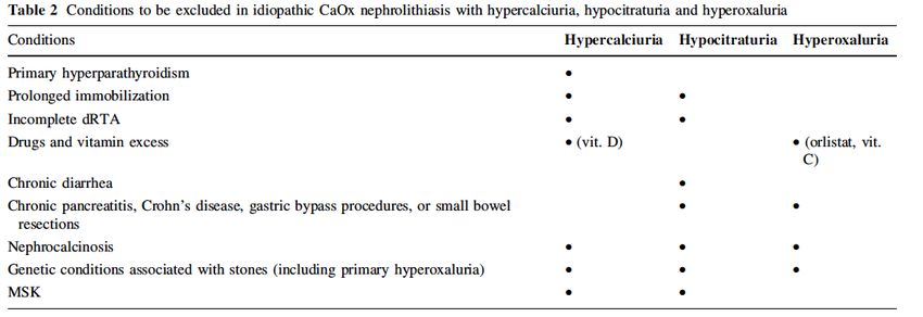
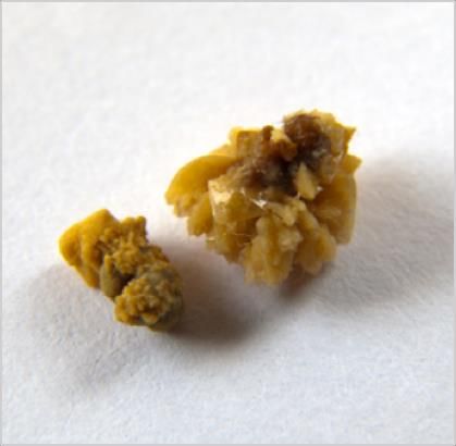
Composition and prevalence of main renal
   stone types

Calcium-based stones   Struvite/infection stones   Uric acid stones     Cystine stones
60 – 80%               10 - 15%                    5 – 10 %             1%
Predominantly          “triple phosphate stones”   Acidic urine         Hereditary
calcium oxalate        (Calcium/magnesium and      Associated with
                       ammonium phosphate)
                                                                        Young patients
                                                   metabolic syndrome   Often recur
                       Urease splitting bacteria                                         Other stones
                       Alkaline urine from                                               1%
                       ammonia                                                           Drug stones
                                                                                         Xanthine crystals
                       Staghorn calculi - large
Recurrent stone formers
• Indication of metabolic activity
• No markers to distinguish between single and recurrent stone formers
• In a population cohort study (N=2,239), developed as a prediction tool for
  assessing risk in first-time stone formers
   • 10-year recurrence of symptomatic stone disease was 30% on the whole
   • 56% risk for second symptomatic episode in high risk patients
   • Recurrence of Kidney Stone (ROKS) nomogram
The Recurrence of Kidney Stone (ROKS) nomogram can be easily applied in first time
                                              symptomatic stone formers.

Andrew D. Rule et al. JASN 2014;25:2878-2886                                                       ©2014 by American Society of Nephrology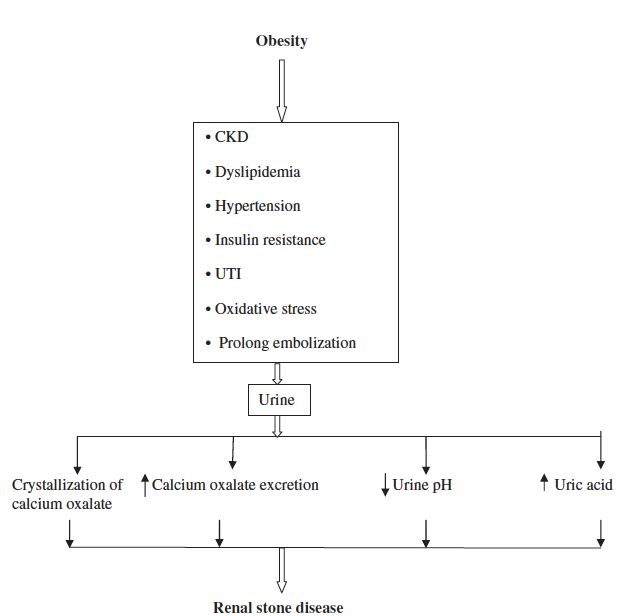
QxMD calculator
ROKS – Recurrence Of Kidney Stone (2018)                         11.Any prior stone found to be mostly calcium oxalate
Predict the risk of a future symptomatic kidney stone after      monohydrate with or without calcium oxalate dehydrate or
the last symptomatic stone.                                      hydroxyapatite?
                                                                 12.Was imaging (CT scan, abdominal X-ray, or ultrasound)
Questions                                                        performed at the last symptomatic stone episode?
1.How many confirmed symptomatic kidney stone episodes           13.Number of stones in both kidneys?
with a passed or obstructing stone on imaging has this           14.Diameter of largest kidney stone?
patient had (including the last episode)?                        15.Symptomatic stone seen at the ureterovesical junction?
2.Number of years since last confirmed symptomatic kidney        16.Stone seen in the renal pelvis or in the lower renal pole?
stone episode?
3.Age in years at last confirmed symptomatic stone episode?
4.Body mass index in kg/m2 at last confirmed symptomatic
stone episode?
5.Gender?
6.Any family history of kidney stones?
7.Incidental (asymptomatic) stone on imaging prior to first
confirmed symptomatic stone episode?
8.Suspected kidney stone event (no stone seen) before first
confirmed symptomatic kidney stone episode?
9.Pregnant during last confirmed symptomatic stone
episode?
10.Any prior stone found to contain any uric acid, brushite or              Vaughn LE et al. Mayo Clin Proc. 2019 Feb; 94(2): 202–210.
struvite?
Case 1 40 yo lady with recent AKI History of renal colic and haematuria, managed conservatively. No fevers, no flank tenderness What is your next investigation? a. CT KUB b. Urine microscopy & c. Spot urine calcium/creatinine ratio d. Renal biopsy
Medullary nephrocalcinosis
• Diffuse calcium deposits in localized at the renal medulla
• Increased risk of calcium stone formation
• Associated with underlying conditions which cause hypercalcemia
  and/or hypercalciuria
• Treat underlying cause
• Further investigations
Urine calcium/Cr ratio 0.73 (normal 0.06 – 0.4)
Creatinine 91 umol/L; eGFR 68 ml/min/1.73m2
Adjusted calcium 2.83 mmol/L (2.1 – 2. 55); PTH 9.6 pmol//L (1.7 – 7.3)
ALP 186 U/L (40 – 120)

Parathyroid scintiscan: localized left inferior parathyroid adenoma
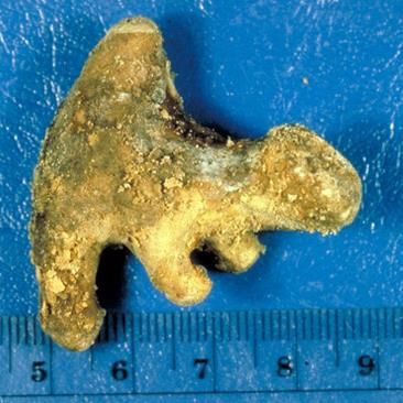
Q2
• Which crystals are characteristic for struvite stones?

                                   c.
a.

      Calcium phosphate crystals           Cystine crystals

b.                                 d.

      Uric acid crystals                Triple phosphate crystals
Struvite stones • Associated with UTIs • Urease-producing bacteria leads to the hydrolysis of urea into ammonium and hydroxyl ions. • Alkalotic urine (pH >7.2) secondary to increase in ammonium & phosphate concentrations • More frequent in woman and older people
Gram-positive bacteria             Gram-negative bacteria                      Yeasts
Staphylococcus aureus              Bacteroides corrodens                       Cryptococcus species
Staphylococcus epidermidis         Helicobacter pylori                         Rhodotorula species
Corynebacterium species (ie, C     Bordetella pertussis                        Sporobolomyces species
ulcerans, C renale, C ovis, C      Bordetella bronchiseptica                   Trichosporon cutaneum
hofmannii, C murium, C equi)       Haemophilus influenzae                      Candida humicola
Mycobacterium rhodochrous group    Haemophilus parainfluenzae
Micrococcus varians                Proteus species (ie, P mirabilis, P
Bacillus species                   morganii, P rettgeri)
Clostridium tetani                 Providencia stuartii
Peptococcus asaccharolyticus       Klebsiella species (K pneumoniae, K
                                   oxytoca)
                                   Pasteurella species
                                   Pseudomonas aeruginosa
                                   Aeromonas hydrophilia
                                   Yersinia enterocolitica
                                   Brucella species
                                   Flavobacterium species
                                   Serratia marcescens
                                   Ureaplasma urealyticum
                                   Mycoplasma T-strain

                          https://emedicine.medscape.com/article/439127-overview#a8
Management • Complete removal key as residual stone material serve as nidus • Recurrence up to 85% • Suppressive antibiotic therapy – prophylaxis and inhibiting stone growth • Urease inhibitors - acetohydroxamic acid (not available in NZ) • Reduces urine alkalinity and urinary ammonium concentration • Impaired effectiveness and increased toxicity in renal impairment • Low phosphorus, low calcium diet
Case 3
A 27-year-old woman has recurrent renal stones. Her serum electrolytes were
normal. She has a history of Sjogren’s syndrome.
Two 24-hour urine collections were similar:
Volume 1.7 L (> 2 – 2.5 L)
pH 6.9 (5.8 – 6.2)
Urinary calcium 5.6 mmol (
• What is the likely stone composition?
a. Calcium oxalate
b. Calcium urate
c. Calcium phosphate
d. Cystine stones
• Further investigations confirmed fasting urine pH 6.9 and venous bicarbonate 19
  mmol/L and potassium 3.5 mmol/L = distal RTA
• Nephrocalcinosis and nephrolithiaisis are frequently associated with distal RTA
  (type 1)
• Associated with hypercalciuria and hypocitraturia
• Higher urine pH and hypocitraturia promote calcium phosphate supersaturation
Acidemia with distal RTA

                                                                 Increase
                                                                 absorption
                                                                 of Ca 2+                             urine Ca 2+
                       Ca 2+
                       PO4 2-                                                                         urine citrate

                                                                                                      urine pH
                                ECF calcium and
                                  phosphate

                                                                                              CaPO4
Management: Increase fluid intake; correction with potassium citrate; concomitant thiazides
           DEXA scan
Q4. • Which is associated with an increased risk for calcium oxalate stone formation? a. Roux-en-Y gastric bypass b. Extracorporeal shock wave lithotripsy (ESWL) c. Sleeve gastrectomy d. Ileostomy
Calcium oxalate stones
• Idiopathic calcium oxalate stones most common
• Diseases affecting the small intestine or pancreas, including Crohn’s
disease, malabsorptive types of bariatric surgery, or chronic pancreatitis, lead to fat
malabsorption and enteric hyperoxaluria
J Nephrol (2016) 29:715–734
24 hour urine interpretation
• Stone analysis still gold standard
    Urine composition                Targets
        Volume (L)                     >2L
            pH                      5.8 – 6.2
    Creatinine (mmol)     0.16 – 0.21 mmol/kg (males)
                         0.13 – 0.18 mmol/kg (females)
     Calcium (mmol)
Metabolic evaluation • Renal stone composition for exclusion of cystinuria, APRT deficiency and struvite stones arising from urease-positive organism • Initial urine microscopy and protein • Serum biochemistry and bicarbonate • Initial and follow up 24 hour urine biochemical evaluation • Evaluation of GI diseases and systemic causes • Periodic imaging to assess stone burden
Empiric therapies
- Increased oral fluids
- Dietary manipulation and low salt intake

      Metabolic evaluation

            Specific management of secondary cause
            Targeted dietary manipulation
            Pharmacological therapies
Dietary therapies • Empiric dietary management for renal stone disease: • Fluids to produce 2 – 2.5L urine /day; appropriately spaced • Low dietary salt (
Pharmacological therapies
• Thiazides in hypercalciuria
   •   Chlorthalidone 25–50 mg daily
   •   Bendroflumethiazide 2.5 mg TDS
   •   Hydrochlorothiazide 25 mg BD, 50 – 100 mg daily
   •   Indapamide 2.5 mg daily
• Alkali therapy in hypocitriuira or recurrent calcium oxalate stone or uric acid
  stone formers
   • Potassium citrate 30 – 80 mmol/day
• Allopurinol in hyperuricosuria
Metabolic evaluation – Who to refer?
• Any patient with or at risk of recurrent stone disease
   • Positive family history
   • Recurrent UTIs
   • Obesity
   • Primary or secondary intestinal malabsorption disorder, or patients with malabsorptive type
     bariatric surgeries
   • Medical conditions predisposing to stone disease (eg cystinuria, struvite stones, Randall’s
     plaques)
   • History of urinary tract abnormalities of reconstruction

• Any patient with solitary kidney

• Any patient who desires further information or insight into preventable and
  reversible causes
You can also read