Morphological characterization of the proventriculus of Gry/lus assimilis Fabricius (Orthoptera, Gryllidae)
←
→
Page content transcription
If your browser does not render page correctly, please read the page content below
Morphological characterization of the proventriculus
       of Gry/lus assimilis Fabricius (Orthoptera, Gryllidae)

                                                                  Carmem s. Fontanetti 1
                                                                        Edison Zefa 1,2

       ABSTRACT. The proventriculus morphology of the cricket G/y//us assimilis Fabri-
       cius, 1775 is described using scanning electron microscopy and the con'elation ofthis
       structure with the feeding habits briefly considered.
       KEY WORDS. Orthoptera, Glyllidae, Gry//us, proventriculus

        ln general, the digestive tube ofthe insects can be divided into three areas:
the foregut or stomodeum, the midgut or ventriculus and the hindgut or proctodeum.
Its morphological diversity has lead several authors to use it as a systematic and
phylogenetic character in several groups (MORTINER 1965; SINGH & JUDD 1966;
GRANT & RENTZ 1967; DELIGNE 1971; BORDAS 1896, 1898).
        The foregut is made up ofthe pharynx, esophagus, crop, and proventriculus
or gizzard. The last structure is of great taxonomic interest, since it is intimately
associated with the insect feeding habit, that can be highly species specific.
        The proventriculus is a transition area between the foregut and midgut and
like the remainder ofthe anterior intestine, has an ectodermal origin and possesses
a sclerotized intima. It presents ali the stages of development, varying from a simple
valve formed by fine cuticle, to a strong muscular organ, armed with spines and
teeth, having as a main function the grinding of food .
        Many studies have been accomplished to recognize relationships between
the morphology ofthe proventriculus and the alimentary habits ofinsects. LEBRUN
(1985) and LEBRUN & LEQUET (1985), showed that the proventriculus of termites
that feed on wood has a cuticular armor formed by strong teeth and sclerotized plates,
while those that feed on decomposed wood do not present teeth and the plates are
less developed. ln the humus eating termites, the armor ofthe proventriculus is weak
and only small plates are present. GIBBS (1967) also observed variations in the
structure of the proventriculus of nine species of Trichoptera, demonstrating that
the proventriculus can perform in the grinding or filtering of food.
        JUDD (1948) studied comparatively several families of ensiferans Orthoptera
and elaborated a key to identify the families and orders, using the proventriculus
and the gastric caecum as characters oftaxonomic value.

1) Departamento de Biologia, Instituto de Biociências, Universidade Estadual Paulista.
   13506-900 Rio Claro, São Paulo, Brazil. E-mail: fontanet@rc.unesp.br
2) CNPq fellow.

                                                     Revta bras. Zool. 17 (1): 193 -198, 2000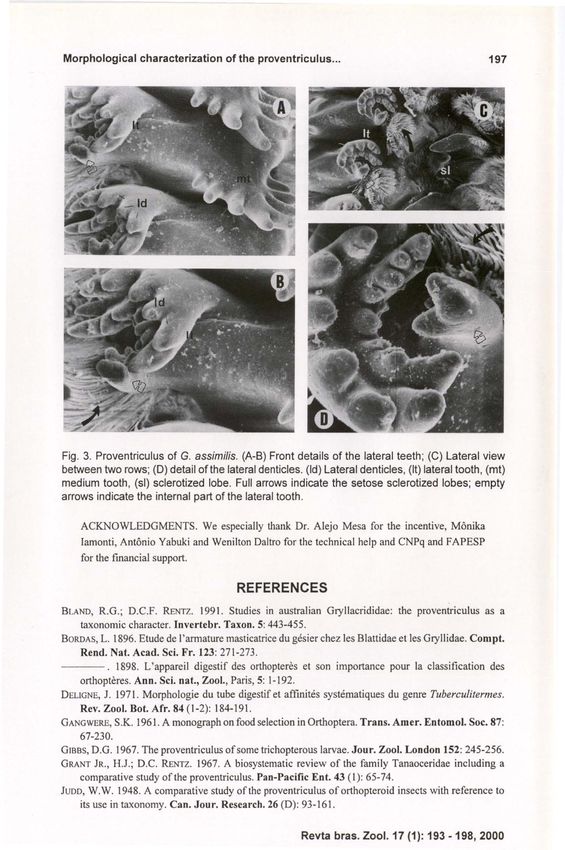
194                                                                    Fontanetti & Zefa

        BLAND & RENTZ (1991) studied 17 species of Australian grilacridids,
discussing the use of the proventriculus as a character and more briefly, the
correlations between the structure of the proventriculus and the alimentary habits
ofthe species. Here in we describe the morphology ofthe proventriculus of Gryllus
assimilis Fabricius, 1775 is described.
        The results ofthis study may eventually be used as a taxonomic resource for
the genus Gryllus Linnaeus, 1758, which includes a great number of cosmopolitan
and sibling species, where the recognition through their external morphology is
difficult.
                           MATERIAL ANO METHOOS
        Seven adult males and six females were collected from grassland and lawns
by E. Zefa at Universidade Estadual Paulista, Campus of Rio Claro, São Paulo, in
the months ofMarch and April, 1998. The specimens were dissected in physiolo-
gical solution and the digestive tubes were removed. The proventriculi were fixed
in a solution of KARNoWSKY (1965) and processed for observation under the
scanning electron microscope. Diagram of the alimentary canal was obtained with
aid of a camera lucida coupled to a stereomicroscope.
                          RESUL TS ANO OISCUSSION
        The digestive tube, especially the proventriculi, of several adult male and
femaIe specimens of G. assimilis was compared and no difference was observed
between the two sexes. It is composed of a short pharynx, that opens into the buccal
cavity, proceeded by the esophagus, the crop and the proventriculus; this segment
form the foregut. The midgut or ventriculus, presents a pair oflarge and voluminous
gastric ceca that line the whole proventriculus. The hindgut is then observed, ending
in the rectum, which opens into the anus. Numerous Malpighian tubes are observed
inserted into the transition between midgut and hindgut (Fig. 1).
        The proventriculus measures approximately 2.5 mm in length and 2.0 mm
in diameter and presents a flat wall in the external part. lnternally, the proventriculus
presents six rows formed by groups of sclerotized appendices united to each other
by sclerotized partitions (Fig. 2A). Twelve sclerotized appendices were observed
composing each row. The sclerotized appendix (Fig. 2B,C) is formed by a central
projection, the medium tooth and two lateral expansions, the lateral teeth. The
medium tooth presents, on average, seven denticles facing the lumen, called medi um
denticles (Fig. 2C). Two projections extend from the medium tooth in a lateral
direction; these are called lateral teeth. They possess several denticles on their apical
face, called lateral denticles (Figs 2C, 3A,B,D). Both the medium denticles and the
lateral ones have a rounded apex. The apical projection ofthe lateral tooth partially
surrounds the basal projection (empty arrows in figures 3A, B). The latter is
therefore located more internally (empty arrows in figures 3C,D). The apical
projection of the lateral tooth is formed by two semicircular rows made up, on
average, of seven denticles (Figs 3C,D). On either side of the rows of sclerotized
appendices, small expansions can be observed, called sclerotized lobes (Figs 2A,B,
3C), with setose in their more internal portion (full arrows in figures 2B,C, 3B,C,D).
These structures correspond to the inner barbed lobes mentioned by JUDO (1948).

Revta bras. Zool. 17 (1): 193 -198, 2000
Morphological characterization of the proventriculus ...                                       195

                                    I
                                    fg

                                   mg

                                   1-
                                     L
Fig . 1. General outline of the digestive tube of Gry/Jus assimílis. (e) Crop, (es) esophagus, (fg)
foregut, (g) gut, (ge) gastrie caeeum, (hg) hindgut, (mg) midgut, (Mt) Malpighian tubes, (p)
pylorus, (ph) pharynx, (pv) proventrieulus, (v) ventrieulus.

        Comparing our data with those of JUDD (1948), the proventriculus of G.
asstmilis resembles that of other species namely Oecanthus nigricornis Walker,
 1869, Neoxabea bipunctata DeGeer, 1773 (Oecanthidae), Falcicula hebardi Rehn,
 1903 ,Anaxipha exigua Say, 1825, Phyllopalpus pulchellus Uhler, 1864, Cyrtoxipha
columbiana Caudell, 1907 (Trigonidiidae), Hapithus brevipennis Saussure, 1897,
Orocharis saltator Uhler, 1864, Tafalisca lurida Walker, 1869 (Eneopteridae),
Nemobius fasciatus DeGeer, 1773 (Nemobiidae) and Cycloptilum squamosum
Scudder, 1868 (Mogoplistidae). ln these species, the main differences were the form
ofthe medium tooth and denticles, and the lateral teeth.
        For the family Gryllidae, JUDD (1948) studied Gryllus domesticus Linnaeus,
 1758, Gryllus assimilis Fabricius, 1775 and Anurogryllus muticus DeGeer, 1773.
Although he mentioned G. assimilis, he did not present a description of its proven-
triculus.
        BLAND & RENTZ (1991), compared several species of Australian grilacri-
dids, and suggested that in the herbivorous ones predominate long lateral denticles,
when compared to predatory ones. Moreover, they comment that the internal
structure ofthe proventriculus is different among genera.
        The morphology ofthe proventriculus observed in G. assimilis, agrees with
the observations ofBLAND & RENTZ (1991), about the correlation between proven-
tricular morphology and alimentary habits of orthopteran species. According au-
thors, species preferentially herbivorous shows long teeth and lateral denticles, like
G. assimilis.

                                                     Revta bras. Zoo I. 17 (1): 193 -198, 2000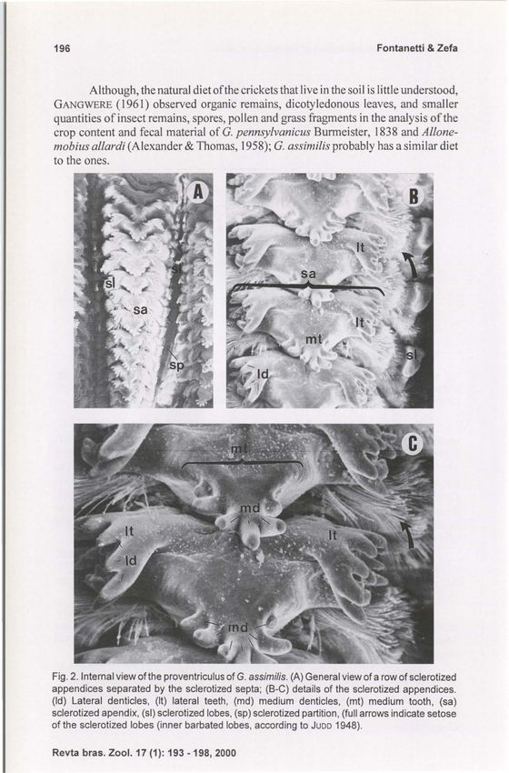
196 Fontanetti & Zefa
Although, the natural diet ofthe crickets that live in the soil is little understood,
GANGWERE (1961) observed organic remains, dicotyledonous leaves, and smaller
quantities ofinsect remains, spores, pollen and grass fragments in the analysis ofthe
crop content and fecal material of G. pennsy/vanicus Burmeister, 1838 and AI/one-
mobius aI/ardi (Alexander & Thomas, 1958); G. assimilis probably has a similar diet
to the ones.
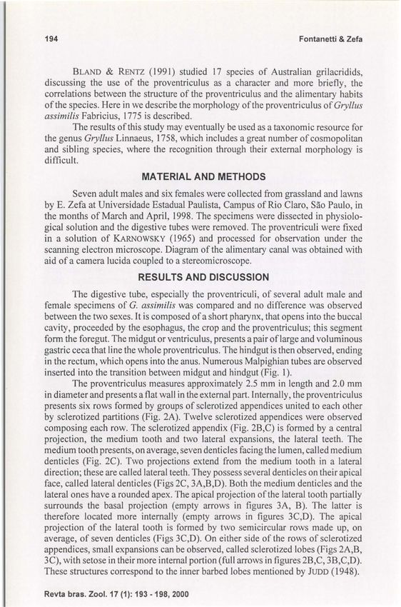
Fig. 2. Internal view ofthe proventriculus of G. assimilis. (A) General view of a row of sclerotized
appendices separated by the sclerotized septa; (B-C) details of the sclerotized appendices.
(Id) Lateral denticles, (It) lateral teeth, (md) medium denticles, (mt) medium taoth, (sa)
sclerotized apendix, (51) sclerotized lobes, (sp) sclerotized partition, (fuI! arrows indicate setose
af the sclerotized lobes (inner barbated lobes, according to JUDD 1948).
Revta bras. Zool. 17 (1): 193 -198, 2000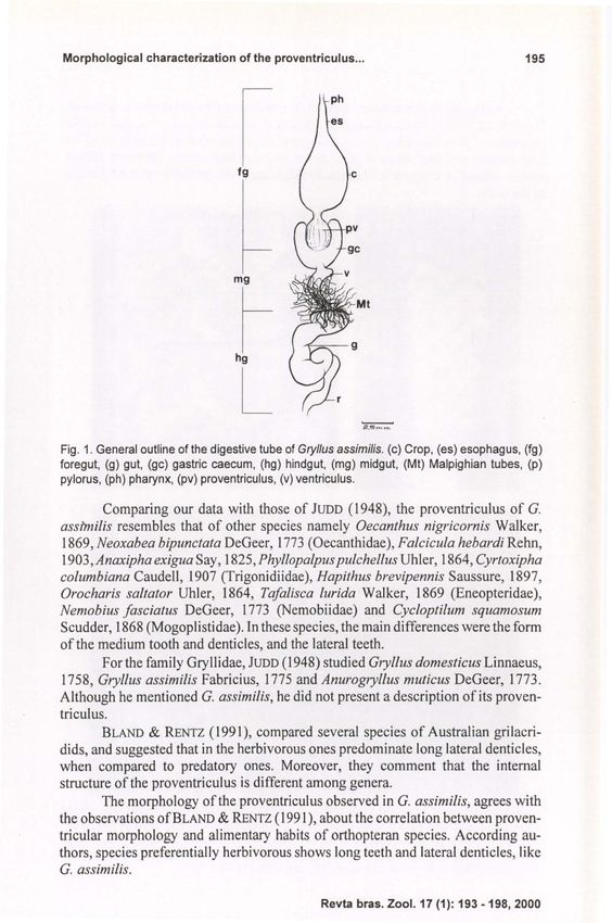
Morphological characterization of the proventriculus ...                                             197

Fig. 3. Proventriculus of G. assimilis. (A-B ) Front details of the lateral teeth; (C) Lateral view
be!ween !wo rows; (D) detail ofthe lateral denticles. (Id) Lateral denticles, (It) lateral tooth , (mt)
medium tooth, (si) sclerotized lobe. Full arrows indicate the setose sclerotized lobes; empty
arrows indicate the internal pari of the lateral tooth .

    ACKNOWLEDGMENTS. We especially thank Dr. Alejo Mesa for the incentive, Mônika
    lamonti , Antônio Yabuki and Wenilton Daltro for the techni cal help and CNPq and FAPESP
    for the [mancial support.

                                         REFERENCES
BLAND, R.G .; D.C.F. RENTZ. 199 1. Studies in australian Gryllacrididae: the proventriculus as a
    taxonomic character. Invertebr. Taxon. 5: 443-455.
BORDAS, L. 1896. Etude de I' armature masticatrice du gésier chez les Blattidae et les Gryllidae. Com pt.
    Rend. Nat. Aead. Sei. Fr. 123: 271 -273 .
- - - . 1898. L'appareil digestif des orthopteres et son importance pour la classification des
    orthopteres. Ann . Sei. nat., Zool., Paris, 5: 1- 192.
DELlGNE, J. 1971. Morphologie du tube digestif et affinités systématiques du genre Tuberculitermes.
    Rev. Zool. BoI. Afr. 84 (1-2): 184-191.
GANGWERE, S.K. 196 1. A monograph on food selection in Orthoptera. Trans. Amer. Entomol. Soe. 87:
    67-230.
GIBBS, D.G. 1967. The proventriculus of some trichopterous larvae. J our. Zool. London 152: 245-256.
GRANT JR., H.J.; D.e. RENTZ. 1967. A biosystematic review of the family Tanaoceridae inc1uding a
    comparative study ofthe proventriculus. Pan-Pacifie Ent. 43 ( 1): 65- 74.
JUDD, W. W. 1948. A comparative study ofthe proventriculus of orthopteroid insects with reference to
    its use in taxonomy. Cano J our. Research . 26 (D): 93 -161 .

                                                        Revta bras. Zool. 17 (1): 193 -198, 2000
198 Fontanetti & Zefa
KARNOWSKY, M.J. 1965. A fOlmal dehyde-glutaraldehyde fixative at high osmolarity for use in electron
microscopy. Jour. Cell Biol. 11: 137-140.
LEBRUN, D. 1985. Structures digestives et régimes alimentaires des Termites. Aetes Coll. Inseets Soe.,
Diepenbeek, 2: 43-44.
LEBRUN, D.; A . LEQUET. 1985. Relations entre le régimes alimentaires et la structure du gésier des
termites. Buli. Soe. Se. Nat. Quest Franee, n.s., 7 (3): 126-139.
MORTIMER, T.J. 1965. The alimentary canal ofsome adult Lepidoptera and Trichoptera. Trans. r. ent.
Soe. London 117 (3): 67-93 .
SlNGH, S.B.; W.W. JUDD. 1966. A comparative study ofthe alimentaty canal ofadult calyptrate Diptera.
Proe. Entornol. Soe. Ontario 96: 29-80.
Recebido em 15.IV.1999; aceito em 08.11.2000.
Revta bras. Zoo I. 17 (1): 193 -198, 2000You can also read