Retroperitoneal ganglioneuroma presenting as an obstructive pyelonephritis: a case report
←
→
Page content transcription
If your browser does not render page correctly, please read the page content below
Journal of Surgical Case Reports, 2021;00, 1–4
doi: 10.1093/jscr/rjab104
Case Report
CASE REPORT
Retroperitoneal ganglioneuroma presenting
Downloaded from https://academic.oup.com/jscr/article/2021/4/rjab104/6248516 by guest on 06 May 2021
as an obstructive pyelonephritis: a case report
Christine Kora1 ,*, Asmae Oulad Amar1 , Soumia El Arabi1 , Obed Rockson2 ,
Siham Nasri1 and Imane Skiker1
1
Department of Radiology, Mohammed VI University Hospital Oujda, Oujda, Morocco and 2 Department of
Surgical Oncology, Mohammed VI University Hospital Oujda, Oujda, Morocco
*Correspondence address. Department of Radiology, Mohammed VI University Hospital Oujda, Résidence Doha Ennasr Immeuble 171 appartement 3
Oujda, Oujda, Morocco. Tel: 00212681537702; E-mail: bonnakora@gmail.com
Abstract
Ganglioneuroma is a nerve tumor arising from the sympathetic neural crest. It is a rare benign tumor. Retroperitoneum is
its second location after the posterior mediastinum. Usually asymptomatic, it is discovered incidentally on imaging. Surgical
resection is the sole treatment. The prognosis is good if the diagnosis is made early with quality R0 surgical excision. We
report a case in a 14-year-old female admitted to the emergency department for obstructive pyelonephritis. Imaging features
found a retroperitoneal mass with characteristics suggestive of a retroperitoneal ganglioneuroma, which was confirmed by
histological study. Ganglioneuroma should be a part of differential diagnoses for any retroperitoneal mass in children and
young adults.
INTRODUCTION exploration was admitted to the emergency department for
exacerbation of the pain associated with fever. Physical exam-
Ganglioneuroma (GN) is a benign nerve tumor in children and
ination found a lumbar contact, guarding in the flank and a
young adults arising from the sympathetic neural crest. It is a
temperature of 40◦ C. Laboratory workup performed showed uri-
rare, usually asymptomatic tumor. It is considered as a radio-
nary tract infection, and parenteral antibiotic therapy based on
logical incidentaloma with increasing incidence due to multiple
ceftriaxone 2 g/day for 10 days was introduced.
imaging performed.
Contrast computed tomography (CT) scan revealed a
We report a case of GN revealed by obstructive pyelonephritis
voluminous (173 × 67 × 59 mm), well-limited and lobulated,
in a 14-year-old patient. We discuss radiological features and
right retroperitoneal tumor, hypodense, with delayed and low
therapeutics strategies of this rare entity based on the recent
enhancement (Fig. 1).
literature.
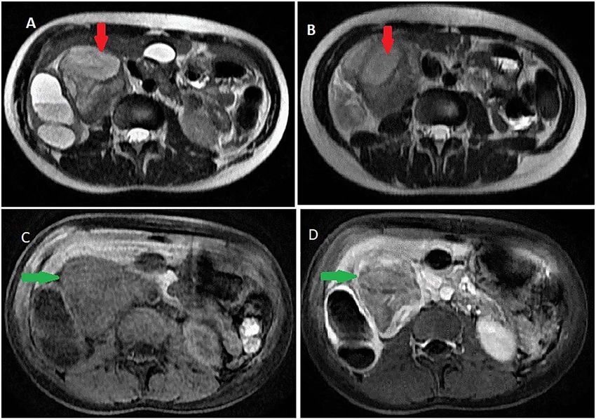
On magnetic resonance imaging (MRI), tumor signal was
heterogeneously high in T2 -weighted images, while iso to low in
T1 -weighted images delayed heterogeneous enhancement with
CASE REPORT some septa and areas of necrosis (Fig. 2).
A 14-year-old female patient with a history of intermittent right It was abutting the abdominal aorta, inferior vena cava
flank pains treated symptomatically without any radiological and right renal pedicle. It extended toward the liver and right
Received: January 4, 2021. Revised: February 28, 2021. Accepted: March 2, 2021
Published by Oxford University Press and JSCR Publishing Ltd. All rights reserved. © The Author(s) 2021.
This is an Open Access article distributed under the terms of the Creative Commons Attribution Non-Commercial License (http://creativecommons.org/
licenses/by-nc/4.0/), which permits non-commercial re-use, distribution, and reproduction in any medium, provided the original work is properly cited.
For commercial re-use, please contact journals.permissions@oup.com
1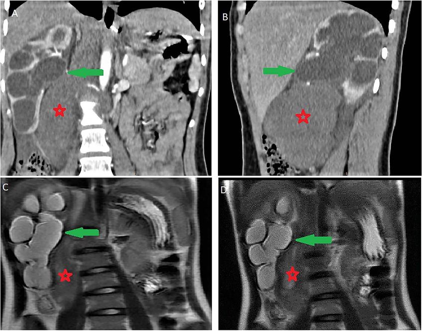
2      C. Kora et al.

Figure 1: Abdominal CT scan showing a retroperitoneal mass; retroperitoneal ganglioneuroma (red spark) before (A) and after (B) heterogeneous enhancement (green

                                                                                                                                                                         Downloaded from https://academic.oup.com/jscr/article/2021/4/rjab104/6248516 by guest on 06 May 2021
arrow).

Figure 2: Abdominal MRI showing a retroperitoneal mass; (A and B) axial T2 -weighted images show heterogeneous high signal in the lesion; (C and D) axial T1 -weighted
image, before (C) and after (D) dynamic sequences with contrast showing a delayed heterogeneous enhancement with some septa and areas of necrosis within the mass.

diaphragmatic pillar. It was compressing the kidney and ureter                        DISCUSSION
with ureterohydronephrosis and cortical thinning. Renal calyces
and pelvis contents were dense and their walls enhanced (Fig. 3).                     GN is a rare benign tumor with an incidence in a population
The adrenal gland was normal.                                                         estimated to one per million person-years, and it constitutes
    Based on this observation, obstructive pyelonephritis due to                      only 0.72–1.6% of primary retroperitoneal tumor [1]. A higher
compression by GN was suspected. Other differential diagnoses                         frequency has been reported in patients with multiple endocrine
of tumors, such as neuroblastoma, ganglioneuroblastoma, ger-                          neoplasia type II and neurofibromatosis type 1 [2].
minal tumor and paraganglioma, were also suspected.                                       GN is a well-differentiated tumor of the sympathetic ner-
    Alpha-fetoprotein and methoxylated derivatives levels were                        vous system, arising from the neural crest [3] which belongs to
normal. Subsequently, a CT-guided biopsy was performed to                             the spectrum of neuroblastic tumors, including neuroblastoma
confirm the diagnosis of GN (Fig. 4).                                                 and ganglioneuroblastoma [4]. It can also come from the mat-
    A laparotomy surgical resection was performed, after a multi-                     uration of pre-existent neuroblastoma spontaneously or after
disciplinary team decision. The post-operative was uneventful                         chemotherapy [5]. It is composed of gangliocytes and mature
and renal function remained normal. She was discharged on day                         stroma [6]. This tumor can grow at any site of the sympathetic
seven of post-operative.                                                              nervous tissue and it is mainly distributed in the posterior medi-
    Histologic work-up of the surgical specimen had confirmed                         astinum, retroperitoneum, neck and adrenal gland in descending
the absence of malignancy especially an immature neuroblastic                         order [7].
tissue.                                                                                   First described by Loretz in 1870, GN is mostly seen in pedi-
    A clinical 2 months follow-up after surgery was satisfactory.                     atric populations, with 60% of cases before 20 years [8]. The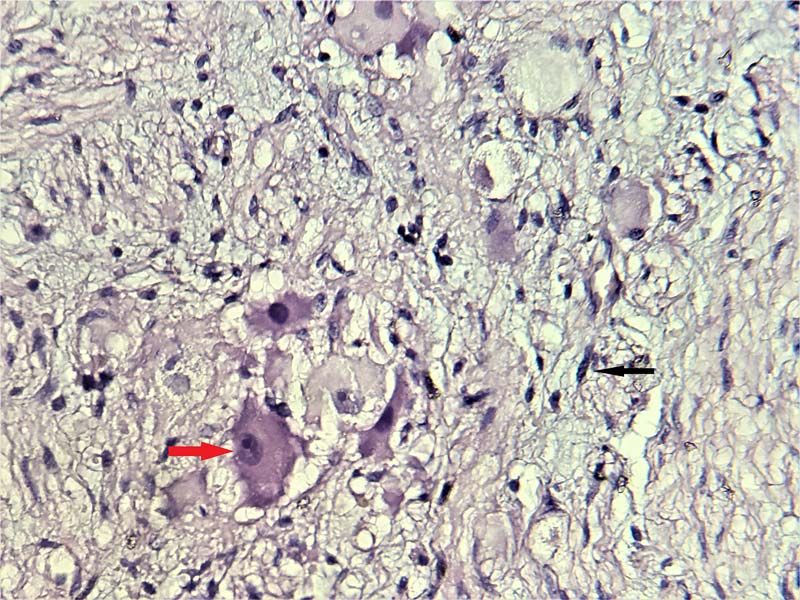
Retroperitoneal ganglioneuroma               3

                                                                                                                                                                          Downloaded from https://academic.oup.com/jscr/article/2021/4/rjab104/6248516 by guest on 06 May 2021
Figure 3: Ureterohydronephrosis due to the mass effect; ureterohydronephrosis (green arrow) due to the mass effect (red spark) on coronal (A), sagittal (B) Ct scan and
coronal MRI T2 -weighted image (C and D).

                                                                                      enhancement in a range of 10–20 HU. GN appears homogeneous
                                                                                      and can be sometimes heterogeneous with circumscribed or
                                                                                      spotted calcifications (in 20% of the patients) [9]. On MRI, T1 -
                                                                                      weighted images show a low or intermediate signal intensity,
                                                                                      whereas T2 -weighted images show a heterogeneous interme-
                                                                                      diate or high-signal intensity. This variability of the signal on
                                                                                      T2 -weighted images is due to two factors: the ratio of myxoid
                                                                                      stroma to cellularity and the amount of collagen present within
                                                                                      the tumor [3]. MRI contrast enhancement is most often slight and
                                                                                      heterogeneous [1].
                                                                                          There is no distinct pathognomonic radiological feature of
                                                                                      GN, but the lack of enhancement is quite suggestive. Before the
                                                                                      histologic study, clinical and radiological diagnosis is challenging
                                                                                      with several differential diagnoses. Among these diagnoses,
                                                                                      secreting paraganglioma must be eliminated before any biopsy
Figure 4: Microscopic scrutiny of the resected specimen; the observed ganglion
cells (red arrow) are mature, having a compact, eosinophilic cytoplasm and a          by measuring methoxylated derivatives. A CT-guided biopsy
single eccentric nucleus with prominent nucleolus; Schwann cells (black arrow)        can then be carried out to confirm the histological nature of
are also mature—HE;400X.                                                              the mass.
                                                                                          Histologically, it is characterized by large ganglions cells
                                                                                      proliferation with eosinophilic cytoplasm, large clear nucleolus
median age of the diagnosis is ∼7 years [4]. A female predom-                         without atypia, in a loose fibrillar background. This was the
inance with a sex ratio of 3:2 has been described [8].                                case of our patient; no immature neuroblastic component
   GN is usually an incidental asymptomatic finding on abdom-                         was noted. Histological study of the surgical specimen is
inal imaging. Sometimes when its size increases, it can cause                         essential, to eliminate a contingent of neuroblastoma and
some non-specific symptoms due to the compression on neigh-                           pheochromocytoma within the GN, which can be missing on
boring organs, as was reported in our case. Some GN secretes                          a core biopsy [10].
catecholamine and vasoactive intestinal peptide causing hyper-                            Resection is the sole treatment. Sometimes in a large tumor,
tension, sweating and diarrhea.                                                       surgery can be challenging because it tends to extent to neigh-
   CT and MRI are the best imaging techniques for the diagnosis                       boring anatomical vital structures. After complete resection, the
and characterization of GN. On imaging, it appears as an oval,                        prognosis is good. However, some cases of progression, late
well-delineated mass and tends to grow around major blood                             recurrence or malignant transformation have been reported,
vessels without narrowing them [3]. On CT, it is a hypodense                          especially after incomplete resection [1]. Furthermore, a long-
mass with a density between 20 and 40 HU and delayed contrast                         term follow-up including imaging control is necessary to prevent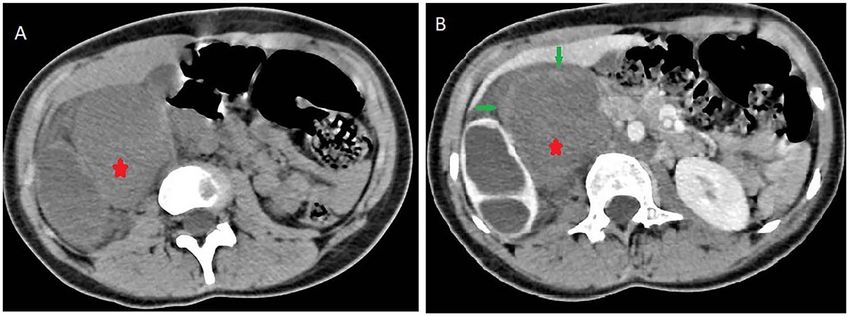
4     C. Kora et al.

potential relapse. There is no need for neoadjuvant or adjuvant         2. Paasch C, Harder A, Gatzky EJ, Ghadamgahi E, Spuler A, Siegel
treatment [8].                                                             R. Retroperitoneal paravertebral ganglioneuroma: a multi-
   In conclusion, retroperitoneal ganglioneuroma should be a               disciplinary approach facilitates less radical surgery. World
part of differential diagnoses for any retroperitoneal mass in             J Surg Oncol 2016;14:194.
children and young adults. Its radiological appearance without          3. Vasiliadis K, Papavasiliou C, Fachiridis D, Pervana S,
being pathognomonic is quite suggestive and needs histologic               Michaelides M, Kiranou M, et al. Retroperitoneal extra-
confirmation. Surgery with a long-term clinical and mostly radi-           adrenal ganglioneuroma involving the infrahepatic inferior
ological follow-up is the sole treatment.                                  vena cava, celiac axis and superior mesenteric artery: a case
                                                                           report. Int J Surg Case Rep 2012;3:541–3.
                                                                        4. Decarolis B, Simon T, Krug B, Leuschner I, Vokuhl C, Kaatsch
CONFLICT OF INTEREST STATEMENT
                                                                           P, et al. Treatment and outcome of Ganglioneuroma and
None declared.                                                             Ganglioneuroblastoma intermixed. BMC Cancer 2016;16:542.
                                                                        5. Ambros IM, Hata J, Joshi VV, Roald B, Dehner LP, Tüchler H,
FUNDING                                                                    et al. Morphologic features of neuroblastoma (Schwannian

                                                                                                                                           Downloaded from https://academic.oup.com/jscr/article/2021/4/rjab104/6248516 by guest on 06 May 2021
                                                                           stroma-poor tumors) in clinically favorable and unfavorable
All authors have declared that no financial support was received
                                                                           groups. Cancer 2002;94:1574–83.
from any organization for the submitted work.
                                                                        6. Arab N, Alharbi A. Retroperitoneal Ganglioneuroma (GN):
                                                                           case report in 14 years old boy. Int J Surg Case Rep
CONSENT                                                                    2019;60:130–2.
                                                                        7. Luo L, Zheng X, Tao KZ, Zhang J, Tang YY, Han FG. Imaging
Written informed consent was obtained from the patient for
                                                                           analysis of ganglioneuroma and quantitative analysis of
publication of this case report and any accompanying images. A
                                                                           paraspinal ganglioneuroma. Med Sci Monit 2019;25:5263–71.
copy of the written consent is available for review by the Editor-
                                                                        8. Kirchweger P, Wundsam HV, Fischer I, Rösch CS, Böhm G,
in-Chief of this journal on request.
                                                                           Tsybrovskyy O, et al. Total resection of a giant retroperi-
                                                                           toneal and mediastinal ganglioneuroma-case report and
AUTHOR CONTRIBUTIONS                                                       systematic review of the literature. World J Surg Oncol
                                                                           2020;18:248.
All the authors testified to the care of the patient and the writing
                                                                        9. Shi C, Li F, Wang Y, Pei L, Wang T. Retroperitoneo-
of the manuscript. The authors have read and agreed with the
                                                                           scopic resection of retroperitoneal nonadrenal ganglioneu-
contents of the manuscript.
                                                                           romas: our technique and clinical outcomes. Int Braz J Urol
                                                                           2018;44:1166–73.
REFERENCES                                                             10. Rebai N, Chaabouni A, Bouassida M, Fourati M, Chabchoub
 1. Dąbrowska-Thing A, Rogowski W, Pacho R, Nawrocka-                      K, Hadj Slimen N, et al. Retroperitoneal, ganglioneuroma:
    Laskus E, Nitek Ż. Retroperitoneal Ganglioneuroma mimick-             report of five cases and review of the literature. Afr J Urol
    ing a kidney tumor. Case report. Pol J Radiol 2017;82:283–6.           2013;19:215–8.
You can also read