Review Article Germinal Matrix-Intraventricular Hemorrhage: A Tale of Preterm Infants
←
→
Page content transcription
If your browser does not render page correctly, please read the page content below
Hindawi
International Journal of Pediatrics
Volume 2021, Article ID 6622598, 14 pages
https://doi.org/10.1155/2021/6622598

Review Article
Germinal Matrix-Intraventricular Hemorrhage: A Tale of
Preterm Infants

          Walufu Ivan Egesa ,1 Simon Odoch,1 Richard Justin Odong,1 Gloria Nakalema,1
          Daniel Asiimwe ,2 Eddymond Ekuk,3 Sabinah Twesigemukama,1
          Munanura Turyasiima ,1 Rachel Kwambele Lokengama,1 William Mugowa Waibi ,1
          Said Abdirashid,1 Dickson Kajoba,1 and Patrick Kumbowi Kumbakulu1
          1
            Department of Paediatrics and Child Health, Faculty of Clinical Medicine and Dentistry, Kampala International University, Uganda
          2
            Department of Surgery, Faculty of Clinical Medicine and Dentistry, Kampala International University, Uganda
          3
            Department of Surgery, Faculty of Medicine, Mbarara University of Science and Technology, Uganda

          Correspondence should be addressed to Walufu Ivan Egesa; ivanwalufu@kiu.ac.ug

          Received 20 December 2020; Accepted 26 February 2021; Published 16 March 2021

          Academic Editor: Somashekhar Marutirao Nimbalkar

          Copyright © 2021 Walufu Ivan Egesa et al. This is an open access article distributed under the Creative Commons Attribution
          License, which permits unrestricted use, distribution, and reproduction in any medium, provided the original work is
          properly cited.

          Germinal matrix-intraventricular hemorrhage (GM-IVH) is a common intracranial complication in preterm infants, especially
          those born before 32 weeks of gestation and very-low-birth-weight infants. Hemorrhage originates in the fragile capillary
          network of the subependymal germinal matrix of the developing brain and may disrupt the ependymal lining and progress into
          the lateral cerebral ventricle. GM-IVH is associated with increased mortality and abnormal neurodevelopmental outcomes such
          as posthemorrhagic hydrocephalus, cerebral palsy, epilepsy, severe cognitive impairment, and visual and hearing impairment.
          Most affected neonates are asymptomatic, and thus, diagnosis is usually made using real-time transfontanellar ultrasound. The
          present review provides a synopsis of the pathogenesis, grading, incidence, risk factors, and diagnosis of GM-IVH in preterm
          neonates. We explore brief literature related to outcomes, management interventions, and pharmacological and
          nonpharmacological prevention strategies for GM-IVH and posthemorrhagic hydrocephalus.

1. Introduction                                                         found no significant improvement in survival without neona-
                                                                        tal and long-term morbidity among VLBW infants between
Germinal matrix-intraventricular hemorrhage (GM-IVH)                    1997 and 2002.
remains a devastating neurological complication with con-
siderable mortality [1] and neurodevelopmental disability               2. Anatomy and Pathogenesis of GM-IVH
[2]. Hemorrhage originates in the capillary network of the
subependymal germinal matrix (GM) of the developing brain               The GM is located in the subependyma of the ventricular
and may disrupt the ependymal lining and progress into the              walls. It gives origin to the cerebral neuroblasts and glia, is
lateral cerebral ventricle [3, 4]. Although significant strides in       highly cellular and gelatinous, and is richly vascularized by
obstetrics and neonatal medicine have led to improved sur-              capillaries that are poorly supported by muscle or collagen
vival of preterm infants with lower gestational age and birth           [11]. Vascularization of the GM is prominent from 7-8
weight [5–7], we seem to have reached the nib of our ability            weeks of gestation and persists into the beginning of the
to ensure morbidity-free survival of very-low-birth-weight              third trimester [12, 13]. The thickness of the GM decreases
(VLBW) infants in advanced care settings [8, 9]. In the                 after 24 weeks of gestation and almost disappears by 36–37
United States, for example, Fanaroff and colleagues [10]                 weeks [11]. Animal studies showed that the characteristic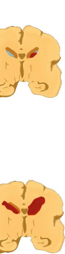
2                                                                                                 International Journal of Pediatrics

architecture of the subependymal matrix as the border zone            lation in neonatal intensive care units (NICUs), later declin-
between cerebral arteries and the collection zone of the              ing by three quarters to 12.5% in 2005, probably as a result of
deep cerebral veins makes it susceptible to focal hypoxic             improvements in ventilators, and the introduction of surfac-
changes [13].                                                         tant and corticosteroids. Based on age at onset, almost 40.6%
     The pathogenesis of GM-IVH is intricate and multifacto-          of low-birth-weight (C) polymorphism [24, 41, 42]. These risk
(CUS). Grade I refers to hemorrhage confined to the sube-              factors are summarized in Table 1.
pendymal GM, and grade II as hemorrhage within the lateral                A proportion of preterm neonates with previously diag-
ventricle without ventricular dilation and/or hemorrhage              nosed mild GM-IVH may deteriorate to severe GM-IVH.
occupying less than 50% of the ventricle. Grade III hemor-            Several risk factors including maternal lower genital tract
rhage is defined by ventricular dilation and/or hemorrhage             infection, lower gestational age [43], necrotizing enterocolitis
occupying more than 50% of the ventricle, while grade IV              (NEC), and thrombocytopenia [44] have been documented.
is ventricular hemorrhage extending into the surrounding
parenchyma [20]. This is illustrated in Figure 1. Mild                6. Clinical and Laboratory
GM-IVH refers to grade I and II hemorrhage, while severe                 Characteristics of GM-IVH
GM-IVH is a term used to refer to grade III and IV hem-
orrhage [21].                                                         The majority of cases of GM-IVH are clinically silent [23, 45]
                                                                      and only detectable by routine brain imaging. Symptomatic
4. Incidence of GM-IVH                                                neonates may manifest with convulsions, bulging fontanel,
                                                                      recurrent apnea, unexplained pallor, respiratory distress,
The global incidence of GM-IVH among preterm infants                  and temperature instability [46, 47]. Clinically identifiable
ranges from 14.7% to 44.7% [22–25], with considerable vari-           seizures are reported more often among neonates with grade
ation across gestational age groups, neonatal intensive care          IV GM-IVH [48].
units, and countries [6, 22, 25, 26]. Hefti et al. [27] examined          A significant reduction in the hematocrit may occur in
for GM-IVH in 345 preterm neonates autopsied from 1914                the presence of a large hemorrhage [17]. Biomarkers for early
to 2015 at Boston Children’s Hospital in the United States            prediction and detection of neuronal injury in neonates have
of America. The incidence of GM-IVH was 4.7% before the               gained clinical value in recent decades. This is because early
1960s and increased to 50% from 1975 to 1980 following                diagnosis may provide a crucial window for implementation
the introduction of novel positive pressure mechanical venti-         of neuroprotective interventions which may translate into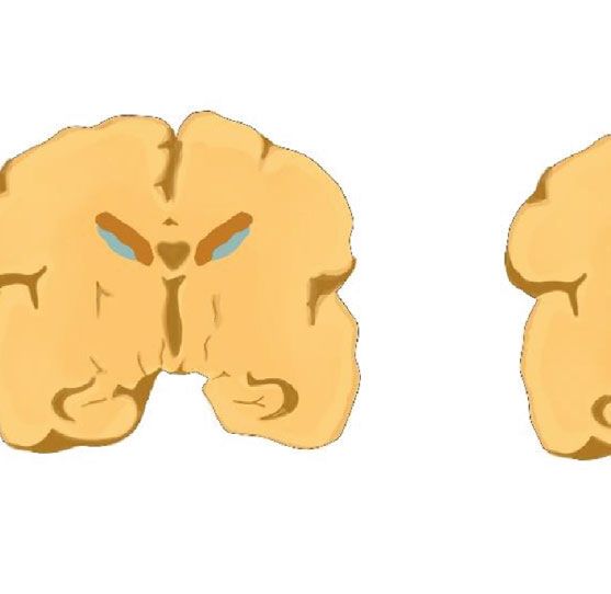
International Journal of Pediatrics                                                                                                      3

                         Lateral ventricle
                                                    Lateral        Germinal           Brain              Hemorrhage confined to
                                                   ventricles       matrix         parenchyma             the germinal matrix

                         Normal sagittal view                   Normal coronal view                         Grade I
                          through the brain                      through the brain

                                                       Extension of                 Intraventricular      Extension of hemorrhage
                  Intraventricular extension                             or       extension involving       into the surrounding
                                                    hemorrhage into a
                    involving 50% of the ventricle          parenchyma
                                                     dilated ventricle
                          ventricle

                                Grade II                             Grade III                               Grade IV

                                                        Figure 1: Grades of GM-IVH.

                                             Table 1: Risk factors for GM-IVH in preterm infants.

                                                                      (i) In vitro fertilization [30, 33]
                                                                       (ii) No antenatal care [31, 32]
                                             (iii) Lack of prenatal corticosteroid administration [25, 29, 31, 33, 34]
                                                                       (iv) Chorioamnionitis [35, 36]
Prenatal
                                                                         (v) Multiple gestation [30]
                                                                         (vi) Low gestation age [32]
                                                                           (vii) Maternal HIV [28]
                                                       (viii) Inherited coagulation abnormalities [24, 41, 42]
                                                                             (i) Fetal distress [22]
                                                                        (ii) Vaginal delivery [25, 38]
                                                                 (iii) Extreme prematurity [25, 28, 36]
Perinatal                                                          (iv) Very low birth weight [28, 36]
                                           (v) Low 5-minute APGAR score and resuscitation at birth [25, 31, 36, 38]
                                                    (vi) Intubation and mechanical ventilation [25, 31, 32, 38]
                                                                            (vii) Male sex [22, 26]
                                                      (i) Neonatal transfer after birth [22, 25, 28, 34, 38, 40]
                    (ii) Medication (e.g., inotropes, hydrocortisone, sodium bicarbonate, normal saline boluses, and opioids) [29, 36, 38]
                                                                               (iii) Anemia [29]
                                                                      (iv) Blood transfusion [28, 32]
                                                                      (v) Neonatal sepsis [31, 33, 36]
                                                               (vi) Patent ductus arteriosus [29, 31, 36]
                                                             (vii) Respiratory distress syndrome [32, 36]
Postnatal
                                                                         (viii) Hypercapnia [36, 38]
                                               (ix) High fraction of inspired oxygen during the first 24 hours [33]
                                                                         (x) Pneumothorax [33, 37]
                                                                          (xi) Hypotension [34, 38]
                                                                           (xii) Hyponatremia [32]
                                                                          (xiii) Hyperglycemia [32]
                                                                     (xiv) Metabolic acidosis [34, 39]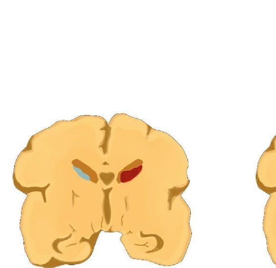
4                                                                                             International Journal of Pediatrics

improved outcomes. Investigators have proposed several bio-        CUS result was abnormal [53]. In 2020, the American Acad-
markers including S100β, activin A, adrenomedullin, eryth-         emy of Pediatrics [58] recommended CUS for all preterm
ropoietin, neuron-specific enolase, oxidative stress markers,       infants born at ≤30 weeks or >30 weeks of gestation with sig-
glial fibrillary acidic protein, and creatine phosphokinase         nificant risk factors. The initial CUS should be performed
BB (CPK-BB). Among these metabolites, elevated S100β               within the first 7-10 days, with subsequent scans at 4-6 weeks
levels in the blood and urine and activin A levels in the blood    of life and at term corrected age or prior to discharge. Serial
are the most promising [49, 50].                                   CUS should be performed for infants with abnormal CUS
                                                                   findings, adjusted according to the clinical state.
7. Cranial Ultrasound
                                                                   8. Magnetic Resonance Imaging
7.1. The Role of CUS. Since the late 1970s, high-resolution
real-time cranial ultrasound (CUS) has been the cornerstone        Magnetic resonance imaging (MRI) is superior to ultrasound
for diagnosis of GMH-IVH [51], with a sensitivity and spec-        at detecting white matter abnormalities, hemorrhagic, and
ificity of 96% and 94%, respectively, for detecting intracranial    cystic lesions [61]. Although MRI is increasingly being uti-
hemorrhage [52]. Worldwide, CUS remains the most readily           lized, it is not readily available, requires the neonate to be
available and widely used neuroimaging modality in NICUs           sedated, and may be unsuitable for unstable severely ill
[53, 54]. Most importantly, CUS is portable, reliable, cost-       infants. Nonetheless, some institutions have demonstrated
effective, noninvasive, and radiation-free, and does not            that MRIs may be performed without sedation of the neonate
require any special preparation [53, 55, 56]. However, the         at term equivalent age [62, 63]. MRI may be performed at
findings are operator-dependent, and subtle lesions may be          term corrected age for infants whose CUS reveals moderate
missed [53]. The anterior fontanelle is the most commonly          to severe abnormalities such as grade III/IV GM-IVH, post-
used site, but an acoustic window through the posterior and        hemorrhagic ventricular dilatation (PHVD), or grade III/IV
mastoid fontanelles can significantly augment the findings           periventricular leukomalacia (PVL), when clinical risk for
[57, 58]. CUS can be performed at the bedside and in the           white matter infarction (WMI) is increased or when parental
incubator, within less than 5 minutes and without significant       reassurance is needed [12, 53].
manipulation of the infant [55].
    Sonographic abnormalities should be correlated on both         9. Clinical Outcomes
coronal and parasagittal views, and findings on the left and
right sides should be graded separately, and the location, size,   According to Wu et al. [43], 8.2% of preterm neonates (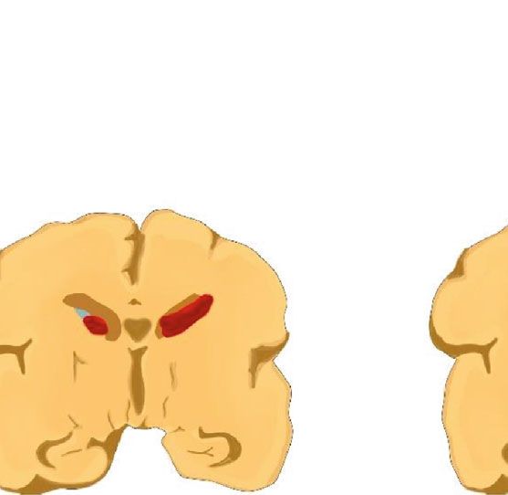
International Journal of Pediatrics                                                                                             5

occur due to acute obstruction of the foramen of Monro or the     no difference between conservative management and serial
aqueduct by a blood clot or due to subependymal scarring [75].    tapping of CSF via lumbar puncture or ventricular tapping
                                                                  as regards to reduced risk of major disability, multiple dis-
10. Management of GM-IVH                                          ability, death, or need for permanent shunt placement [85].
                                                                  Needless to say, repeated ventricular punctures inflict a new
10.1. General Strategies. Management of GM-IVH is focused         injury to the frontal lobe with each puncture and may
on addressing systemic issues of the neonate such as blood        increase infection risk [86].
pressure and respiratory status, which might influence pro-
gression of hemorrhage. Screening for sequelae of GM-IVH          11.2. Surgical Strategies
should be performed, and necessary interventions are
done, including management of hypotension, shock, ane-            11.2.1. DRIFT. DRIFT involves the insertion of right frontal
mia, and metabolic acidosis through judicious use of intra-       and left occipital catheters, with intraventricular injection of
venous fluids and blood transfusion. Continuous EEG or             tissue plasminogen activator (e.g., urokinase) that is insuffi-
amplitude-integrated EEG monitoring is indicated in the           cient to produce a systemic effect [87, 88]. After 8 hours of
presence of seizures [17].                                        TPA injection, irrigation with artificial CSF is commenced
                                                                  at a rate of 20 ml/hour, under ICP monitoring, with the goal
10.2. Mesenchymal Stem Cell Therapy. Animal models [76]           of maintaining a pressure < 7 mmHg. The drainage fluid
and phase I randomized controlled trials (RCTs) involving         clears over about 72 hours, from a dark-colored thick fluid
extremely preterm infants [77] have documented the promis-        to straw-colored CSF [87]. The DRIFT approach is associated
ing therapeutic potential of intraventricular transplantation     with secondary hemorrhage and does not reduce mortality
of allogenic mesenchymal stem cells (MSCs) in severe GM-          neither does it alter the need for permanent shunt placement
IVH. This novel therapy is thought to attenuate brain injury      [89, 90]. Contrastingly, studies have shown a reduction in
following GM-IVH and prevent the development of PHH.              severe cognitive disability among survivors at 2 years of life
Current evidence is weak, and thus, more human clinical tri-      [90] and at 10 years of life [91]. When performed within
als are needed to provide a stronger body of evidence regard-     three weeks of IVH onset in extremely-low-birth-weight
ing the therapeutic benefits and harms of MSCs [78].               (ELBW) neonates, fibrinolytic therapy followed by external
Nevertheless, a phase 2 RCT [79] to evaluate the efficacy           ventricular drainage may significantly reduce the need for
and safety of umbilical cord blood-derived MSCs (Pneumos-         permanent shunt surgery, without increasing the risk of
tem®) in 23 to
6 International Journal of Pediatrics
Table 2: Strategies for prevention of GM-IVH in preterm neonates.
Prenatal Perinatal Postnatal
Avoid interhospital transport
Elevated midline head positioning
Delivery at a tertiary hospital Minimize handling and stimulation
Prevent preterm birth Prompt delivery upon recognition Fluid therapy for hypotension
Corticosteroids of fetal distress Near-infrared spectroscopy monitoring of cerebral oxygenation
Delayed cord clamping Prevent and treat NEC and sepsis
Erythropoiesis stimulation agents
(e.g., erythropoietin and darbepoetin)
such as skin breakdown, ventricular hemorrhage, CSF infec- infection [96, 106] predominantly caused by Staphylococcus
tion, and leak [99]. Apnea and ventriculitis have also been species [105].
documented [98]. Repeated tapping from a VR has been
shown not to increase the risk of reservoir infection [95]. A 12. Prevention of GM-IVH
prospective multicenter cohort of VLBW neonates with
severe GM-IVH observed no difference in infection rates To protect the preterm brain from GM-IVH, a multifaceted
between VR and ventriculosubgaleal shunts (17% versus approach addressing specific antenatal, delivery room, post-
14%, p = 0:71) [92]. natal, and NICU factors should be implemented (Table 2)
[111, 112]. Since GM-IVH is primarily linked to increased
(2) Ventriculosubgaleal Shunt. Ventriculosubgaleal shunt vascular fragility and disturbance in CBF, strategies are
(VSGS) placement provides a temporary treatment of PHH directed to strengthening the GM microvasculature and to
in medically unstable infants and also averts the need for stabilizing the CBF.
repeated tapping of CSF [100]. Through a small scalp inci-
12.1. Prevent Preterm Birth. Measures that target prevention
sion near the anterior fontanelle, under local anesthesia and
of preterm birth are the most important strategies for mini-
mild sedation, a ventricular catheter is carefully placed into
mizing the occurrence of GM-IVH [21]. Preterm birth may
the lateral ventricle and anchored to the dura. Blunt dissec-
be spontaneous or induced in situations such as eclampsia.
tion is performed to create a pouch between the periosteum
Unless medically indicated, preterm birth can be delayed by
and galea, creating a subgaleal pouch where the outermost
evidence-based approaches such as antenatal progesterone
(proximal) end of the ventricular catheter is placed to allow
supplementation from 16 to 24 weeks through 34 weeks of
for CSF drainage [86, 101, 102]. The procedure is described
gestation in women with a current singleton pregnancy and
in a recent publication by Kuo [86] and can be safely accom-
previous spontaneous delivery, and those with a short cervi-
plished in the NICU or the operating theatre [101, 103]. Col-
cal length (≤20 mm before 24 weeks’ gestation). Other inter-
lection of CSF in the subgaleal space can result in a
ventions such as avoidance of tobacco smoking during
cosmetically unappealing scalp swelling [104]. VSGS has
pregnancy, cervical cerclage for cervical incompetence, toco-
been associated with recurrent meningitis, subgaleal adhe-
lytics for preterm labour, and dedicated preterm birth pre-
sions, shunt obstruction requiring ventricular catheter revi-
vention clinics have been utilized [113, 114].
sion or renewal, CSF leakage, and slippage of the catheter
into or out of the ventricle [101, 102, 105]. It is estimated that 12.2. Prenatal Corticosteroids. The World Health Organiza-
12% of patients with VSGS require a permanent ventriculo- tion [115] strongly recommends prenatal corticosteroid use
peritoneal shunt [101], which if needed is often placed when for all women at 24 to 34 weeks’ gestation for whom preterm
the CSF protein content decreases toInternational Journal of Pediatrics 7
conducted between 1995 and 2004 provided evidence that there was no difference in adverse neurosensory outcomes at
MgSO4 administered to women at high risk of preterm 18 months of life. In addition, a multicenter double-blind
labor provides significant neuroprotection against moderate RCT showed that administration of prophylactic ibuprofen
to severe CP, without causing adverse effects on the infants within the first 6 hours of birth was ineffective against pre-
[121]. The World Health Organization, American College venting grade II to IV GM-IVH [143]. Therefore, both indo-
of Obstetricians and Gynecologists (ACOG), and the Soci- methacin and ibuprofen are not recommended for
ety for Maternal-Fetal Medicine currently recommend the prevention of GM-IVH, but are reserved for treatment of
use of MgSO4 for women at risk of imminent preterm birth patent ductus arteriosus.
before 32 weeks of gestation for prevention of cerebral
palsy during infancy and childhood [122, 123]. Compared 12.7. Midline Head Positioning and Head Tilting. Midline
to controls, MgSO4 has not been found to reduce the rates (neutral) head positioning is thought to optimize cerebral
of GM-IVH [124]. venous drainage through the internal jugular veins, which
are the major outflow paths for cranial blood. Head rotation
12.4. Delivery at Tertiary Center and Avoidance of to either side may result in ipsilateral occlusion or obstruc-
Interhospital Transport. Evidence from a large retrospective tion of the jugular venous drainage system [144]. Near-
analysis of 67,596 VLBW preterm neonates found a correla- infrared spectroscopy (NIRS) shows that midline head
tion between interhospital transport and increased incidence position and head tilting (elevating the head of the incubator
and severity of GM-IVH [40], which has been linked to upwards by 15–30°) facilitates hydrostatic cerebral venous
increased head and torso vibrations during neonatal trans- outflow in preterm infants [145, 146]. Moreover, Doppler
port [125]. A cohort study of 5,712 infants born at 24–30 ultrasonography studies showed that occlusion of the jugular
weeks in the Australian and New Zealand Neonatal Network venous system by changes in head position results in large
from 1995–97 found that infants transferred to another hos- alterations in blood flow velocities in the superior sagittal
pital after birth had 1.60 times higher odds of developing sinus, increased cerebral blood volume, and ICP [145, 147,
severe GM-IVH (95% CI: 1.15 to 2.21, p < 0:01) [22]. There- 148] which may result in GM-IVH. Head positioning and
fore, when high-risk preterm delivery is anticipated, it should tilting has been reported to have no effect on cerebral hemo-
be conducted in a tertiary center [38, 126]. dynamics and oxygenation in preterm infants [149] which
contrasts the findings of other studies [148]. Recent system-
12.5. Delayed Cord Clumping. Delayed cord clamping (DCC) atic reviews and meta-analyses [149, 150] reported inconclu-
results in a higher hematocrit [127–129], superior vena cava sive evidence that head positioning prevents the occurrence
blood flow, right ventricle output, and right ventricular and extension of GM-IVH. However, a single-center study
stroke volume [130], higher blood pressure and admission [151] found that placing8 International Journal of Pediatrics
findings. Thus, cerebral oximetry can be used to monitor which survival of smaller infants was low. As such, further
high-risk infants such that timely interventions are taken to research is required, before a recommendation can be made.
improve cerebral oxygenation [162].
13. Follow-Up of Survivors of GM-IVH
12.10. Ethamsylate. Ethamsylate is thought to promote plate-
let adhesion and increase capillary basement membrane sta- Outpatient follow-up should be done to identify morbidities
bility through hyaluronic acid polymerization [163]. A and provide appropriate guidance and treatment through
Cochrane Database Systematic Review [164] of 1410 preterm comprehensive neurorehabilitation programs [102]. Given
infants from seven trials showed that infants < 35 weeks of the increased risk of PHH, head circumference should be
gestation with ethamsylate are significantly less likely to continually monitored [64, 72]. Children with neuropsycho-
develop GM-IVH compared to controls. While a significant logical deficits require special support while in school [73]
reduction in severe GM-IVH was observed (RR 0.67, 95% with regard to writing, reading, and mathematics.
CI 0.49 to 0.94), the review did not show a significant differ-
ence in neonatal mortality or neurodevelopmental outcome 14. Conclusion
at two years between infants treated with ethamsylate and
controls. Thus, routine use of ethamsylate for prevention of In recent years, considerable advances in perinatal-neonatal
GM-IVH in preterm infants is not recommended. care have resulted in improved survival outcomes of babies
born at the threshold of viability. This has been paralleled
12.11. Phenobarbitone. Earlier observations showed that phe- by a rising number of infants who develop complications
nobarbitone may dampen fluctuations in systemic blood such as GM-IVH, a multifactorial neuropathology that exclu-
pressure [165] and also protect the brain after hypoxia- sively affects infants of ≤32 weeks’ gestation or those who
ischemia. A 2013 Cochrane review conducted by Smit et al. weighInternational Journal of Pediatrics 9
Conflicts of Interest cally ill premature infants,” Pediatrics, vol. 106, no. 4,
pp. 625–632, 2000.
The authors declare no competing interests. [16] D. Yang, J. M. Baumann, Y. Y. Sun et al., “Overexpression of
vascular endothelial growth factor in the germinal matrix
induces neurovascular proteases and intraventricular hemor-
References rhage,” Science Translational Medicine, vol. 5, no. 193, article
193ra90, 2013.
[1] C. H. Pieper, J. Smith, D. Maree, and F. C. Pohl, “Is nCPAP of
[17] A. Whitelaw, “Core concepts: intraventricular hemorrhage,”
value in extreme preterms with no access to neonatal inten-
NeoReviews, vol. 12, no. 2, pp. e94–e101, 2011.
sive care?,” Journal of Tropical Pediatrics, vol. 49, no. 3,
pp. 148–152, 2003. [18] L. A. Papile, J. Burstein, R. Burstein, and H. Koffler, “Inci-
dence and evolution of subependymal and intraventricular
[2] S. Bolisetty, A. Dhawan, M. Abdel-Latif, B. Bajuk, J. Stack,
hemorrhage: a study of infants with birth weights less than
and K. Lui, “Intraventricular hemorrhage and neurodevelop-
1,500 gm,” The Journal of Pediatrics, vol. 92, no. 4, pp. 529–
mental outcomes in extreme preterm infants,” Pediatrics,
534, 1978.
vol. 133, no. 1, pp. 55–62, 2014.
[3] P. Ballabh, “Intraventricular hemorrhage in premature [19] K. Kuban and R. L. Teele, “Rationale for grading intracranial
infants: mechanism of disease,” Pediatric Research, vol. 67, hemorrhage in premature infants,” Pediatrics, vol. 74, no. 3,
no. 1, pp. 1–8, 2010. pp. 358–363, 1984.
[4] J. J. Volpe, “Impaired neurodevelopmental outcome after [20] T. E. Inder, J. M. Perlman, and J. J. Volpe, “Preterm IVH/-
mild germinal matrix-intraventricular hemorrhage,” Pediat- posthemorrhagic hydrocephalus,” in Volpe’s neurology of
rics, vol. 136, no. 6, pp. 1–3, 2015. the newborn, pp. 637–698, Elsevier, Philadelphia, 6th edition,
2018.
[5] J. D. Horbar, G. J. Badger, J. H. Carpenter et al., “Trends in
mortality and morbidity for very low birth weight infants, [21] J. Lim and E. Hagen, “Reducing germinal matrix-
1991–1999,” Pediatrics, vol. 110, no. 1, pp. 143–151, 2002. intraventricular hemorrhage: perinatal and delivery room
[6] X. Kong, F. Xu, R. Wu et al., “Neonatal mortality and morbid- factors,” NeoReviews, vol. 20, no. 8, pp. e452–e463, 2019.
ity among infants between 24 to 31 complete weeks: a multi- [22] A. M. Heuchan, N. Evans, D. J. Henderson Smart, and J. M.
center survey in China from 2013 to 2014,” BMC Pediatrics, Simpson, “Perinatal risk factors for major intraventricular
vol. 16, no. 1, p. 174, 2016. haemorrhage in the Australian and New Zealand Neonatal
[7] B. J. Stoll, N. I. Hansen, E. F. Bell et al., “Neonatal outcomes of Network, 1995–97,” Archives of Disease in Childhood. Fetal
extremely preterm infants from the NICHD Neonatal and Neonatal Edition, vol. 86, no. 2, pp. 86F–890, 2002.
Research Network,” Pediatrics, vol. 126, no. 3, pp. 443–456, [23] H. Kadri, A. A. Mawla, and J. Kazah, “The incidence, timing,
2010. and predisposing factors of germinal matrix and intraventric-
[8] J. G. Anderson, R. J. Baer, J. C. Partridge et al., “Survival and ular hemorrhage (GMH/IVH) in preterm neonates,” Child's
major morbidity of extremely preterm infants: a population- Nervous System, vol. 22, no. 9, pp. 1086–1090, 2006.
based study,” Pediatrics, vol. 138, no. 1, article e20154434, [24] L. A. Ramenghi, M. Fumagalli, M. Groppo et al., “Germi-
2016. nal matrix hemorrhage: intraventricular hemorrhage in
[9] H. Inoue, M. Ochiai, K. Yasuoka et al., “Early mortality and very-low-birth-weight infants - the independent role of
morbidity in infants with birth weight of 500 grams or less inherited thrombophilia,” Stroke, vol. 42, no. 7, pp. 1889–
in Japan,” Journal of Pediatrics, vol. 190, article 112e117.e3, 1893, 2011.
pp. 112–117.e3, 2017. [25] K. T. Yeo, R. Thomas, S. S. Chow et al., “Improving incidence
[10] A. A. Fanaroff, B. J. Stoll, L. L. Wright et al., “NICHD Neona- trends of severe intraventricular haemorrhages in preterm
tal Research Network. Trends in neonatal morbidity and infants10 International Journal of Pediatrics
[30] A. Bordbar and M. Farjadnia, “Maternal morbidities and [44] H. C. Jen, J. J. Graber, J. L. Hill, S. M. Alaish, R. W. Voigt, and
occurrence of intraventricular hemorrhage in preterm E. D. Strauch, “Surgical necrotizing enterocolitis and intra-
infants,” Journal of Pediatric Intensive Care, vol. 4, no. 3, ventricular hemorrhage in premature infants below 1000 g,”
pp. 156–161, 2015. Journal of Pediatric Surgery, vol. 41, no. 8, pp. 1425–1430,
[31] A. Ghoor, G. Scher, and D. E. Ballot, “Prevalence of and risk 2006.
factors for cranial ultrasound abnormalities in very-low- [45] J. M. Perlman and N. Rollins, “Surveillance protocol for the
birth-weight infants at Charlotte Maxeke Johannesburg Aca- detection of intracranial abnormalities in premature neo-
demic Hospital,” African Journal of Child Health, vol. 11, nates,” Archives of Pediatrics & Adolescent Medicine,
no. 2, pp. 66–70, 2017. vol. 154, no. 8, pp. 822–826, 2000.
[32] E. A. Guzman, J. R. D. Bertagnon, and Y. Juliano, “Frequency [46] S. A. Adegoke, A. O. Olugbemiga, K. P. Bankole, and O. A.
of peri-intraventricular hemorrhage and its associated factors Tinuade, “Intraventricular hemorrhage in newborns weigh-
in premature newborns,” Einstein, vol. 8, no. 3, pp. 315–319, ingInternational Journal of Pediatrics 11
SECTION ON RADIOLOGY, “Routine neuroimaging of the [73] T. M. Luu, L. R. Ment, K. C. Schneider, K. H. Katz, W. C.
preterm brain,” Pediatrics, vol. 146, no. 5, article Allan, and B. R. Vohr, “Lasting effects of preterm birth and
e2020029082, 2020. neonatal brain hemorrhage at 12 years of age,” Pediatrics,
[59] R. M. Jones, E. M. Clark, K. Broad, and E. Smit, “Outcome vol. 123, no. 3, pp. 1037–1044, 2009.
following preterm intraventricular haemorrhage - what to tell [74] A. Hill, G. D. Shackelford, and J. J. Volpe, “A potential mech-
the parents,” Paediatr Child Health (Oxford), vol. 28, no. 9, anism of pathogenesis for early posthemorrhagic hydroceph-
pp. 431–435, 2018. alus in the premature newborn,” Pediatrics, vol. 73, no. 1,
[60] M. I. Levene, “Measurement of the growth of the lateral ven- pp. 19–21, 1984.
tricles in preterm infants with real-time ultrasound,” Archives [75] J. Strahle, H. J. L. Garton, C. O. Maher, K. M. Muraszko, R. F.
of Disease in Childhood, vol. 56, pp. 900–904, 1981. Keep, and G. Xi, “Mechanisms of hydrocephalus after neona-
[61] M. Hinojosa-Rodríguez, T. Harmony, C. Carrillo-Prado et al., tal and adult intraventricular hemorrhage,” Translational
“Clinical neuroimaging in the preterm infant: diagnosis and Stroke Research, vol. 3, no. S1, pp. 25–38, 2012.
prognosis,” Neuroimage Clin., vol. 16, no. 16, pp. 355–368, [76] S. Y. Ahn, Y. S. Chang, D. K. Sung et al., “Mesenchymal stem
2017. cells prevent hydrocephalus after severe intraventricular
[62] A. M. Mathur, J. J. Neil, R. C. McKinstry, and T. E. Inder, hemorrhage,” Stroke, vol. 44, no. 2, pp. 497–504, 2013.
“Transport, monitoring, and successful brain MR imaging [77] S. Y. Ahn, Y. S. Chang, S. I. Sung, and W. S. Park, “Mesenchy-
in unsedated neonates,” Pediatric Radiology, vol. 38, no. 3, mal stem cells for severe intraventricular hemorrhage in pre-
pp. 260–264, 2008. term infants: phase I dose-escalation clinical trial,” Stem Cells
[63] V. Neubauer, E. Griesmaier, K. Baumgartner, A. Mallouhi, Translational Medicine, vol. 7, no. 12, pp. 847–856, 2018.
M. Keller, and U. Kiechl-Kohlendorfer, “Feasibility of cere- [78] O. Romantsik, M. Bruschettini, A. Moreira, B. Thébaud, and
bral MRI in non-sedated preterm-born infants at term- D. Ley, “Stem cell-based interventions for the prevention and
equivalent age: report of a single centre,” Acta Paediatrica, treatment of germinal matrix-intraventricular haemorrhage
vol. 100, no. 12, pp. 1544–1547, 2011. in preterm infants,” Cochrane Database of Systematic
[64] V. Gilard, A. Chadie, F. Ferracci et al., “Post hemorrhagic Reviews, vol. 9, no. 9, article CD013201, 2019.
hydrocephalus and neurodevelopmental outcomes in a con- [79] ClinicalTrialsgov, “Efficacy and safety of pneumostem for
text of neonatal intraventricular hemorrhage: an institutional IVH in premature infants (phase 2a),” NCT02890953.
experience in 122 preterm children,” BMC Pediatrics, vol. 18, https://clinicaltrials.gov/ct2/show/NCT02890953.
no. 1, p. 288, 2018. [80] P. V. Sandoval, P. H. Rosales, D. G. Q. Hernández, E. A. C.
[65] T. Schindler, L. Koller-smith, K. Lui, B. Bajuk, and Naranjo, and V. G. Navarro, “Intraventricular hemorrhage
S. Bolisetty, “Causes of death in very preterm infants cared and posthemorrhagic hydrocephalus in preterm infants:
for in neonatal intensive care units: a population-based retro- diagnosis, classification, and treatment options,” Child's Ner-
spective cohort study,” BMC Pediatrics, vol. 17, no. 1, p. 59, vous System, vol. 35, pp. 917–927, 2019.
2017. [81] A. J. Brouwer, F. Groenendaal, M. J. N. L. Benders, and L. S.
[66] E. A. Christian, D. Jin, F. Attenello et al., “Trends in hospital- De Vries, “Early and late complications of germinal matrix-
ization of preterm infants with intraventricular hemorrhage intraventricular haemorrhage in the preterm infant: what is
and hydrocephalus in the United States, 2000-2010,” Fluids new?,” Neonatology, vol. 106, no. 4, pp. 296–303, 2014.
and Barriers of the CNS., vol. 12, Suppl 1, p. O1, 2015. [82] M. N. Cizmeci, F. Groenendaal, K. D. Liem et al., “Random-
[67] Y. Futagi, Y. Toribe, K. Ogawa, and Y. Suzuki, “Neurodeve- ized Controlled Early versus Late Ventricular Intervention
lopmental outcome in children with intraventricular hem- Study in Posthemorrhagic Ventricular Dilatation: Outcome
orrhage,” Pediatric Neurology, vol. 34, no. 3, pp. 219–224, at 2 Years,” The Journal of Pediatrics, vol. 226, pp. 28–35.e3,
2006. 2020.
[68] K. Klebermass-Schrehof, C. Czaba, M. Olischar et al., “Impact [83] L. Mahoney, K. Luyt, D. Harding, and D. Odd, “Treatment
of low-grade intraventricular hemorrhage on long-term neu- for post-hemorrhagic ventricular dilatation: a multiple-
rodevelopmental outcome in preterm infants,” Child's Ner- treatment meta-analysis,” Frontiers in Pediatrics, vol. 8,
vous System, vol. 28, no. 12, pp. 2085–2092, 2012. p. 238, 2020.
[69] A. Mukerji, V. Shah, and P. S. Shah, “Periventricular/intra- [84] C. R. Kennedy, C. Kennedy, M. Campbell, and International
ventricular hemorrhage and neurodevelopmental outcomes: PHVD Drug Trial Group, “International randomised con-
a meta-analysis,” Pediatrics, vol. 136, no. 6, pp. 1132–1143, trolled trial of acetazolamide and furosemide in posthaemor-
2015. rhagic ventricular dilatation in infancy,” Lancet, vol. 352,
[70] R. L. Sherlock, P. J. Anderson, and L. W. Doyle, “Victorian no. 9126, pp. 433–440, 1998.
Infant Collaborative Study Group. Neurodevelopmental [85] A. Whitelaw and R. Lee-Kelland, “Repeated lumbar or ven-
sequelae of intraventricular haemorrhage at 8 years of age tricular punctures in newborns with intraventricular haemor-
in a regional cohort of ELBW/very preterm infants,” Early rhage,” Cochrane Database of Systematic Reviews, vol. 4,
Human Development, vol. 81, pp. 909–916, 2005. no. 4, article CD000216, 2017.
[71] A. Whitelaw, M. Thoresen, and I. Pople, “Posthaemorrhagic [86] M. F. Kuo, “Surgical management of intraventricular hem-
ventricular dilatation,” Archives of Disease in Childhood. Fetal orrhage and posthemorrhagic hydrocephalus in premature
and Neonatal Edition, vol. 86, no. 2, pp. 72F–774, 2002. infants,” Biomed Journal, vol. 43, no. 3, pp. 268–276,
[72] C. R. Kennedy, S. Ayers, M. J. Campbell et al., “Randomized, 2020.
controlled trial of acetazolamide and furosemide in posthem- [87] K. Aquilina, “Intraventricular haemorrhage of the newborn,”
orrhagic ventricular dilation in infancy: follow-up at 1 year,” Advances in clinical neuroscience & rehabilitation : ACNR,
Pediatrics, vol. 108, no. 3, pp. 597–607, 2001. vol. 11, no. 5, pp. 22–24, 2011.12 International Journal of Pediatrics
[88] Y. S. Park, Y. Kotani, T. K. Kim et al., “Efficacy and safety of hemorrhagic hydrocephalus,” Child's Nervous System,
intraventricular fibrinolytic therapy for post-intraventricular vol. 26, no. 11, pp. 1505–1515, 2010.
hemorrhagic hydrocephalus in extreme low birth weight [102] A. Nagy, L. Bognar, I. Pataki, Z. Barta, and L. Novak, “Ventri-
infants: a preliminary clinical study,” Child's Nervous System, culosubgaleal shunt in the treatment of posthemorrhagic and
vol. 37, no. 1, pp. 69–79, 2020. postinfectious hydrocephalus of premature infants,” Child's
[89] A. Whitelaw, D. Evans, M. Carter et al., “Randomized clinical Nervous System, vol. 29, no. 3, pp. 413–418, 2013.
trial of prevention of hydrocephalus after intraventricular [103] C. S. Karas, M. N. Baig, and S. W. Elton, “Ventriculosubgaleal
hemorrhage in preterm infants: brain-washing versus tap- shunts at Columbus Children’s Hospital: neurosurgical
ping fluid,” Pediatrics, vol. 119, no. 5, pp. e1071–e1078, 2007. implant placement in the neonatal intensive care unit,” Jour-
[90] A. Whitelaw, S. Jary, G. Kmita et al., “Randomized trial of nal of Neurosurgery, vol. 107, pp. 220–223, 2007.
drainage, irrigation and fibrinolytic therapy for premature [104] R. K. Kutty, S. B. Sreemathyamma, P. Korde, R. B. Prabhakar,
infants with posthemorrhagic ventricular dilatation: develop- A. Peethambaran, and G. K. Libu, “Outcome of ventriculo-
mental outcome at 2 years,” Pediatrics, vol. 125, no. 4, subgaleal shunt in the management of infectious and non-
pp. e852–e858, 2010. infectious hydrocephalus in pre-term infants,” Journal of
[91] K. Luyt, S. Jary, C. Lea et al., “Ten-year follow-up of a rando- Pediatric Neurosciences, vol. 13, no. 3, pp. 322–328, 2018.
mised trial of drainage, irrigation and fibrinolytic therapy [105] B. K. Willis, C. R. Kumar, E. L. Wylen, and A. Nanda, “Ven-
(DRIFT) in infants with post-haemorrhagic ventricular dila- triculosubgaleal shunts for posthemorrhagic hydrocephalus
tation,” Health Technology Assessment, vol. 23, no. 4, pp. 1– in premature infants,” Pediatric Neurosurgery, vol. 41, no. 4,
116, 2019. pp. 178–185, 2005.
[92] J. C. Wellons 3rd, C. N. Shannon, R. Holubkov et al., “Shunt- [106] A. Reinprecht, W. Dietrich, A. Berger, G. Bavinzski,
ing outcomes in posthemorrhagic hydrocephalus: results of a M. Weninger, and T. Czech, “Posthemorrhagic hydrocepha-
hydrocephalus clinical research network prospective cohort lus in preterm infants: long-term follow-up and shunt-
study,” Journal of Neurosurgery. Pediatrics, vol. 20, no. 1, related complications,” Child’s Nerv Syst., vol. 17, no. 11,
pp. 19–29, 2017. pp. 663–669, 2001.
[93] E. A. Christian, E. F. Melamed, E. Peck, M. D. Krieger, and [107] S. Kazan, A. Güra, T. Uçar, E. Korkmaz, H. Ongun, and
J. G. McComb, “Surgical management of hydrocephalus sec- M. Akyuz, “Hydrocephalus after intraventricular hemor-
ondary to intraventricular hemorrhage in the preterm rhage in preterm and low-birth weight infants: analysis of
infant,” Neurosurgery: Pediatrics, vol. 17, no. 3, pp. 278– associated risk factors for ventriculoperitoneal shunting,”
284, 2016. Surgical Neurology, vol. 64, no. S2, pp. 77–81, 2005.
[94] S. Robinson, “Neonatal posthemorrhagic hydrocephalus [108] I. C. Lee, H. S. Lee, P. H. Su, W. J. Liao, J. M. Hu, and J. Y.
from prematurity: pathophysiology and current treatment Chen, “Posthemorrhagic hydrocephalus in newborns: clinical
concepts: a review,” Journal of Neurosurgery. Pediatrics, characteristics and role of ventriculoperitoneal shunts,” Pedi-
vol. 9, no. 3, pp. 242–258, 2012. atrics and Neonatology, vol. 50, no. 1, pp. 26–32, 2009.
[95] K. Kormanik, J. Praca, H. J. Garton, and S. Sarkar, “Repeated [109] M. Vassilyadi, Z. Tataryn, M. F. Shamji, and E. C. G. Venture-
tapping of ventricular reservoir in preterm infants with post- yra, “Functional outcomes among premature infants with
hemorrhagic ventricular dilatation does not increase the risk intraventricular hemorrhage,” Pediatric Neurosurgery,
of reservoir infection,” Journal of Perinatology, vol. 30, no. 3, vol. 45, pp. 247–255, 2009.
pp. 218–221, 2010. [110] A. Whitelaw and K. Aquilina, “Management of posthaemor-
[96] P. Chittiboina, H. Pasieka, A. Sonig et al., “Posthemorrhagic rhagic ventricular dilatation,” Archives of Disease in Child-
hydrocephalus and shunts: what are the predictors of multi- hood - Fetal and Neonatal Edition, vol. 97, no. 3, pp. F229–
ple revision surgeries?,” Journal of Neurosurgery: Pediatrics, F233, 2012.
vol. 11, no. 1, pp. 37–42, 2013. [111] S. C. Handley, M. Passarella, H. C. Lee, and S. A. Lorch, “Inci-
[97] P. Peretta, P. Ragazzi, C. F. Carlino, P. Gaglini, and G. Cinalli, dence trends and risk factor variation in severe intraventricu-
“The role of Ommaya reservoir and endoscopic third ventri- lar hemorrhage across a population based cohort,” Journal of
culostomy in the management of post-hemorrhagic hydro- Pediatrics, vol. 200, pp. 24–29.e3, 2018.
cephalus of prematurity,” Child's Nervous System, vol. 23, [112] D. McLendon, J. Check, P. Carteaux et al., “Implementation
pp. 765–771, 2007. of potentially better practices for the prevention of brain
[98] T. T. Rhodes, W. H. Edwards, R. L. Saunders et al., “External hemorrhage and ischemic brain injury in very low birth
ventricular drainage for initial treatment of neonatal posthem- weight infants,” Pediatrics, vol. 111, no. 4, pp. e497–e503,
orrhagic hydrocephalus: surgical and neurodevelopmental 2003.
outcome,” Pediatric Neurosurgery, vol. 13, pp. 255–262, 2004. [113] J. P. Newnham, J. E. Dickinson, R. J. Hart, C. E. Pennell, C. A.
[99] L. Jian, S. Hang-song, L. Zheng-lang, Y. Li-sheng, W. Heng, Arrese, and J. A. Keelan, “Strategies to prevent preterm
and Z. Nu, “Implantation of Ommaya reservoir in extremely birth,” Frontiers in Immunology, vol. 5, p. 584, 2014.
low weight premature infants with posthemorrhagic hydro- [114] K. Rundell and B. Panchal, “Preterm labor: prevention and
cephalus: a cautious option,” Child's Nervous System, management,” American Family Physicians, vol. 95, no. 6,
vol. 28, no. 10, pp. 1687–1691, 2012. pp. 366–372, 2017.
[100] B. B. Fulmer, P. A. Grabb, W. J. Oakes, and T. B. Mapstone, [115] World Health Organisation, “WHO recommendations on
“Neonatal ventriculosubgaleal shunts,” Neurosurgery, interventions to improve preterm birth outcomes,” 2015.
vol. 47, no. 1, pp. 80–84, 2000. Retrieved from: https://www.who.int/reproductivehealth/
[101] V. Köksal, “Ventriculosubgaleal shunt procedure and its publications/maternal_perinatal_health/preterm-birth-
long-term outcomes in premature infants with post- guideline/en/.International Journal of Pediatrics 13
[116] B. H. Lee, B. J. Stoll, S. A. McDonald, R. D. Higgins, and [130] R. Sommers, B. S. Stonestreet, W. Oh et al., “Hemodynamic
National Institute of Child Health and Human Development effects of delayed cord clamping in premature infants,” Pedi-
Neonatal Research Network, “Adverse neonatal outcomes atrics, vol. 129, no. 3, pp. e667–e672, 2012.
associated with antenatal dexamethasone versus antenatal [131] C. H. Backes, B. K. Rivera, U. Haque et al., “Placental transfu-
betamethasone,” Pediatrics, vol. 117, no. 5, pp. 1503–1510, sion strategies in very preterm neonates: a systematic review
2006. and meta-analysis,” Obstetrics and Gynecology, vol. 124,
[117] T. M. O’Shea and L. W. Doyle, “Perinatal glucocorticoid ther- no. 1, pp. 47–56, 2014.
apy and neurodevelopmental outcome: an epidemiologic per- [132] A. Chiruvolu, V. N. Tolia, H. Qin et al., “Effect of delayed
spective,” Seminars in Neonatology, vol. 6, no. 4, pp. 293–307, cord clamping on very preterm infants,” American Journal
2001. of Obstetrics and Gynecology, vol. 213, no. 5, pp. 676.e1–
[118] H. S. Bada, “Prevention of intracranial hemorrhage,” NeoRe- 676.e7, 2015.
views, vol. 1, no. 3, pp. 48e–452, 2000. [133] B. D. Garg, N. S. Kabra, and A. Bansal, “Role of delayed cord
[119] T. Xiong, A. Maheshwari, J. Neu, A. EIs-aie, and M. Pammi, clamping in prevention of necrotizing enterocolitis in preterm
“An overview of systematic reviews of randomized- neonates: a systematic review,” The Journal of Maternal-Fetal
controlled trials for preventing necrotizing enterocolitis in & Neonatal Medicine, vol. 32, no. 1, pp. 164–172, 2019.
preterm infants,” Neonatology, vol. 117, pp. 46–56, 2020. [134] P. Jegatheesan, E. Belogolovsky, M. Nudelman, D. Song, and
[120] G. Vinukonda, K. Dummula, S. Malik et al., “Effect of prena- B. Govindaswami, “Neonatal outcomes in preterm multiples
tal glucocorticoids on cerebral vasculature of the developing receiving delayed cord clamping,” Archives of Disease in
brain,” Stroke, vol. 41, no. 8, pp. 1766–1773, 2010. Childhood - Fetal and Neonatal Edition, vol. 104, no. 6,
[121] X. Zeng, Y. Xue, Q. Tian, R. Sun, and R. An, “Effects and pp. F575–F581, 2019.
safety of magnesium sulfate on neuroprotection: a meta- [135] American College of Obstetricians and Gynecologists,
analysis based on PRISMA guidelines,” Medicine, vol. 95, “Delayed umbilical cord clamping after birth. Committee
no. 1, pp. 1–12, 2016. opinion No. 684,” Obstetrics & Gynecology, vol. 129, pp. e5–
[122] American College of Obstetricians and Gynecologists Com- 10, 2017.
mittee on Obstetric Practice; Society for Maternal-Fetal Med- [136] World Health Organisation, “Delayed umbilical cord clamp-
icine, “Committee Opinion No. 455: Magnesium sulfate ing for improved maternal and infant health and nutrition
before anticipated preterm birth for neuroprotection,” outcomes: guideline,” 2014. Retrieved from: https://www
Obstetrics and Gynecology, vol. 115, no. 3, pp. 669–671, 2010. .who.int/nutrition/publications/guidelines/cord_clamping/
[123] World Health Organization, “WHO recommendation on the en/ Accessed on September 15, 2020.
use of magnesium sulfate for fetal protection from neurolog- [137] L. R. Ment, W. B. Stewart, T. A. Ardito, E. Huang, and J. A.
ical complications. World Health Organization,” 2015. Madri, “Indomethacin promotes germinal matrix microves-
Retrieved from: https://extranet.who.int/rhl/topics/ sel maturation in the newborn beagle pup,” Stroke, vol. 23,
newborn-health/who-recommendation-use-magnesium- no. 8, pp. 1132–1137, 1992.
sulfate-fetal-protection-neurological-complications.
Accessed on November 8, 2020. [138] L. R. Ment, B. Vohr, W. Allan et al., “Outcome of children in
the indomethacin intraventricular hemorrhage prevention
[124] L. García Alonso, M. Pumarada Prieto, E. González Colme-
trial,” Pediatrics, vol. 105, no. 3, pp. 485–491, 2000.
nero et al., “Prenatal treatment with magnesium sulphate:
Initial clinical outcomes in pre- term infants less than 29 [139] the Neocosur Neonatal Network, M. J. Luque, J. L. Tapia
weeks and correlation with neonatal magnesium levels,” Ana- et al., “A risk prediction model for severe intraventricular
les de Pediatría (Barcelona, Spain), vol. 86, no. 3, pp. 135–141, hemorrhage in very low birth weight infants and the effect
2017. of prophylactic indomethacin,” Journal of Perinatology,
vol. 34, no. 1, pp. 43–48, 2014.
[125] L. Blaxter, M. Yeo, D. McNally et al., “Neonatal head and
torso vibration exposure during inter-hospital transfer,” Jour- [140] L. R. Ment, C. C. Duncan, R. A. Ehrenkranz et al., “Random-
nal of Engineering in Medicine, vol. 231, no. 2, pp. 99–113, ized low-dose indomethacin trial for prevention of intraven-
2017. tricular hemorrhage in very low birth weight neonates,” The
[126] M. Gleißner, G. Jorch, and S. Avenarius, “Risk factors for Journal of Pediatrics, vol. 112, no. 6, pp. 948–955, 1988.
intraventricular hemorrhage in a birth cohort of 3721 prema- [141] H. Mirza, W. Oh, A. Laptook, B. Vohr, R. Tucker, and B. S.
ture infants,” Journal of Perinatal Medicine, vol. 28, no. 2, Stonestreet, “Indomethacin prophylaxis to prevent intraven-
pp. 104–110, 2000. tricular hemorrhage: association between incidence and tim-
[127] N. K. Dipak, R. N. Nanavat, N. K. Kabra, A. Srinivasan, and ing of drug administration,” Journal of Pediatrics, vol. 163,
A. Ananthan, “Effect of delayed cord clamping on hemato- no. 3, pp. 706–710.e1, 2013.
crit, and thermal and hemodynamic stability in preterm neo- [142] B. Schmidt, P. Davis, D. Moddemann et al., “Long-term
nates: a randomized controlled trial,” Indian Pediatrics, effects of indomethacin prophylaxis in extremely-low-birth-
vol. 54, no. 2, pp. 112–115, 2017. weight infants,” The New England Journal of Medicine,
[128] J. W. Kaempf, M. W. Tomlinson, A. J. Kaempf et al., “Delayed vol. 344, no. 26, pp. 1966–1972, 2001.
umbilical cord clamping in premature neonates,” Obstetrics [143] C. Dani, G. Bertini, M. Pezzati et al., “Prophylactic ibuprofen
and Gynecology, vol. 120, 2, Part 1, pp. 325–330, 2012. for the prevention of intraventricular hemorrhage among
[129] J. S. Mercer, B. R. Vohr, M. M. Mcgrath, J. F. Padbury, preterm infants: a multicenter, randomized study,” Pediat-
M. Wallach, and W. Oh, “Delayed cord clamping in very pre- rics, vol. 115, no. 6, pp. 1529–1535, 2005.
term infants reduces the incidence of intraventricular hemor- [144] G. H. Watson, “Effect of head rotation on jugular vein blood
rhage and late-onset sepsis: a randomized, controlled trial,” flow,” Archives of Disease in Childhood, vol. 49, no. 3,
Pediatrics, vol. 117, no. 4, pp. 1235–1242, 2006. pp. 237–239, 1974.14 International Journal of Pediatrics
[145] A. Pellicer, F. Gayá, R. Madero, J. Quero, and F. Cabañas, [159] H. E. Elser, D. Holditch-Davis, and D. H. Brandon, “Cerebral
“Noninvasive continuous monitoring of the effects of head oxygenation monitoring: a strategy to detect IVH and PVL,”
position on brain hemodynamics in ventilated infants,” Pedi- Newborn and Infant Nursing Reviews, vol. 11, no. 3, pp. 153–
atrics, vol. 109, no. 3, pp. 434–440, 2002. 159, 2011.
[146] G. Pichler and M. van Boetzelar, “Effect of tilting on cerebral [160] J. D. Tobias, “Cerebral oxygenation monitoring: near-
hemodynamics in preterm and term infants,” Biology of the infrared spectroscopy,” Expert Review of Medical Devices,
Neonate, vol. 80, no. 3, pp. 179–185, 2001. vol. 3, pp. 235–243, 2014.
[147] F. Cowan and M. Thoresen, “Changes in superior sagittal [161] L. M. Dix, F. van Bel, and P. M. Lemmers, “Monitoring cere-
sinus blood velocities due to postural alterations and pressure bral oxygenation in neonates: an update,” Frontiers in Pediat-
on the head of the newborn infant,” Pediatrics, vol. 75, no. 6, rics, vol. 5, p. 46, 2017.
pp. 1038–1047, 1985. [162] V. Y. Chock, S. H. Kwon, N. Ambalavanan et al., “Cerebral
[148] J. R. Emery and J. L. Peabody, “Head position affects intracra- oxygenation and autoregulation in preterm infants (early
nial pressure in newborn infants,” The Journal of Pediatrics, NIRS study),” Journal of Pediatrics, vol. 227, pp. 94–100.e1,
vol. 103, no. 6, pp. 950–953, 1983. 2020.
[149] K. A. de Bijl-Marcus, A. J. Brouwer, L. S. de Vries, and G. van [163] H. J. McCrea and R. L. Ment, “The diagnosis, management
Wezel-Meijler, “The effect of head positioning and head tilt- and postnatal prevention of intraventricular hemorrhage in
ing on the incidence of intraventricular hemorrhage in very the preterm neonate,” Clinics in Perinatology, vol. 35, no. 4,
preterm infants: a systematic review,” Neonatology, vol. 111, pp. 777–792, 2008.
pp. 267–279, 2017. [164] R. Hunt and E. Hey, “Ethamsylate for the prevention of mor-
[150] O. Romantsik, M. G. Calevo, M. Bruschettini, and Cochrane bidity and mortality in preterm or very low birth weight
Neonatal Group, “Head midline position for preventing the infants,” Cochrane Database of Systematic Reviews, vol. 20,
occurrence or extension of germinal matrix-intraventricular no. 1, article CD004343, 2010.
hemorrhage in preterm infants,” Cochrane Database of Sys- [165] P. D. WIMBERLEY, H. C. LOU, H. PEDERSEN, M. HEJL,
tematic Reviews, vol. 7, no. 7, article CD012362, 2017. N. A. LASSEN, and B. FRIIS-HANSEN, “Hypertensive peaks
[151] M. Kochan, B. Leonardi, A. Firestine et al., “Elevated midline in the pathogenesis of intraventricular hemorrhage in the
head positioning of extremely low birth weight infants: effects newborn. Abolition by phenobarbitone sedation,” Acta Pae-
on cardiopulmonary function and the incidence of periven- diatrica Scandinavica, vol. 71, no. 4, pp. 537–542, 1982.
tricular- intraventricular hemorrhage,” Journal of Perinatol- [166] E. Smit, D. Odd, and A. Whitelaw, “Postnatal phenobarbital
ogy, vol. 39, no. 1, pp. 54–62, 2019. for the prevention of intraventricular haemorrhage in pre-
[152] C. Howarth, J. Banerjee, T. Leung, S. Eaton, J. K. Morris, and term infants,” Cochrane Database of Systematic Reviews,
N. Aladangady, “Cerebral oxygenation in preterm infants vol. 8, article CD001691, 2013.
with necrotizing enterocolitis,” Pediatrics, vol. 146, no. 3, arti- [167] J. C. Fauchère, B. M. Koller, A. Tschopp et al., “Safety of early
cle e20200337, 2020. high-dose recombinant erythropoietin for neuroprotection in
[153] E. A. Cristofalo, R. J. Schanler, C. L. Blanco et al., “Random- very preterm infants,” The Journal of Pediatrics, vol. 167,
ized trial of exclusive human milk versus preterm formula no. 1, pp. 52–57.e3, 2015.
diets in extremely premature infants,” The Journal of Pediat- [168] J. C. Fauchere, C. Dame, R. Vonthein et al., “An approach to
rics, vol. 163, no. 6, pp. 1592–1595.e1, 2013. using recombinant erythropoietin for neuroprotection in
[154] J. Sun, G. Marwah, M. Westgarth, N. Buys, D. Ellwood, and very preterm infants,” Pediatrics, vol. 122, no. 2, pp. 375–
P. H. Gray, “Effects of probiotics on necrotizing enterocolitis, 382, 2008.
sepsis, intraventricular hemorrhage, mortality, length of hospi- [169] A. P. Neubauer, W. Voss, M. Wachtendorf, and
tal stay, and weight gain in very preterm infants: a meta-anal- T. Jungmann, “Erythropoietin improves neurodevelopmental
ysis,” Advances in Nutrition, vol. 8, no. 5, pp. 749–763, 2017. outcome of extremely preterm infants,” Annals of Neurology,
[155] P. Manzoni, M. Meyer, I. Stolfi et al., “Bovine lactoferrin sup- vol. 67, no. 5, pp. 657–666, 2010.
plementation for prevention of necrotizing enterocolitis in [170] J. Song, H. Sun, F. Xu et al., “Recombinant human erythro-
very-low-birth-weight neonates: a randomized clinical trial,” poietin improves neurological outcomes in very preterm
Early Human Development, vol. 90, Suppl 1, pp. S60–S65, infants,” Annals of Neurology, vol. 80, no. 1, pp. 24–34, 2016.
2014.
[171] L. P. Brion, E. F. Bell, and T. S. Raghuveer, “Vitamin E sup-
[156] M. Pammi and G. Suresh, “Enteral lactoferrin supplementa- plementation for prevention of morbidity and mortality in
tion for prevention of sepsis and necrotizing enterocolitis in preterm infants,” Cochrane Database of Systematic Reviews,
preterm infants,” Cochrane Database of Systematic Reviews, vol. 4, no. 4, article CD003665, 2003.
vol. 6, article CD007137, 2017.
[157] A. Martin, A. Ghadge, P. Manzoni et al., “Protocol for the
Lactoferrin Infant Feeding Trial (LIFT): a randomised trial
of adding lactoferrin to the feeds of very-low birthweight
babies prior to hospital discharge,” BMJ Open, vol. 8, article
e023044, 2018.
[158] E. V. Asztalos, K. Barrington, A. Lodha, W. Tarnow-Mordi,
and A. Martin, “Lactoferrin infant feeding trial_Canada
(LIFT_Canada): protocol for a randomized trial of adding
lactoferrin to feeds of very-low-birth-weight preterm
infants,” BMC Pediatrics., vol. 20, no. 1, p. 40, 2020.You can also read