Serum protein electrophoretic pattern in piglets during the early postnatal period
←
→
Page content transcription
If your browser does not render page correctly, please read the page content below
www.nature.com/scientificreports
OPEN Serum protein electrophoretic
pattern in piglets during the early
postnatal period
Csilla Tóthová1, Robert Link2, Petronela Kyzeková2 & Oskar Nagy1*
The pattern of serum proteins, the typical features of the electrophoretogram in newborn piglets
and during their postnatal development is not completely described. Therefore, the aim of this
study was to characterize the changes in serum protein electrophoretic pattern and features of the
electrophoretograms during the early postnatal period. Significant changes during the monitored
period were found in all evaluated parameters (P < 0.001). The most marked changes were observed
mainly in the period before weaning. The concentrations of total proteins, albumin and γ-globulins
were before colostrum intake low, γ-globulins represented the smallest proportion of protein
fractions. The proportion of α1-globulins was after birth a dominant protein fraction. Significant
increase of total proteins, α2-, β- and γ-globulins and decrease of α1-globulins was found 2 days after
colostrum intake. The albumin and A/G values increased after birth gradually until weaning. After
weaning a significant changes were found in absolute concentrations of total protein and albumin, and
in relative values of β-globulin fractions. Presented results showed marked developmental alterations
in the serum protein pattern in piglets along with the age. The study also brings new knowledge in the
field of description of typical features of electrophoretograms in the observed period of piglet’s life.
At birth, the piglets are physiologically not mature and their growth involves several morphological, metabolic
and physiological changes, which may be reflected also by biochemical changes. Glucogenesis, lipogenesis, as
well as the liver functions are not fully developed, which is related also to limited protein synthesis1,2. Thus,
the concentrations of serum proteins in piglets during most of the fetal period and immediately after birth are
markedly lower compared to those in adult pigs, and the measured values are only around 20 g/l3,4. The main
components of serum proteins in newborn piglets are glycoproteins (especially α1-acid glycoprotein, fetuin, α1-
antitrypsin, α1-fetoprotein), while albumin is only a minor component (in contrast to nearly 60% of total serum
proteins in newborn calves) and immunoglobulins are practically a bsent5–8. The placenta of sows is classified as
epitheliochorial type, which does not allow maternal immunoglobulins to be transferred to the fetal circulation.
Therefore, the survival of piglets and their sufficient growth and development depend on the intake of colostrum,
which provides them not only with energy for growth and heat production, but also with passive immunity due
to the intestinal uptake of immunoglobulins9,10. Colostrum is the ‘first milk’ and an essential source of energy,
nutrients and is critical for development of the piglets’ immune system and optimum lifetime performance.
Providing satisfactory colostrum intake to piglets is a major challenge. Because piglets are born with little energy
and very few protecting antibodies, colostrum intake is the main determinant of their survival through provi-
sion of energy and immune protection and has potential long-term effects. As the transfer of macromolecules
across the intestinal mucosa is possible only for a short time after birth, the period of colostrum intake is the
most important11. During this period the piglets obtain the immune defenses necessary for a sustained growth
and development12. Weaning and the immediate post-weaning period are critical stressful life periods for piglets,
when they need to be adapted to the separation from the sow and to receive complex solid feed. At this time,
piglets are very susceptible to gastrointestinal disorders and respiratory infections.
The parameters related to protein metabolism belong to relevant markers, which may reflect the changes in the
organism associated with growth and development, as well as changes in nutrition. During the postnatal develop-
ment, especially in the first days of life, the serum protein pattern of piglets may undergo profound changes, the
importance of which is not yet completely understood. Some studies performed to evaluate the developmental
changes in the serum proteins were conducted many years ago, and were oriented predominantly to the evalua-
tion of separate serum proteins, especially acute phase proteins, not the complete serum protein pattern6,13,14. The
1
Clinic of Ruminants, University of Veterinary Medicine and Pharmacy, Komenského 73, 041 81 Košice, Slovak
Republic. 2Clinic of Swine, University of Veterinary Medicine and Pharmacy, Komenského 73, 041 01 Košice, Slovak
Republic. *email: oskar.nagy@uvlf.sk
Scientific Reports | (2021) 11:17539 | https://doi.org/10.1038/s41598-021-96957-6 1
Vol.:(0123456789)www.nature.com/scientificreports/
Blood sampling—age of piglets
Parameters 0 2d 1w 2w 4w 6w P value
x 16.74a 16.12a 30.99b 49.67c 58.53d 55.74d
Albumin < 0.001
± SD 3.06 3.06 5.23 7.12 3.39 4.59
x 68.04a 24.67b 26.52c 21.34d 20.28d 21.85d
α1-Globulins < 0.001
± SD 4.24 3.51 3.76 2.65 1.85 1.85
x 4.32a 5.54b 6.50c 5.99b,c 6.09b,c 5.42b,d
α2-Globulins < 0.001
± SD 1.02 1.31 1.15 1.08 1.30 1.17
x 8.48a 17.57b 15.26b 11.42c 9.53a 11.23c
β-Globulins < 0.001
± SD 1.62 3.80 4.33 2.14 1.52 1.31
x 2.42a 36.11b 20.53b 11.38c 5.56d 5.63d
γ-Globulins < 0.001
± SD 1.07 6.12 5.93 4.25 1.33 1.47
x 0.20a 0.19a 0.46b 1.03c 1.43d 1.28d
A/G < 0.001
± SD 0.05 0.04 0.11 0.26 0.19 0.24
Table 1. Changes of the relative concentrations of serum protein fractions (%) and albumin/globulin ratios
(A/G) in piglets during the evaluated period (mean ± SD). 0 after birth, d days, w weeks, P value results of the
analysis of variance (Friedman test); a,b,c,dDifferent superscripts indicate the significance of the differences in
means (P < 0.05).
findings of these studies showed that in porcine fetuses, fetuin, α-fetoprotein and α1-antitrypsin were the major
serum proteins, but albumin was not detected. The serum concentrations of α-fetoprotein, α1-antitrypsin and
transferrin fell progressively till birth, whereas those of fetuin, albumin and α1-glycoprotein started to increase.
At birth, α1-glycoprotein and fetuin were present in very high concentrations, but albumin still represented only
about 7% of serum proteins. Serum protein electrophoresis has been successfully applied in clinical laboratories
for the separation and analysis of blood serum proteins, as well as to detect protein a bnormalities15,16. However,
there are no published papers available describing the electrophoretogram in piglets to show the typical features,
nature and type of protein zones in this animal species. Since many physiological and functional changes occur in
piglets during intensive growth and maturation in the neonatal period, we expected that these processes may lead
to changes also in the composition of serum proteins. Therefore, the objective of this study was to characterize
the neonatal piglet serum protein electrophoretic pattern, and to evaluate the changes in the concentrations of
main serum protein fractions before and after natural sucking and during the postnatal development including
the period from immediately after birth (pre-colostral) to period after weaning. The aim of the study was also to
describe the typical features of the electrophoretograms of piglets during this evaluated period.
Results
Serum protein electrophoresis in piglets identified five distinct bands comprising albumin, α1-, α2-, β- and
γ-globulins. The results of protein analyses are presented in Tables 1 and 2. Representative example of the
electrophoretograms showing the protein fractionation during the evaluated postnatal period is presented in
Figs. 1 and 2.
The analysis of the relative concentrations of all serum protein fractions showed significant changes during
the evaluated period of postnatal development (Table 1; P < 0.001).
The mean value of albumin obtained immediately after birth (day 0), as well as 48 h after birth were low mak-
ing up on average only 16.74% and 16.12% of total proteins, respectively and was among the protein fractions
the second prominent fraction. Albumin values increased significantly approximately two-fold at the age of
1 week, followed with a further gradual significant increase of values in the next monitored period. The highest
relative mean values of albumin were found 4 weeks after birth before weaning and its values decreased non
significantly again after weaning. The relative concentrations of α1-globulins recorded were immediately after
birth very high and the most prominent fraction with an average value of 68.04%. Subsequently, from day 2
after birth, a significant decrease (more than 40%) in the relative values of α1-globulins was observed. The low-
est and non-significantly different mean values of this fraction were found in 2 to 6 week old piglets. Significant
variations were found also in the relative concentrations of α2-globulins. They showed an opposite tendency of
changes, a significant increase in values from 2nd day after birth. The highest relative mean values of α2-globulins
were recorded in piglets between 1 and 4 weeks of age and after weaning the values decreased. The mean relative
concentration of β-globulins was the lowest immediately after birth and the values were significantly the highest
(about two times) 48 h after birth. From the age of 1 week, a gradual decrease in β-globulin values was observed
up to the period before weaning and after weaning the values increased again. Gamma globulins represented
the smallest proportion of protein fractions after birth with an average value of 2.42%. A significant increase of
values was found 2 days after birth. The mean proportion of γ-globulins in total proteins was almost 40% and
the values were this time the highest. From the 1st week of age, a significant gradual decrease in the proportion
of γ-globulins was observed and the lowest and similar mean values as before colostrum intake were found
before (4 weeks) and after weaning (6 weeks). The lowest A/G values were recorded immediately after birth and
48 h after birth. The mean A/G values increased significantly approximately two-fold in the age of 1 week with a
Scientific Reports | (2021) 11:17539 | https://doi.org/10.1038/s41598-021-96957-6 2
Vol:.(1234567890)www.nature.com/scientificreports/
Blood sampling—age of piglets
Parameters 0 2d 1w 2w 4w 6w P value
x 26.1a 56.8b 54.1c 49.1d 49.6d 47.8e
TP < 0.001
± SD 2.3 5.5 6.1 4.3 3.8 2.7
x 4.4a 9.1b 16.9c 24.5d 29.1e 26.6f.
Albumin < 0.001
± SD 1.0 1.5 3.8 4.0 2.9 2.4
x 17.8a 13.9b 14.2b 10.4c 10.0c 10.4c
α1-globulins < 0.001
± SD 2.0 1.5 1.5 1.2 1.0 1.1
x 1.1a 3.1b 3.5b 3.0c 3.0b,c 2.7c
α2-Globulins < 0.001
± SD 0.3 0.8 0.6 0.6 0.7 0.5
x 2.2a 10.0b 8.5c 5.7d 4.8d 5.4d
β-Globulins < 0.001
± SD 0.5 2.4 3.1 1.4 1.0 0.7
x 0.6a 20.7b 11.1c 5.6d 2.7e 2.7e
γ-Globulins < 0.001
± SD 0.3 4.8 3.3 2.0 0.6 0.8
Table 2. Changes of the absolute concentrations of serum total proteins (TP) and protein fractions (g/l) in
piglets during the evaluated period (mean ± SD). 0 after birth, d days, w weeks, P value results of the analysis
of variance (Friedman test); a,b,c,d,e,fDifferent superscripts indicate the significance of the differences in means
(P < 0.05).
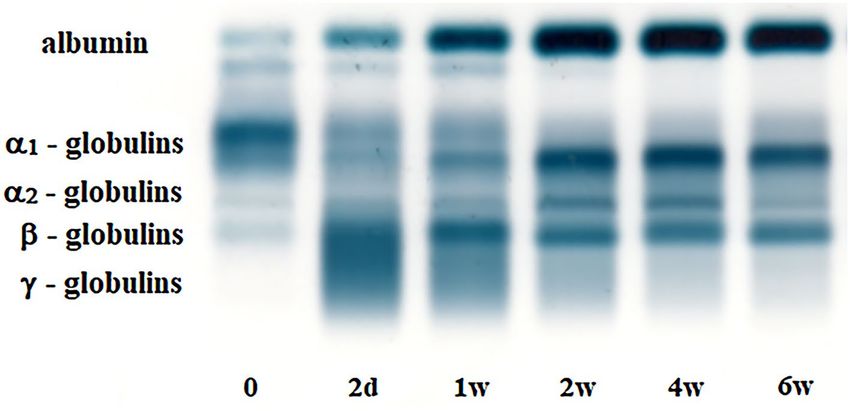
Figure 1. Electrophoretograms showing the protein fractions and changes in their proportion in a piglet during
the early postnatal period—before colostrum inteke (0), on day 2 (2d), 1 week (1w), 2 weeks (2w), 4 weeks (4w)
and 6 weeks (6w) after birth. (The display of the original gel is included in a Supplementary Information file).
further gradual significant increase until the week 4 after birth. After weaning the mean A/G values were non-
significantly lower compared to the blood sampling before weaning.
The absolute concentrations of total serum proteins and all serum protein fractions showed significant changes
during the evaluated period of postnatal development (Table 2; P < 0.001). The concentrations of TP were the
lowest immediately after birth and increased markedly after the sucking. The mean value was significantly the
highest 2 days after birth (more than two times higher). Subsequently, from the week 1 of age the mean TP con-
centrations started gradually significantly decrease until the end of the evaluated postnatal period with the lowest
mean values found after weaning. Compared to globulins, the absolute concentrations of albumin immediately
after birth were very low. In the following period, a significant gradual increase of its concentrations was found
until the pre-weaning period (4th week), when the mean albumin concentration was the highest (approx. sixfold
higher than the pre-colostral mean value). After weaning the mean albumin value was again significantly lower.
An opposite trend was found in the α1-globulins showing the significantly highest mean value immediately
after birth and decreasing values until the age of two weeks. Significant alterations were observed also in the
absolute concentrations of α2-globulins. Their concentrations increased significantly on day 2 and 1 week after
birth and a subsequent significant decrease of values was recorded in the pre-weaning and post-weaning period.
Similar tendency in the pre-weaning period was found in the changes of β-globulin concentrations. Their absolute
concentrations increased significantly about 4.5-fold two days after birth and then started gradually to decrease
until the 4st week of age. One week after weaning a slight non-significant increase of the values was observed.
Immediately after birth, only minimal concentrations of γ-globulins were found in piglets. After sucking and
colostrum intake the mean concentration of γ-globulins significantly increased more than 30-fold two days after
Scientific Reports | (2021) 11:17539 | https://doi.org/10.1038/s41598-021-96957-6 3
Vol.:(0123456789)www.nature.com/scientificreports/
Figure 2. Representative electrophoretograms in a piglet showing the protein fractionation of serum proteins
into five bands: albumin, α1-, α2-, β- and γ-globulins during the monitored period—(a) precolostral serum, (b)
on day 2, (c) 1 week, (d) 2 weeks, (e) 4 weeks and (f) 6 weeks after birth.
birth. From the age of 1 week the values started markedly decrease until the age of 4 weeks and then remained
at the same low levels also after weaning.
Electrophoresis (Fig. 1) showed in newborn piglets a double-peak in the albumin zone with two approxi-
mately similarly sized lower peaks (Fig. 2a). In two days old piglets was the albumin zone characterized by a
more prominent higher peak in the anodal side and a minor peak on the cathodal side (Fig. 2b). In 1 week old
piglets this peak appeared only as a shoulder after the main albumin fraction (Fig. 2c) and from the second week
of life only a typical one peak of albumin was observed. The α-globulins migrated in α1- and α2-fractions and the
α1-globulin fraction was the most prominent in newborn piglet’s serum observable as a wide and high peak on
the electrophoretogram (Fig. 2a). Its size decreased with advancing age from the second week of life the shape
and size stayed relatively stable (Fig. 2d,e). The α2-globulin zone in newborn piglets was characterized by one
peak, but after weaning it showed a separation in two small peaks (Fig. 2f). Serum protein electrophoresis identi-
fied in the β-globulin zone one overall distinct band, except of the period 48 h after the first sucking, when the
Scientific Reports | (2021) 11:17539 | https://doi.org/10.1038/s41598-021-96957-6 4
Vol:.(1234567890)www.nature.com/scientificreports/
β-globulins increased markedly and formed with the γ-globulins the β-γ fusion (Fig. 2b). The γ-globulin peak
in neonatal piglets was very low (Fig. 2a), and a sharp increase 48 h after the first sucking was found forming
with the β-globulin peak the aforementioned β-γ bridging (Fig. 2b). Its shape and size started gradually decrease
from the 1st week of life (Fig. 2c–f). The γ-globulin peaks were during the pre-weaning and post-weaning period
again low and clearly distinguishable from β-globulin zone.
Discussion
The physiology of young animals is different from adults and may be markedly affected by growth, development,
stressful conditions, as well as feeding, especially of colostrum or milk, which may have a bigger impact on the
metabolism and health state in comparison with the adults12. It was found that the neonatal period in piglets
is characterized by intensive changes in carbohydrate and fat metabolism17,18. In this study presented results
showed important alterations also in the protein composition of blood serum in piglets during the postnatal
development. The concentrations of total serum proteins in piglets immediately after birth were very low (around
25 g/l) and increased approximately two-fold two days after the first sucking. Bengtsson19 found in newborn
piglets also low concentrations of total serum proteins and their values increased about two-fold after 3 days
of sucking. Similarly, Dvorák20 and Martin et al.10 presented the lowest concentrations of total proteins in the
newborn piglets before colostrum intake, but shortly after the ingestion of colostrum, the total proteins reached
values of more than 70 g/l (approximately a three-fold increase). These higher concentrations decreased slightly
until weaning, which is in agreement with our results.
The increase of total serum protein concentrations was accompanied by important qualitative, as well as quan-
titative changes in the protein fractions. At birth, the serum concentrations of albumin in piglets are relatively low,
because its synthesis during the fetal development starts later when compared to other serum proteins, and due to
the epitheliochorial type of the placenta the molecule transfer is not possible not only for immunoglobulins, but
also for albumin21,22. According to Ingvarsson et al.3 and Martin et al.10, albumin represents only approximately
6–7% of total proteins in the serum of newborn piglets, which represents its significantly lower ratio compared
to the results recorded in the presented work. The average albumin values obtained in newborn piglets by these
authors were lower (3 g/l and 2.4 g/l) compared to the concentration recorded in our study. Slightly higher
values were presented in the study conducted by Szymeczko et al.23, in which albumin constituted in average
14.2% of the total proteins. These results are similar to findings recorded in our study. The hepatic synthesis of
albumin is markedly activated from the first day of postnatal life, which is reflected by rapid increase of serum
concentrations24. Similarly, Bengtsson19 obtained in newborn piglets low concentrations of albumin, which was
followed by a significant increase of values after sucking for 4 h. According to Page25, the hepatic synthesis of
albumin increase about tenfold from birth to approximately 10 days of age, while Dvorák20 observed a gradual
increase of albumin concentrations from birth until the beginning of the third month of age.
Albumin is one of the major proteins in the colostrum of pigs26. The concentration of albumin in the sow´s
colostrum is high at farrowing and decrease rapidly during the first day of lactation27. The piglets may absorb
albumin from the colostrum during the first hours of life, and its uptake by mucosal cells ceases 24–36 h after
birth28. Thus, the increase of the concentrations of albumin in the piglets 48 h after the first sucking (from 4.4
to 9.1 g/l) may be partly the result of its absorption from the colostrum and partly by de novo synthesis by the
piglets3. On the other hand, some authors stated that the intake of colostrum has no great influence on the
increase of serum albumin concentrations in the piglets after birth and the increased synthesis of albumin in the
liver of newborn suckled piglets is sufficient to account for its increased concentrations p ostnatally20,25.
On the electrophoretogram presented in the study the albumin zone in newborn piglets was characterized
by two peaks. The shape and size of these peaks changed with advancing age, showing a more prominent, major
and higher peak in the anodal side (moving faster) and a minor peak on the cathodal side of the albumin zone
(moving slower), which probably represents the so called postalbumin fraction. This pattern of very low albumin
concentrations in newborn piglets differs markedly from those obtained in other animal species and the presence
of double albumin zone was previously not described in any other species. A minor postalbumin fraction was
described only in equine serum as a shoulder on the cathodal side of the albumin peak29. The existence of serum
albumin, prealbumin and postalbumin was previously suggested also in perinatal pigs by Lardinois and Page30,
but their functions were not clearly described. Postalbumin could be a fetal-specific protein that disappear with
advancing age31. Because up to now there is in the literature no exact explanation for the specific findings in
albumin zone pattern in the first week of piglets life reported many years ago as well as in the presented study,
further research would be helpful to clarify the biochemical and immunological properties of postalbumin in
newborn piglets.
The predominant serum protein in newborn piglets before sucking is α1-acid glycoprotein, which represents
approximately 50% of total serum proteins at birth, while in adult pigs decreases approximately 30-times and
constitutes only 0.3% of blood proteins. Its concentrations in newborn piglets are around 12 mg/ml, whereas in
adult pigs it presents only values about 0.25 mg/ml14,32. Similarly, Heegaard et al.33 showed higher serum con-
centrations of α1-glycoprotein in perinatal piglets compared to one month old ones. They found 2–5 days old
piglets to have a mean serum concentration of α1-glycoprotein of 6.6 mg/ml, possibly reflecting a rapid decrease
of its concentrations in the first few days after birth, while in 1 month old pigs a mean serum concentration
of 1.1 mg/ml was found. According to Lampreave and Piñeiro7, the concentrations of α1-glycoprotein in this
critical period may be an adaptive response to prepare the newborn pig for the extrauterine life. Furthermore,
α1-glycoprotein was described as general acute phase protein in pigs32,34. The blood serum of newborn piglets is
rich also in α1-antitrypsin and fetuin (may represent approximately 18% of total serum proteins at birth), which
play important role by the inhibition of the effect of proteases absorbed by the gastrointestinal tract in the first
days of life7,13. Although α1-fetoprotein is a major serum protein in fetal life, in contrast to other animal species,
Scientific Reports | (2021) 11:17539 | https://doi.org/10.1038/s41598-021-96957-6 5
Vol.:(0123456789)www.nature.com/scientificreports/
its concentrations in porcine fetuses and neonatal piglets are lower than those of fetuin and α1-antitrypsin35,36.
The aforementioned proteins belong to the α1-globulin fraction and their high concentrations in newborn piglets
might account for very high α1-globulin values observed in our study in piglets immediately after birth.
The protein pig major acute phase protein (pig-MAP) shows similarity with the proteins from the inter-
α-trypsin inhibitor f amily37,38, therefore, it may also migrates into the α1-zone or α1-α2 interzone. In the first
days after birth, a marked increase of serum pig-MAP concentrations was observed in piglets10, indicating that
colostrum could be a source of this protein. Furthermore, the activated synthesis of pig-MAP by the liver may
be another cause of its increased serum concentrations during the postnatal development. On the other hand,
pig-MAP belong to acute phase proteins, thus, the rapid increase of its concentrations may suggest some kind
of acute phase response to the rapid growth and various environmental f actors39. According to Piñeiro et al.40,
pig-MAP is considered an important stress marker, which concentrations increase in stressful conditions or due
to changes in the composition of diet and in the pattern of feed administration.
Szymeczko et al.23 stated also that in newborn piglets α-globulins are the dominant fractions, but from these
α2-globulins account for a larger proportion (about 50% of total proteins). In contrast to these data, our results
suggest that α1-globulins compose the largest fraction of serum proteins and by visual estimation represent the
most marked zone on the electrophoretogram, observable as broad and high peak in newborn piglets. In context
of the literature data, according to which α1-glycoprotein is the predominant protein in neonatal piglets, we
assumed that the markedly high peak observable on the electrophoretogram is part of the α1-fraction.
The α2-globulin fraction includes some acute phase proteins such as haptoglobin, α2-macroglobulin, as well
as ceruloplasmin41. From these, α2-macroglobulin, ceruloplasmin, haptoglobin, as well as lipoproteins, have
been found in fetal pig serum at the end of fetal development and in neonatal piglets, but they are all minor
components42,43. Martin et al.10 observed in piglets a marked increase of haptoglobin concentrations during the
first days of life, while these values were more than two-fold higher than those recorded in 6-month old pigs.
Piñeiro et al.44 recorded in growing pigs a trend of increasing haptoglobin concentrations with age reaching a
maximum at 12 weeks of life, while the higher values observed in the nursery period might be due to stress caused
by weaning, separation from the mother, regrouping, changes of environment, as well as diet. Pié et al.45 described
also an acute phase response after weaning, which was associated with increased expression of proinflammatory
cytokines at days 0–2 post weaning. On the other hand, in the study conducted by Perri et al.46, haptoglobin
was not influenced by the age and weaning, but the values of pig-MAP were higher in 3 weeks old piglets than
in those after weaning. Transferrin and complement are the main components of the β-globulin f raction47. The
major function ascribed to transferrin is the transport of iron ions from the intestine and the reticuloendothelial
system to all proliferating cells in the b ody48. According to Thoren-Tolling and M artinsson49, its concentrations
at birth are low (approximately 1 mg/ml), which is consistent with the transitory iron deficiency state in new-
born piglets. The increase of β-globulins observed in our study in piglets from the second day of life may in part
reflect the aforementioned increase of serum transferrin concentrations. Furthermore, some immunoglobulins
absorbed from the colostrum, mainly IgA and IgM5,50, may migrate into the beta-fraction and contribute to the
increase of β-globulins in piglets 48 h after colostrum intake and their subsequent gradual decrease associated
with the degradation of these immunoglobulins.
The very low γ-globulin fraction in piglets immediately after birth reflect the lack of immunoglobulins in
their serum before sucking, indicating (similarly to newborn calves) the existence of a barrier between the sow
and the fetus against large molecules, including maternal immunoglobulins. The placenta of sows isolates the
fetus from the maternal plasma p roteins51, and therefore the maternal immunoglobulins, some other physi-
ologically active proteins and bioactive peptides with different functions must be absorbed from the colostrum
due to the intestinal m ucosa52,53. The newborn piglets are reliant on these immunoglobulins absorbed from
colostrum for passive humoral immune protection until their own immune system has sufficiently matured to
produce antibodies against foreign a ntigens11. A very small amount of immunoglobulins may be detected in the
blood of newborn piglets as result of selective transport. Piglets are functionally undeveloped and are dependent
on maternal immunoglobulins for immune protection at least during the period from birth until w eaning54,55.
Furthermore, trace quantities of immunoglobulins may be synthesized de novo, predominantly by the thymus,
spleen, liver and bone marrow, which belong to the main lymphopoetic tissues during the fetal d evelopment56.
After the first intake of colostrum the immunoglobulins (mainly IgG) rapidly enter the systemic blood circulation
and their serum concentrations increase m arkedly57,58. This pattern was observable in our study in the γ-globulin
fraction, manifested by significantly increased concentrations 48 h after the first sucking. B engtsson19 stated also
that sucking for 4 h is accompanied by a sharp increase of β- and γ-globulins, while Szymeczko et al.23 observed
in the 12th h of life an over 100-fold increase of γ-globulins compared to the approximately 34-fold increase
obtained in our study.
The analysis of the obtained results showed relatively high standard deviations, which suggests a wider range
and greater variability of values in the evaluated piglets. This pattern might be influenced by a variety of fac-
tors, among which the amount of IgG available to individual piglets and consumed may be the most important.
Bland and R ooke59 stated that the initial concentrations of IgG in colostrum are very variable even within sows
in the same unit. Differences were found also between different parts of the udder, while caudal teats tend to
have lower IgG concentrations compared with the cranial t eats60. Other factors, which influence the intake of
immunoglobulins by the piglet are related to its ability to compete successfully and suckle, the position of the
piglet in the birth order, as well as to the intestinal permeability and efficiency of transmission of IgG from the
lumen of the intestine to the bloodstream prior to the gut closure11,61. This substantial variability between piglets
to transfer IgG from the gut to the bloodstream may be explained by different birth weight, prematurity and
several genetic factors62,63.
From immunoglobulins, IgG is the most clinically important globulin for piglets during the first weeks of life.
The immunoglobulins absorbed from the colostrum and then from milk provide in piglets the passive immunity,
Scientific Reports | (2021) 11:17539 | https://doi.org/10.1038/s41598-021-96957-6 6
Vol:.(1234567890)www.nature.com/scientificreports/
until the organism is capable to initiate their synthesis de novo. According to Rooke et al.64, this process in piglets
is possible from the 7th day of life. Curtis and B ourne65 stated that the half-lives of IgG in the blood serum of
piglets range from 6.5 to 22.5 days, and that any IgG production by young piglets in the first 2–3 weeks of life
contributes little to serum IgG concentrations. In the study of Martin et al.10, the IgG concentrations decreased
progressively due to catabolism until the age of 8 weeks, when they reached minimum values. According to
Curtis and Bourne5, the serum concentrations of immunoglobulins after this period start to increase again, as
the active synthesis of IgG surpasses its breakdown. In our study, the concentrations of γ-globulins decreased
markedly from the 2nd day of life until the age of 4 weeks reaching very low values, and this finding remained in
piglets also 1 week after weaning. Due to this status of the immune system, the weaning and early post-weaning
period are the most critical periods in the life of piglets, when they are susceptible to many important diseases
resulting in growth retardation, even death. Furthermore, many stress factors associated with this period, such
as removal from the sow, dietary changes, adapting to a new environment, mixing of piglets from different litters
intestine, may negatively affect the immunological responses of the organism and result in serious diseases66.
The aforementioned alterations in the composition of serum proteins during the postnatal development resulted
also in marked changes of A/G ratio, and were primarily caused by changing globulin pattern during the growth
and development.
Conclusions
The presented study significantly expands the current knowledge in the analysis of the serum protein fractions in
piglets and showed marked differences in the composition of serum proteins in newborn piglets before colostrum
intake and during their postnatal development including the period after weaning. The results suggest marked
developmental alterations in the serum protein pattern along with the age. Among these alterations observed,
the changes in albumin, α- and γ-globulin fractions are of note. The serum protein electrophoretic pattern of
newborn piglets before the intake of colostrum were characterized by very low concentrations of total proteins,
albumin and γ-globulins and very high proportion of α1-globulins. Forty-eight hours after the first sucking there
was a significant increase of total proteins and γ-globulins and significant decrease of α1-globulins. The changes
observed in further periods of postnatal development were progressive and were characterized by a gradual
decrease of total proteins, β- and γ-globulins, and further increase of albumin concentrations until the period
before weaning. The changes in protein profile after weaning as a critical period for piglets was characterized by
a decrease of total protein and albumin fraction and increase of β-globulin fraction. No significant changes were
found in γ-globulin values. The aforementioned changes in the serum protein pattern may be associated with
physiological changes during rapid growth and development, intensive metabolic processes, as well as changes
in feeding and stress due to weaning. The study also brings new knowledge in the field of description of typical
features of electrophoretograms in piglets in the observed period of their life. Seeing that the serum protein
electrophoretic pattern in newborn piglets is not completely understood, there is a need for further investiga-
tions to improve understanding of changes occurring in the serum protein pattern and the obtained specificities
in piglets during postnatal development.
Methods
Ethics declarations. This study was based on the standard clinical examination and blood sample col-
lection. The blood samples were collected as per standard sampling procedure used without any harm to the
animals. All procedures with animals in the study were conducted in accordance with the ethical standards and
relevant quidelines and regulations. The experimental protocols were approved by the institutional Ethical Com-
mittee of the University of Veterinary Medicine and Pharmacy in Košice on protection of animals used for scien-
tific purposes (26/11/2019) and complied with the institutional requirements of the Code of Ethics for Scientists
(Directive 74/2019/UVLF). The study was carried out in compliance with ARRIVE guidelines.
Animals and sample collections. A total of 50 newborn crossbreed piglets Large White x Landrace of
both sexes (21 males and 29 females) from five sows (first to third gestation) were included into the study. The
study was performed in the university experimental facility. The clinically healthy sows were housed individu-
ally in straw bedded farrowing crates, fed twice daily a commercial diet, parturitions were watched and occurred
naturally. Environmental temperature of the farrowing room ranged between 20 and 22 °C and the farrowing
pens were equipped with the heating lamp for piglets. After birth the piglets had their umbilical cord cut, roughly
dried and weighted. The average number of piglets born alive was 11 per sow. The mean body weight of piglets at
birth was 1.66 ± 0.28 kg. They were housed together with their mother until weaning in standard farrowing pens
with straw bedding and space warming using a heat lamp to 28–31 °C. Each pen had a non-lidded hopper and
a nipple water to ensure ad libitum feeding and free water access. The weaning of piglets was done at the age of
35 days (5 weeks), when they were moved from the farrowing to the nursery unit within the same object without
transport. Piglets were fed from the day 10 of their life until the end of the evaluated period with the commercial
complete feed intended for piglets from day 10 after birth until the age of the 14th day after weaning (Weaning
pellets PP+, DeHeus, Czech Republic). The main chemical composition of the pellets and additives in the diet
are presented in Table 3. The health status of the animals was evaluated daily until the end of the study using
routine clinical diagnostic procedures, and was oriented to the observation of general health state, feed intake,
weight gain and behaviour. The piglets were during the whole monitored period in a good general health status
without any obvious clinical signs of diseases. The first blood samples from piglets were collected immediately
after birth before colostrum sucking (day 0). The piglets were subsequently returned to the nursing sows allowed
to suckle. Further blood sample collections were performed on day 2 (2d), 7 (1w), 14 (2w), 28 (4w) after birth
(1 week before weaning), and 7 days after weaning (6w). Piglets were weighted individually before each blood
Scientific Reports | (2021) 11:17539 | https://doi.org/10.1038/s41598-021-96957-6 7
Vol.:(0123456789)www.nature.com/scientificreports/
Components of feed Additives
Crude protein (%) 17.4 Vitamin A (IU/kg) 16 000
Crude fibre (%) 4.5 Vitamin E (mg/kg) 160
Fat (%) 5.4 Vitamin D3 (IU/kg) 1 200
Ash (%) 4.7 Cu (as copper sulphate, mg/kg) 139.0
Lysine (%) 1.22 Zn (as zinc sulphate, mg/kg) 109.0
Methionine (%) 0.44 Fe (as ferrous sulphate, mg/kg) 99.0
Ca (%) 0.5 Mn (as manganese oxide, mg/kg) 40.0
P (%) 0.52 I (as calcium iodate, mg/kg) 1.0
Na (%) 0.25 Se (as sodium selenite, mgkg) 0.2
Se (as selenomethionine, mgkg) 0.1
Table 3. Chemical composition of the used piglet pellets and additives in the feed (in 1 kg of dry matter).
sample collection. The average body weights in the after birth subsequent monitored periods were 1.81 ± 0.32 kg
(2 days), 2.86 ± 0.69 kg (1 week), 4.74 ± 1.35 kg (2 weeks), 9.46 ± 1.97 kg (4 weeks) and 15.4 ± 2.68 kg (6 weeks).
Blood samples of approximately 1 ml were taken from the sinus ophtalmicus into serum gel separator tubes
without additives and anticoagulants (Sarstedt, Nümbrecht, Germany). Animals for the blood collection from
orbital sinus were manually restrained in dorsal recumbency position placed on V-board table. The head of the
piglets was in front of the edge of the table and both the front and the back legs were held such they were pointed
toward posterior of the pigs. After letting the blood samples to coagulate at room temperature, sera were obtained
by centrifugation at 4000×g for 15 min and then transferred into Eppendorf tubes. The aliquots of sera were kept
frozen at − 20 °C for further laboratory analyses.
Laboratory analyses. To evaluate the changes in the protein profile in piglets serum samples were ana-
lyzed for the concentrations of total proteins and main protein fractions. The concentrations of total serum pro-
teins (TP, g/l) were measured on the automated biochemical analyzer Alizé (Lisabio, Poully en Auxois, France)
according to the biuret method with commercially available diagnostic kits (Randox, Crumlin, United King-
dom). Serum protein electrophoresis was performed according to the application notes of the manufacturer
on agarose gel using an automated electrophoresis system Hydrasys and commercial diagnostic kits Hydragel
7 Proteine for the separation of serum protein fractions (Sebia Corporate, Lisses, Evry Cedex, France). Serum
protein fractions were separated by zone electrophoresis on a buffered agarose gel at pH 8.8. Ten microliters of
each serum sample were applied to performed, numbered sample wells on the agarose gel. Control serum (Con-
trol Serum Human Normal, Sebia Corporate, France) was included in each run of samples. The electrophoretic
migration was performed 15 min at 20 °C constantly at 10 W, 40 mA, and 240 V. After migration, the gels were
stained in amidoblack staining solution and then destained with acidic solutions and dried completely. After
separation and staining of the gels, the staining intensity of individual protein bands was quantified using a
densitometer Epson Perfection V700 (Epson America Inc., California, USA) in conjunction with image analysis
software Phoresis version 5.50 (Sebia Corporate, France)8. The following protein fractions were identified: albu-
min, α1-, α2-, β- and γ-globulins. They were expressed as relative values (%) according to the optical density and
their absolute concentrations (g/l) were quantified from the TP concentrations. Albumin:globulin ratios (A/G)
were calculated as well.
Statistical analyses. Descriptive statistical procedures were performed to calculate the arithmetic means
(x) and standard deviations (SD) for each evaluated variable and sample collection time. The distribution of data
was evaluated using Kolmogorov–Smirnov Test for normality. Not all the evaluated parameters showed normal
distribution. Therefore, the data were subjected to analysis of variance using a non-parametric Friedman test to
assess the age-dependent changes during the monitored period. The significance of differences in values between
the sample collections was evaluated by Tukey’s Multiple Comparisons post-test. Significance was considered at
5% probability level. All calculations were carried out by the GraphPad Prism V5.02 (GraphPad Software Inc.,
California, USA) computer program.
Data availability
The datasets generated and/or analysed during the current study are available from the corresponding author
on reasonable request.
Received: 31 March 2021; Accepted: 18 August 2021
References
1. Svendsen, L. S., Weström, B. R., Svendsen, J., Olsson, A. & Karlsson, B. W. Blood serum characteristics of newborn pigs: Compari-
son of unaffected pigs with pigs belonging to five mortality groups. Acta Vet. Scand. 32, 287–299 (1991).
2. Mota-Rojas, D. et al. Foetal and neonatal energy metabolism in pigs and humans: A review. Vet. Med. 56, 215–225 (2011).
Scientific Reports | (2021) 11:17539 | https://doi.org/10.1038/s41598-021-96957-6 8
Vol:.(1234567890)www.nature.com/scientificreports/
3. Ingvarsson, B. I., Carlsson, R. N. K. & Karlsson, B. W. Synthesis of α-fetoprotein, albumin and total serum protein in neonatal pigs.
Biol. Neonate 34, 259–268 (1978).
4. Cavanagh, M. E. et al. Proteins in cerebrospinal fluid and plasma of fetal pigs during development. Dev. Neurosci. 5, 492–502
(1982).
5. Curtis, J. & Bourne, F. J. Immunoglobulin quantitation in sow serum, colostrum and milk and the serum of young pigs. Biochim.
Biophys. Acta 236, 319–332 (1971).
6. Lampreave, F. & Piñeiro, A. Characterization of a new alpha-glycoprotein as the major serum component in later fetal and newborn
pigs. Comp. Biochem. Physiol. 72B, 215–219 (1982).
7. Lampreave, F. & Piñeiro, A. Concentrations of major plasma proteins in serum and whole-tissue extracts in porcine fetuses during
development. J. Reprod. Fertil. 95, 441–449 (1992).
8. Tóthová, C., Nagy, O., Kováč, G. & Nagyová, V. Changes in the concentrations of serum proteins in calves during the first month
of life. J. Appl. Anim. Res. 44, 338–346 (2016).
9. Le Dividich, J. & Noblet, J. Effect of colostrum intake on metabolic rate and plasma glucose in the neonatal pig in relation to
environmental temperature. Biol. Neonate 46, 98–104 (1984).
10. Martin, M., Tesouro, M. A., González-Ramón, N., Piňeiro, A. & Lampreave, F. Major plasma proteins in pig serum during postnatal
development. Reprod. Fertil. Dev. 17, 437–445 (2005).
11. Rooke, J. A. & Bland, I. M. The acquisition of passive immunity in the new-born piglet. Livest. Prod. Sci. 78, 13–23 (2002).
12. Yu, K. et al. Age-related serum biochemical reference intervals established for unweaned calves and piglets in the post-weaning
period. Front. Vet. Sci. 6, 123 (2019).
13. Weström, B. R., Karlsson, B. W. & Svendsen, J. Levels of serum protease inhibitors during fetal and postnatal development of the
pig. Biol. Neonate 41, 22–31 (1982).
14. Lampreave, F. & Piñeiro, A. The major serum protein of fetal and newborn pigs: Biochemical properties and identification as a
fetal form of α1-acid glycoprotein. Int. J. Biochem. 16, 47–53 (1984).
15. Giot, J. F. Agarose gel electrophoresis: Applications in clinical chemistry. J. Med. Biochem. 29, 9–14 (2010).
16. Coşkun, Ö. & Öztopuz, Ö. Electrophoresis applications used in medicine. Med. Sci. 15, 12–25 (2020).
17. Mersmann, H. J. Metabolic patterns in the neonatal swine. J. Anim. Sci. 38, 1022–1030 (1974).
18. Pégorier, J. P., Duée, P. H., Assan, R., Peret, J. & Girard, J. Changes in circulating fuels, pancreatic hormones and liver glycogen
concentration in fasting or suckling newborn pigs. J. Dev. Physiol. 3, 203–217 (1981).
19. Bengtsson, S. G. Identification and characterization of soluble blood serum proteins in newborn piglets. J. Anim. Sci. 38, 95–99
(1974).
20. Dvořák, M. Changes in blood protein levels in piglets during development and during stress. Vet. Med. 31, 403–414 (1986).
21. Stone, R. T. In vitro liver synthesis and serum levels of alpha-fetoprotein and albumin in the fetal pig. Biol. Reprod. 20, 573–580
(1981).
22. Le Dividich, J., Rooke, J. A. & Herpin, P. Nutritional and immunological importance of colostrum for the new-born pig. J. Agric.
Sci. 143, 469–485 (2005).
23. Szymeczko, R. et al. Changes in the content of major proteins and selected hormons in the blood serum of piglets during the early
postnatal period. Folia Biol. 57, 97–103 (2009).
24. Stone, R. T. Isolation and characterisation of recombinants cDNA clones corresponding to developmentally regulated genes in pig
liver. J. Anim. Sci. 67, 1082–1089 (1989).
25. Page, L. A. In vivo and in vitro studies on the synthesis of albumin by perinatal pigs. Dev. Biol. 20, 125 (1969).
26. Gallagher, D. P., Cotter, P. F. & Mulvihill, D. M. Porcine milk proteins: A review. Int. Dairy J. 7, 99–118 (1997).
27. Karlsson, B. W. Fetoprotein and albumin levels in the blood serum of developing neonatal pigs. Comp. Biochem. Physiol. 34, 535
(1970).
28. Klobasa, F. & Butler, J. E. Absolute and relative concentrations of immunoglobulins G, M and A, and albumin in the lacteal secre-
tions of sows of different lactation numbers. Am. J. Vet. Res. 48, 176–182 (1987).
29. Kaneko, J. J. Serum proteins and the dysproteinemias. In Clinical Biochemistry of Domestic Animals 5th edn (ed. Kaneko, J. J.)
117–138 (Academic Press, 1997).
30. Lardinois, R. & Page, L. A. Serum albumin, prealbumin and postalbumin in perinatal pigs. Dev. Biol. 19, 261–280 (1969).
31. Kirsch, J. A. W., Wise, R. W. & Oliver, I. T. Post-albumin, a foetal-specific rat plasma protein: Purification, physicochemical and
immunological studies. Biochem. J. 102, 763–766 (1967).
32. Itoh, K. et al. The influence of age and health status on the serum α1-acid glycoprotein level of conventional and specific pathogen-
free pigs. Can. J. Vet. Res. 57, 74–78 (1993).
33. Heegaard, P. M. H., Miller, I., Sorensen, N. S., Soerensen, K. E. & Skovgaard, K. Pig α1-glycoprotein: Characterization and first
description in any species as a negative acute phase protein. PLoS ONE 8, e68110 (2013).
34. Tecles, F., Martínez-Subiela, S. & Cerón, J. J. Optimization of a spectrophotometric method for quantification of acid-soluble
glycoprotein in porcine serum. Can. J. Vet. Res. 11, 161–164 (2007).
35. Gitlin, D. & Boesman, M. Sites of serum α-fetoprotein synthesis in the human and in the rat. J. Clin. Invest. 46, 1010–1016 (1967).
36. Calvo, M., Naval, J., Lampreave, F. & Piñeiro, A. Fatty acids bound to α-fetoprotein and albumin during rat development. Biochim.
Biophys. Acta 959, 238–246 (1988).
37. González-Ramón, N. et al. The major acute phase protein in pigs is homologous to human plasma kallikrein sensitive PK-120.
FEBS Lett. 371, 227–230 (1995).
38. Saguchi, K. et al. Cloning and characterisation of cDNA for inter-α-trypsin inhibitor family heavy chain-related protein (IHRP),
a novel human plasma glycoprotein. J. Biochem. 117, 14–18 (1995).
39. Alava, M. A. et al. Pig-MAP, porcine acute phase proteins and standardization of assays in Europe. Comp. Haematol. Int. 7, 208–213
(1997).
40. Piñeiro, M. et al. Characterisation of the pig acute phase protein response to road transport. Vet. J. 173, 669–674 (2007).
41. Bossuyt, X. Advances in serum protein electrophoresis. Adv. Clin. Chem. 42, 43–80 (2006).
42. Weström, B. R. Identification and characterization of trypsin, chymotrypsin and elastase inhibitors in porcine serum. Hoppe-Seylers
Z. Physiol. Chem. 360, 1869–1878 (1979).
43. Johansson, M. B. N. & Karlsson, B. W. Lipoprotein and lipids profiles in the blood serum of the fetal, neonatal and adult pig. Biol.
Neonate 42, 127–137 (1982).
44. Piñeiro, C. et al. Pig-MAP and haptoglobin concentration reference values in swine from commercial farms. Vet. J. 179, 78–84
(2009).
45. Pié, S. et al. Weaning is associated with an upregulation of expression of inflammatory cytokines in the intestine of piglets. J. Nutr.
134, 641–647 (2004).
46. Perri, A. M., O’Sullivan, T. L., Harding, J. C. S., Wood, R. D. & Friendship, R. M. Hematology and biochemistry reference intervals
for Ontario commercial nursing pigs close to the time of weaning. Can. Vet. J. 58, 371–376 (2017).
47. Cerón, J. J., Caldin, M. & Martinez-Subiela, S. Electrophoresis and acute phase protein measurement. In Schalm’s Veterinary
Hematology 6th edn (eds Weiss, D. J. & Wardrop, K. J.) 1157–1161 (Willey-Blackwell, 2010).
48. de Jong, G., van Dijk, J. P. & van Eijk, H. G. The biology of transferrin. Clin. Chim. Acta 190, 1–46 (1990).
Scientific Reports | (2021) 11:17539 | https://doi.org/10.1038/s41598-021-96957-6 9
Vol.:(0123456789)www.nature.com/scientificreports/
49. Thoren-Tolling, V. & Martinsson, K. On the transferrin concentration in serum of sows and growing pigs and in sow colostrum.
Acta Vet. Scand. 15, 1120–1134 (1974).
50. Klobasa, F., Werhahn, F. & Butler, J. F. Composition of sow milk during lactation. J. Anim. Sci. 64, 1458–1466 (1987).
51. Styk, B. & Hána, L. Pecularities of certain components of newborn pig serum. Acta Virol. 7, 285 (1963).
52. Schanbacher, F. L., Talhouk, R. S. & Murray, F. A. Biology and origin of bioactive peptides in milk. Livest. Prod. Sci. 50, 102–123
(1997).
53. Xu, R. J., Wang, F. & Zhang, S. H. Postnatal adaptation of the gastrointestinal tract in neonatal pigs: A possible role of milk-borne
growth factors. Livest. Prod. Sci. 66, 95–107 (2000).
54. Bianchi, A. T. J., Zwart, R. J., Jeurissen, S. H. M. & Moonen-Leusen, H. W. M. Development of the B- and T-cell compartments in
porcine lymphoid organs from birth to adult life: An immunohistochemical approach. Vet. Immunol. Immunopathol. 33, 201–221
(1992).
55. Kanka, T. et al. Structural changes of mucous membrane and the presence of immunoglobulins in small intestines of neonatal
pigs. Acta Vet. Brno 82, 317–322 (2013).
56. Sinkora, J., Rehakova, Z., Sinkora, M., Cukrowska, B. & Tla-Skalova-Hogenova, H. Early development of immune system in pigs.
Vet. Immunol. Immunopathol. 87, 301–306 (2002).
57. Frenyó, V. L., Pethes, G., Antal, T. & Szabó, I. Changes in colostral and serum IgG content in swine in relation to time. Vet. Res.
Commun. 4, 275–282 (1980).
58. Klobasa, F., Werhahn, E. & Butler, J. E. Regulation of humoral immunity in the piglet by immunoglobulins of maternal origin. Res.
Vet. Sci. 31, 195–206 (1981).
59. Bland, I. M. & Rooke, J. A. Effects on colostrum immunoglobulin G (IgG) concentrations and piglet colostrum intake: Of sow,
udder section and time. Proc. Br. Soc. Anim. Sci. 158, 158 (1998).
60. Inoue, T., Kitano, K. & Inoue, K. Possible factors influencing immunoglobulin G concentration in swine colostrum. Am. J. Vet.
Res. 42, 533–536 (1980).
61. de Passille, A. M., Rushen, J. & Pelletier, G. Sucking behaviour and serum immunoglobulin levels in neonatal piglets. Anim. Sci.
47, 447–456 (1988).
62. Svendsen, L. S., Weström, B. R., Svendsen, J., Olsson, A. & Karlsson, B. W. Intestinal macromolecular transmission in underprivi-
leged and unaffected newborn piglets: Implication for survival of underprivileged piglets. Res. Vet. Sci. 48, 184–189 (1990).
63. Sangild, P. T., Holtug, K., Diemaes, L., Schmidt, M. & Skadhauge, E. Birth and prematurity influence intestinal function in the
newborn pig. Comp. Biochem. Physiol. A 118, 359–361 (1997).
64. Rooke, J. A. et al. Relationships between passive absorption of immunoglobulin G by piglet and plasma concentrations of immu-
noglobulin G at weaning. Livest. Prod. Sci. 81, 223–234 (2003).
65. Curtis, J. & Bourne, F. J. Half-lives of immunoglobulins IgG, IgA and IgM in the serum of new-born pigs. Immunology 24, 147–155
(1973).
66. McCracken, B. A., Spurlock, M. E., Roos, M. A., Zuckermann, F. A. & Gaskins, H. R. Weaning anorexia may contribute to local
inflammation in the piglet small intestine. J. Nutr. 129, 613–619 (1999).
Acknowledgements
This research was funded by the Grants No. VEGA 1/0398/18 and VEGA 1/0314/20 from the Ministry of Educa-
tion, Science, Research and Sport of the Slovak Republic.
Author contributions
C.T. and O.N.: Conceptualization, Methodology, Formal analysis, Project administration. R.L. and P.K.: Investi-
gation, Sample collection, Data curation. O.N. Writing—original draft preparation, Visualization, Supervision.
C.T.: Writing—review and editing. All authors read and approved the final version of the manuscript.
Competing interests
The authors declare no competing interests.
Additional information
Supplementary Information The online version contains supplementary material available at https://doi.org/
10.1038/s41598-021-96957-6.
Correspondence and requests for materials should be addressed to O.N.
Reprints and permissions information is available at www.nature.com/reprints.
Publisher’s note Springer Nature remains neutral with regard to jurisdictional claims in published maps and
institutional affiliations.
Open Access This article is licensed under a Creative Commons Attribution 4.0 International
License, which permits use, sharing, adaptation, distribution and reproduction in any medium or
format, as long as you give appropriate credit to the original author(s) and the source, provide a link to the
Creative Commons licence, and indicate if changes were made. The images or other third party material in this
article are included in the article’s Creative Commons licence, unless indicated otherwise in a credit line to the
material. If material is not included in the article’s Creative Commons licence and your intended use is not
permitted by statutory regulation or exceeds the permitted use, you will need to obtain permission directly from
the copyright holder. To view a copy of this licence, visit http://creativecommons.org/licenses/by/4.0/.
© The Author(s) 2021
Scientific Reports | (2021) 11:17539 | https://doi.org/10.1038/s41598-021-96957-6 10
Vol:.(1234567890)You can also read