Sleep parameters improvement in PTSD soldiers after symptoms remission
←
→
Page content transcription
If your browser does not render page correctly, please read the page content below
www.nature.com/scientificreports
OPEN Sleep parameters improvement
in PTSD soldiers after symptoms
remission
P. F. Rousseau1*, R. Vallat2,3, O. Coste4, H. Cadis1, F. Nicolas5, M. Trousselard6, P. Ruby2 &
S. Khalfa1
Eye movement desensitization and reprocessing (EMDR) is a psychotherapy for the treatment of
posttraumatic stress disorder (PTSD). It is still unclear whether symptoms remission through EMDR
therapy is associated with a beneficial effect on one of the PTSD symptoms, sleep disturbance. Our
objective was therefore to study sleep parameters before and after symptom remission in soldiers
with PTSD. The control group consisted of 20 healthy active duty military men who slept in a sleep lab
with standard polysomnography (PSG) on two sessions separated by one month. The patient group
consisted of 17 active duty military with PTSD who underwent EMDR therapy. PSG-recorded sleep
was assessed 1 week before the EMDR therapy began and 1 week after PTSD remission. We found
that the increased REMs density after remission was positively correlated with a greater decrease of
symptoms. Also, the number of EMDR sessions required to reach remission was correlated with intra-
sleep awakenings before treatment. These results confirm the improvement of some sleep parameters
in PTSD after symptoms remission in a soldier’s population and provide a possible predictor of
treatment success. Further experiments will be required to establish whether this effect is specific to
the EMDR therapy.
Abbreviations
EMDR Eye movement desensitization and reprocessing
PSG Polysomnography
PTSD Posttraumatic stress disorder
TST Total sleep time
SPT Sleep period time
SOL Sleep onset latency
REM Rapid eye movement
SE Sleep efficiency
SME Sleep maintenance efficiency
WASO Wake after sleep onset
Sleep disturbances represent a key factor in the genesis and maintenance of psychiatric disorders1. An estimated
90% of people with posttraumatic stress disorder (PTSD) report difficulty initiating and maintaining sleep2,3.
Even in the absence of nightmares, sleep problems remain the most frequently reported complaint in PTSD4.
Self-reported poor sleep quality is only minimally explained by psychiatric comorbidity, age, and sex in these
patients, which suggests an important role of the syndrome in the sleep i mpairment5.
The severity of PTSD symptoms are positively correlated with sleep disturbances6. Sleep problems may in turn
worsen daytime PTSD symptoms7, and contribute to comorbid psychiatric disorders such as major depression8–11,
suicidal behaviour12, and substance abuse13. Sleep disturbances in PTSD are also correlated with a decreased
1
Laboratoire de Neurosciences Cognitives UMR 7291, Aix Marseille Université CNRS, Marseille, France. 2Lyon
Neuroscience Research Center, Brain Dynamics and Cognition Team, INSERM UMR 1028, CNRS UMR 5292,
Université Claude Bernard Lyon 1, Lyon, France. 3Center for Human Sleep Science, Department of Psychology,
University of California, Berkeley, CA 94720‑1650, USA. 4Hôpital d’Instruction des Armées Desgenettes,
Unité de Pathologie du Sommeil, Lyon, France. 5Service de Psychiatrie, Hôpital d’Instruction des Armées
Sainte-Anne, Toulon, France. 6Unité Neurophysiologie du Stress, Département des Neurosciences et des
Contraintes Opérationnelles, Institut de Recherche Biomédicale des Armées, Brétigny sur Orge, France. *email:
rousseaupierrefrancois@gmail.com
Scientific Reports | (2021) 11:8873 | https://doi.org/10.1038/s41598-021-88337-x 1
Vol.:(0123456789)
www.nature.com/scientificreports/
ability to carry out daily activities14–16, again fitting with problems related to insufficient sleep in the general
population, including functional impairment, reduced quality of life17–22, and increased risk of accidents23. Of
note, sleep problems do not always improve after otherwise successful treatment for P TSD4,24–26.
Polysomnography (PSG) studies have yielded inconsistent findings regarding the presence and nature of sleep
disturbance in individuals with P TSD27, with some studies reporting reduction and/or disruption of rapid eye
movement (REM) sleep28–30, whereas others have described a higher percentage of REM sleep31–34 in individuals
with PTSD compared with other clinical groups or good sleepers without PTSD. A recent study that used at-home
sleep recordings in 71 control subjects and 60 military-related PTSD subjects found a significantly reduced per-
centage of REM sleep in the latter group35. Inconsistent results have been found for non-REM (NREM) sleep, with
some studies reporting PTSD-associated NREM disturbances35–38, and several others reporting no effects31,39,40.
Based on these findings, meta-analyses identified a core pattern of sleep disruption in P TSD1,41, consisting of
longer N1 sleep, shorter N2 and N3 sleep, as well as greater rapid eye movements (REMs) density in REM sleep
compared to individuals without PTSD. Nevertheless, more studies are needed to better characterize the core
sleep disturbances in PTSD patients.
The mechanisms and neurophysiological correlates of the attenuation or resolution of such disorders by
pharmacological42 or p sychotherapeutic4,43 treatments are still poorly understood. Increased central nervous
system adrenergic activity might contribute to the pathophysiology of P TSD44,45 by interfering with normal
REM sleep46, so studies have investigated the use of effective pharmacological t reatments42, including prazosin,
a generic alpha-1 adrenergic antagonist, in military veterans with PTSD. Objective sleep assessment studies
involving prazosin and a placebo have shown significantly increased sleep time and REM sleep duration, reduced
REM sleep latency47 and no changes in NREM sleep or sleep onset latency. Cognitive behavioral therapy for
insomnia also increased total sleep time in PTSD individuals compared to a wait-list g roup4, and both prazosin
and imagery rehearsal therapy improved sleep quality and decreased nightmare f requency48.
Eye movement desensitization and reprocessing (EMDR) therapy has been shown to significantly decrease
PTSD symptoms and is therefore among the first-line psychological interventions for PTSD49,50. Beyond a signifi-
cant decrease in PTSD symptoms, EMDR has shown its capacity to modify the brain anatomy after r emission51,52
or the brain’s functional metabolism to the traumatic event s cript53,54. Yet, its precise effect and mechanism
remain unclear. To date, only one study has investigated the effect of EMDR therapy on sleep using P SG43. They
compared mood, anxiety, autonomic activation, as well as subjective and objective sleep parameters before and
after EMDR therapy in a group of 13 PTSD patients and 11 healthy controls. Before treatment, PTSD patients had
significantly lower subjective sleep quality and an increase PSG-defined wake after sleep onset (WASO) compared
to the control group. These parameters were however non-significantly different from the control group after
treatment, which suggests that EMDR therapy was associated with sleep improvements in these patients. Even
though the authors did not find any significant interaction group × time for the other sleep parameters (most
likely because of a low sample size), they did find that the density of REMs during REM sleep was lower after
treatment in the PTSD group (Cohen d = 1.37 of within-group comparison for PTSD patients). The latter find-
ing of therapy-induced changes in REMs density is consistent with Stickgold’s theory that the eye movements
in EMDR therapy would reproduce the rapid eye movements of REM sleep, and would thus trigger some of the
brain mechanisms that are typical of REM sleep55, a stage of sleep known for its role in emotional regulation and
memory processing. In this theory, the state and eye movements induced by the bilateral alternating stimulation
would permit the integration of traumatic memories into associative cortical networks without interference
from hippocampally mediated episodic recall. This mechanism would thus alleviate the need for or replace the
emotional regulation at play during REM sleep which may induce a diminution in REM sleep REMs density.
Although EMDR therapy can use stimuli other than eye movements (tactile or auditory), recent findings have
shown accumulating evidence of a treatment gain of using eye movements as opposed to no eye movements or
other forms of bilateral stimulations56–59. Accordingly, it is possible that EMDR therapy somehow emulates, and/
or compensates for, REM-sleep mechanisms that are supposedly failing in patients with PTSD (as indicated by
the high prevalence of nightmares in these patients). Since only few studies so far have objectified the effect of
psychotherapies on PTSD patients’ sleep, our study proposes to objectively assess sleep thanks to polysomnogra-
phy measures before and after PTSD symptoms remission using EMDR therapy. The patient and control groups
are from the same population (soldiers) and larger than in previous studies, the patient group is homogenous in
origin of trauma (war), sleep has been recorded in an ecological situation (at home), and the sleep parameters
have been assessed thanks to an automatic and thus objective method of sleep scoring60. Our objectives are to
demonstrate and characterize improvement of sleep parameters after PTSD symptoms remission in a soldier’s
population and to investigate whether some pre-treatment sleep parameters could be predictive of the duration/
success of the EMDR therapy used.
Our first goal was therefore to study the effect of PTSD symptoms remission on a wide range of objective
sleep parameters in soldiers with PTSD as compared with healthy military controls. Our second objective was
to test whether some of these sleep parameters before treatment could predict the efficiency of EMDR therapy
in individuals with PTSD, as measured by the number of EMDR sessions needed to achieve remission.
Building on previous findings4,43,47, we hypothesized that remission from PTSD would be associated with
improvements in objective sleep parameters, and we therefore aimed to provide an exhaustive overview of these
sleep parameters, looking not only at sleep macro-structure but also sleep microstructure (e.g. sleep spindles,
REMs) as well as spectral power in pre-defined frequency bands.
Scientific Reports | (2021) 11:8873 | https://doi.org/10.1038/s41598-021-88337-x 2
Vol:.(1234567890)www.nature.com/scientificreports/
Materials and methods
We recruited exclusively male soldiers, divided into two groups: control and EMDR (for participants with PTSD).
The justification for including only male soldiers is mainly practical: there are very few female soldiers present
in the fighting unit of the French Army, and women in the military are therefore less at risk of being exposed to
traumatic experiences.
Participants. Control group. The control group consisted of healthy active duty soldiers recruited by the
Institute of Naval Medicine of the Army Health Service in Toulon, France. Individuals selected for the control
group had no notable medical history and no current or chronic diseases, no history of depression or alcohol,
tobacco or substance abuse or dependence. No participant had any major sleep disorder. Controls were not
receiving any medication and had no history of recent shift work or transmeridian travel for at least 2 months
before the experiment.
EMDR group. Participants were recruited by two psychiatrists in Sainte-Anne military hospital in Toulon,
France. The diagnosis of PTSD was established according to the Diagnostic and Statistical Manual of Mental
Disorders, 4th Edition, Text Revision (DSM-IV TR)61. We excluded individuals with present and past neuro-
logical or psychiatric conditions, with the exception of anxiety and depressive disorders if their occurrence was
related to PTSD. Participants could keep their psychotropic medication only if they took selective serotonin
reuptake inhibitors (SSRIs) as long as the therapy did not change during the trial and if the onset of treatment
was more than 3 months ago. We also excluded individuals with a history of sleep disorder (assessed objectively
or reported by the participant) before the traumatic event. Participants had to have no history of recent shift
work or transmeridian travel for at least 2 months before the experiment. Diagnoses were established and clini-
cal interviews were performed by psychiatrists not otherwise engaged in the study. Participants were assessed
by two psychiatrists for PTSD and other mental health disorders by using the structured Mini-International
Neuropsychiatric Interview62 to check for the absence of a psychiatric disorder before the trauma in PTSD and
to screen for potential comorbid psychiatric disorders.
All soldiers had PTSD related to war and five took SSRIs. Comorbidities included major depression (9/15),
panic disorder (2/15), agoraphobia (12/15) and obsessive–compulsive disorder (1/15); none had an addictive
disorder. None had received exposure therapy or cognitive-behavioural therapy before EMDR procedure.
Participants completed the questionnaires listed below, before and after the end of EMDR therapy. PTSD
symptoms were evaluated with the Posttraumatic Stress Checklist Scale (PCLS)63 and the Clinician-Administered
PTSD Scale (CAPS)64. Subjective sleep quality was measured using the Pittsburgh Sleep Quality Index (PSQI)65,66;
and the PSQI-Addendum for PTSD (PSQI-A), which is specific to sleep disorders in PTSD67,68. To assess PTSD
comorbidity, we used the Beck Depression I nventory69. All questionnaires were validated in French.
EMDR procedure. EMDR therapy involves associations of cognitive, emotional and physical assessments
of actual distress to traumatic scenery as well as imaginal exposure while attending to bilateral alternating
stimulations70. The individual is asked to visualize the most salient aspect of a traumatic memory while the
therapist induces bilateral stimulation (by means of ocular, tactile or auditory stimulations). This process results
in a change in cognitive processing of memory and cessation of trauma-related distress, such as negative emo-
tion disappearance, while eliminating physical discomfort associated with the initial memory and establishing
a positive cognition about the s elf70. The EMDR therapy was performed according to the standard p rotocol71 by
one psychiatrist and one psychologist trained and accredited by EMDR Europe. Sessions were planned every 7
to 15 days according to the availability of patients and the therapist.
All traumatic targets related to the traumatic event were treated until reaching a subjective unit of discomfort
of zero and completely true positive cognitions without any body discomfort. Hence, at the end of the protocol,
participants were symptom-free and no longer had a diagnosis of PTSD after the EMDR therapy, as assessed
again by two psychiatrists according to DSM-IV criteria.
Polysomnography data collection. Control group. The sleep data for the control group were collected
for a previous published study72. In this study, control military participants spent two nights with a delay be-
tween two nights of 1 month in the lab to record baseline sleep before performing a simulated flight in a hypo-
baric chamber. People in the control group were synchronized with diurnal activity (07:00–22:00 h ± 30 min)
and nocturnal rest (22:00–07:00 ± 30 min). They slept on a camp bed in total darkness during a fixed time slot
i.e. from 22:30 to 06:30. Only data for the nights before the simulated flight were analyzed and compared to the
EMDR group. Sleep architecture was assessed from standard polysomnographic recordings including electro-
encephalography (EEG), electro-oculography (EOG) of each eye and chin electromyography (EMG) by using
A10 loggers (EmblaTM, Flaga, Reykjavik, Iceland)73. Details of the methodology for this group were previously
published72.
EMDR group. Sleep was recorded before and after treatment at the participant’s home. The first sleep recording
took place the week before EMDR began (T0) and the last one the week after therapy ended (T1). EEGs (Cz-A2
and Pz-A2) and EOG of each eye were recorded using a tone Actiwave device (Actiwave, Camntech Ltd.,)74 with
a 128-Hz sampling frequency and 10-bit resolution.
For recording, electrodes were placed at about 8 pm, then a calibration procedure was launched, and the
recorded session was programmed to start at 10 pm. The scalp and skin were prepared with Nuprep abrasive
Scientific Reports | (2021) 11:8873 | https://doi.org/10.1038/s41598-021-88337-x 3
Vol.:(0123456789)www.nature.com/scientificreports/
paste. Then the electrodes were fixed with EC2 fixing and conductive paste. In the morning, the participants
removed the electrodes and the devices were returned to the experimenters for EEG sleep analysis.
Of importance, participants did not undergo an adaptation night, mostly for issues of feasibility and accept-
ability to participants. However, sleep recordings were conducted in a natural at-home setting, using a miniature
ambulatory system (with only a limited number of channels), thus minimizing any sleep disturbances caused by
an unfamiliar environment or a cumbersome recording system.
Polysomnography data analysis. Sleep PSG recordings of both groups were analyzed using the ASEEGA
s oftware75. The calculation of sleep parameters was based on an automatic sleep staging algorithm (ASEEGA,
v1.3, Physip, France)60, which uses a single central EEG channel to identify sleep stages, calculate summary
statistics, and perform more advanced analyses such as the calculation of epoch-by-epoch spectral power in
determined frequency bands, as well as the detection of several graphoelements of the sleep microstructure
(e.g. spindles, alpha bursts in REM sleep). This algorithm has been extensively validated in healthy individu-
als (accordance rate with manual scorings 82.9%)60,76 and patients with sleep or psychiatric disorders77–80. The
automatic analysis provided by ASEEGA software involves three steps: preprocessing, analysis and classification.
This analysis pipeline was developed by Physips, France60,75.
Briefly, preprocessing includes downsampling of the raw signal to 100 Hz and automatic artefacts d etection60.
Next, data-driven automated tuning of the frequency bands of interest is performed to take into account the
strong inter-individual variability of EEG signals60. Finally, the EEG signal is filtered by using a non-uniform
filter bank at the previously identified frequency bands. During analysis, the pre-processed signal is analyzed
independently within each frequency band of interest. Depending on the type of EEG features to be estimated,
autoregressive modelling, Fourier transform, or instantaneous frequency measurement is used to extract spectral
and temporal information and detect sleep microstructures (e.g. spindles and alpha bursts). This analysis step
also includes rough temporal localization of awakenings and REM episodes. During classification, because of
the EEG variability, the use of predefined sleep-stage patterns is ill-suited to automatic sleep scoring. ASEEGA
uses an adaptive fuzzy logic iterative system to repeatedly update the sleep stage pattern definitions. The analysis
provides values related to the duration of each phase of sleep (N1, N2, N3, REM and Wake) and the latencies of
falling asleep and reaching each phase (with sleep onset defined as the first three epochs of N1 sleep or the first
epoch of any other sleep stage). It also provides data on sleep microstructures, namely, spindles and alpha burst
(average and total duration, average and total power for each).
REMs density in REM sleep was extracted from the two EOG recordings for each individual and at each
time by using the automatic method described in (Vallat et al.)81 and implemented in the Sleep module of the
Visbrain package82. The EOG data were missing for two EMDR participants and one control, who were therefore
not included in further analyses. For the remaining participants, the following steps were applied. First, the EOG
data were visually inspected by an expert scorer (RV) and participants with too many artefacts on both EOGs at
one or both times were excluded from further analyses. This step resulted in excluding three patients and three
controls. Then, the EOG data were downsampled to 100 Hz and bandpass-filtered between 0.3 and 30 Hz. For
each electrode, the signal was smoothed by using a moving average with a window of 200 ms and the detection
signal was defined as the absolute of the first derivative (50-ms step) of the smoothed signal. Saccades were
detected as events exceeding the mean plus two times the standard deviation of the detection signal, considering
only REM sleep epochs. The REMs density was then defined as the total number of saccades (average of the two
eyes) divided by the duration of REM sleep in minutes. For four EMDR participants, one of the two EOGs had
poor quality data (e.g., the EOG electrode fell off during the night). In those cases, we therefore duplicated the
REM density values of the good EOG to the bad EOG. In other words, we assumed that both EOGs had the same
REM density values, which were calculated only from the EOG with good data quality. As a final outlier detection
step, we computed for each participant the difference in REM density at T1 minus T0. Participants with values
above or below 2 SDs on both eyes were excluded. This step resulted in the exclusion of one control participant.
In total, our final sample size for REMs analysis was composed of 14 controls and 12 EMDR participants.
We list below all the standard sleep parameters that were included in subsequent a nalyses73. First, standard
parameters related to sleep duration and macrostructure such as: the total sleep time (TST), sleep period time
(SPT), sleep onset latency (SOL, with sleep onset defined as the first 3 epochs of N1 or the first epoch of any other
sleep stage), wake after sleep onset (WASO), sleep efficiency (SE, defined as TST divided by the total recording
time), sleep maintenance efficiency (SME, defined as TST divided by SPT), duration and percentage relative to
TST of each sleep stage (N1, N2, N3, REM sleep), latency from sleep onset to first epoch of REM sleep, latency
from lights out to first epoch of N2 sleep, number of awakenings and density of awakenings per hour of SPT,
number of stage shifts and density of stage shifts per hour of SPT, number and duration of REM periods. Second,
parameters obtained from spectral analyses, including the relative power in the delta frequency band (0.1–4 Hz)
in N3 sleep, N2 sleep and REM sleep, the relative power in the sigma frequency band (12–16 Hz) during N2 sleep,
and the relative power in the theta frequency band (4–8 Hz) during REM sleep. Relative powers were calculated
by dividing the absolute power in the frequency band of interest by the total power in the 0.1–50 Hz band. Note
that we did not include all possible combinations of frequency band and sleep stages (5 frequency bands × 5 sleep
stages = 25 parameters), but instead focused on a priori relevant sleep stages and frequency bands, especially in
the context of memory consolidation and emotional regulation. Third, parameters measured using automatic
detection of the sleep microstructure, such as the number of spindles, their average duration and frequency, and
their density per minute of NREM sleep (N2 + N3), the number of alpha bursts during REM sleep and the density
of alpha bursts per minute of REM sleep, and similarly, the number and density of REMs during REM sleep.
Scientific Reports | (2021) 11:8873 | https://doi.org/10.1038/s41598-021-88337-x 4
Vol:.(1234567890)www.nature.com/scientificreports/
EMDR Control
(n = 17) (n = 20) p-value
Socio-demographic characteristics
Age (years) 36.8 ± 5.70 29.4 ± 4.55 < 0.001
Time between T0 and T1 (months) 3.61 ± 1.16 1 < 0.001
Duration of illness (years) 6.45 ± 0.92 /
Duration of therapy (number of sessions) 4.41 ± 2.09 /
Use of psychiatric medications
Selective serotonin reuptake inhibitors 5/15
Psychometric scales T0 T1
PCLS 63.24 ± 8.68 24.06 ± 7.89 < 0.001
CAPS 80.94 ± 14.07 15.25 ± 13.45 < 0.001
Beck Depression Inventory 15.65 ± 5.16 3.19 ± 2.74 < 0.001
PSQI 12.0 ± 2.76 5.62 ± 3.28 < 0.001
PSQI-A 8.82 ± 2.43 2.53 ± 2.20 < 0.001
Comorbidity on the MINI T0 T1
Major depressive disorder 9/15 0/15 0.005
Panic disorder 2/15 0/15 0.16
Agoraphobia 12/15 2/15 0.005
Obsessive compulsive disorder 1/15 0/15 0.33
Table 1. Socio-demographic and clinical characteristics of individuals with post-traumatic stress disorder
receiving eye movement desensitization and reprocessing (EMDR) therapy and controls. Values are mean ± SD.
Significant p-values are in bold. PCLS Posttraumatic Stress Checklist Scale, CAPS Clinician-Administered
PTSD Scale, PSQI Pittsburgh Sleep Quality Index, PSQI-A PSQI-Addendum for PTSD, MINI Mini-
International Neuropsychiatric Interview.
Statistical analysis. Patients’ clinical data before and after symptoms remission were compared by two-
sided paired t-tests and between-group comparisons of clinical and socio-demographic data using two-sided
independent t-tests. Sleep parameters were analyzed using two-way repeated measures ANOVA with Group
(EMDR or Control) as a between factor and Time (T0 or T1) as a within factor. In the event of significant
interactions (Group x Time), uncorrected two-sided t-tests and/or paired t-tests were used for post-hoc com-
parisons. For the patient’s group, pairwise Pearson correlations were calculated between the sleep parameters
and the number of EMDR sessions required to reach remission. All statistical analyses were performed using
the Pingouin 0.3.3 package for Python (https://pingouin-stats.org/) created by Vallat, 2018, with the exception of
age-adjusted ANCOVA, which were conducted using the JAMOVI software (https://www.jamovi.org/).
Ethics. The study protocol was in line with the Declaration of Helsinki and was reviewed and approved by
the local ethics committee (Comité de Protection des Personnes South Mediterranean 2, registration number
2014-002126-12), and all participants provided written informed consent.
Results
Our control group consisted of 20 healthy active duty military (mean age 29.4 ± 4.55 years). One of them was not
included in subsequent analyses because PSG data was not usable at T1. The EMDR group consisted of 17 active
duty military participants with PTSD (mean age 36.8 ± 5.70 years). Two participants—one who did not complete
the EMDR therapy, and another whose PSG recording before therapy was not usable—were not included in the
analyses. EMDR participants underwent a mean of 4.41 ± 2.09 EMDR sessions (range 2–8) before reaching remis-
sion. As shown in Table 1, EMDR therapy significantly reduced symptoms related to PTSD (as measured on the
PCLS, CAPS and BDI clinical scales), and significantly improved subjective sleep quality (PSQI and PSQI-A).
The sleep parameters for both groups and both sessions are presented in Fig. 1, Table 2 and S1. We found a
significant Group × Time interaction for 4 sleep parameters: total sleep time (TST; F(1, 32) = 5.08, p = 0.031),
duration of N2 stage (F(1, 32) = 5.29, p = 0.028), density as well as absolute count of rapid eye movements (REMs)
in REM sleep (averaged across both EOG channels; density: F(1, 25) = 8.26, p = 0.008), count: F(1, 25) = 5.23,
p = 0.03). Post-hoc tests showed that TST and N2 were significantly higher in the control group than in the
EMDR group at baseline (T0, p = 0.009 and p = 0.035 respectively) but not at T1 (both p’s > 0.14). The density
and absolute count of REMs were significantly higher at T1 in the EMDR group compared to the control group
(p < 0.001 and p = 0.004 respectively), with no such difference at T0 (both p’s > 0.32).
Because our patient and control groups significantly differed in age (see Table 1), we ran the analyses again
after adjusting for age. While the interactions for the density and absolute count of REMs remained significant,
the interaction for TST and the duration of N2 sleep were no longer significant (p = 0.18 and p = 0.14, respec-
tively). Interestingly, age-adjusted models also revealed a significant interaction for the relative delta and theta
power in REM sleep (delta: F(1, 31) = 6.15, p = 0.019; theta: F(1, 31) = 4.41, p = 0.044). Post hoc tests revealed a
Scientific Reports | (2021) 11:8873 | https://doi.org/10.1038/s41598-021-88337-x 5
Vol.:(0123456789)www.nature.com/scientificreports/
Figure 1. Sleep parameters. Red lines = EMDR group, grey lines = control group. Black stars indicate significant
group main effects, while blue stars represent significant post-hoc tests when a significant interaction group ×
time was present. F-values are reported in Table 2. *p < 0.05, **p < 0.01, ***p < 0.001. TST total sleep time, SOL
sleep onset latency, SE sleep efficiency.
pattern of significantly higher delta power and lower theta power in EMDR patients compared to controls at T1
(p = 0.007 and p = 0.011, respectively) but not at T0 (both p’s > 0.25).
We also found a main effect of Group (EMDR vs Control) for several sleep parameters. Specifically, compared
to control participants, patients in the EMDR group had significantly shorter TST (all F-values are shown in
Table 2) and sleep period time (SPT), longer sleep onset latency (SOL), lower sleep efficiency (SE) and sleep
maintenance efficiency (SME), longer duration and percentage of N1 sleep and shorter duration of N3 sleep and
REM sleep, longer latency from lights out to N2 sleep, higher number of awakenings and stage transitions (both
absolute and density per hour of SPT), higher relative delta power in N2 sleep and REM sleep, lower relative
theta power in REM sleep, higher number and density of REMs during REM sleep. Age-adjusted models revealed
an overall similar pattern of group differences, though 7 out of the above 19 sleep parameters were no longer
significantly different between groups: SME, duration of N1 and N3 sleep, number and density of awakenings,
number of stage transitions, and relative theta power in REM sleep.
To facilitate the comparison of our findings to Raboni et al. 2014’s study, we report in Table S1 the effect sizes
(Cohen d) of the comparison of all sleep parameters, both within each group and between the two groups at
each time point.
Having tested the main effects of group as well as the interaction between group and time, we next investi-
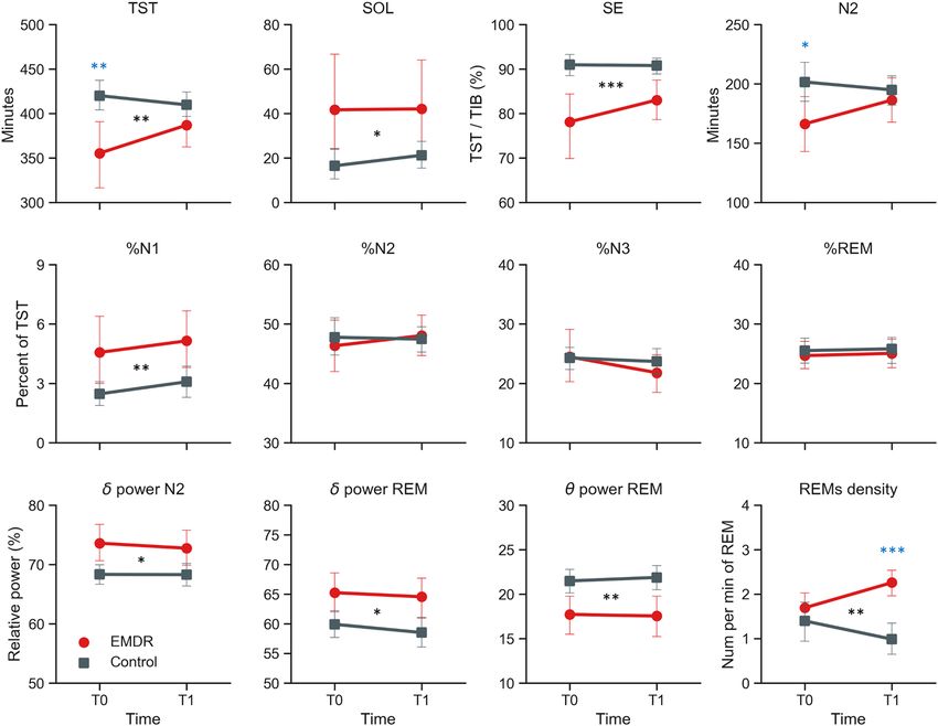
gated whether the number of EMDR sessions in the patient group was predicted by sleep parameters at baseline.
We found significant correlations between the number of EMDR sessions required to reach remission and the
following sleep parameters, the duration and percentage of REM sleep (duration: r = 0.51, p = 0.04; percentage:
r = 0.615, p = 0.01), the number and density of awakenings (number: r = 0.68, p = 0.004; density: r = 0.78, p < 0.001),
and the density of alpha bursts during REM sleep (r = − 0.58, p = 0.01). With the exception of the latter, higher
values for these sleep parameters before therapy predicted a higher number of EMDR sessions required to reach
remission (see Fig. 2 and Table S2). Of note, only the density of awakenings remained significant after applying
a Holm-Bonferroni correction for multiple comparisons.
Scientific Reports | (2021) 11:8873 | https://doi.org/10.1038/s41598-021-88337-x 6
Vol:.(1234567890)www.nature.com/scientificreports/
Group Control EMDR ANOVA (F-values)
Time T0 T1 T0 T1 Group Time Interaction
TST 419.98 ± 38.0 409.63 ± 30.6 355.31 ± 83.5 386.88 ± 52.7 11.62** 1.19 5.08*
SPT 442.12 ± 35.4 424.63 ± 34.3 387.22 ± 99.3 412.09 ± 47.3 5.3* 0.18 4.11
SOL 16.45 ± 16.4 21.18 ± 13.6 41.62 ± 44.0 42.03 ± 41.5 5.43* 0.79 0.05
WASO 15.60 ± 22.0 10.37 ± 9.2 24.62 ± 21.4 18.47 ± 13.7 3.29 3.02 0.2
SE 90.94 ± 6.0 90.76 ± 4.2 78.11 ± 15.1 83.01 ± 9.3 15.25*** 1.22 1.83
SME 95.05 ± 5.2 96.54 ± 2.3 92.54 ± 6.5 93.79 ± 5.1 4.84* 1.72 0.02
N1 10.22 ± 5.5 12.68 ± 7.3 16.47 ± 14.8 19.94 ± 11.9 7.19* 1.63 0.12
N2 201.50 ± 40.0 194.87 ± 30.1 166.19 ± 52.6 186.25 ± 39.8 3.44 1.39 5.29*
N3 101.65 ± 19.4 96.18 ± 17.6 83.50 ± 26.4 83.31 ± 23.6 7.55** 2.72 0.78
REM 106.60 ± 21.3 105.89 ± 22.8 89.16 ± 28.9 97.38 ± 26.4 4.58* 0.86 0.87
%N1 2.47 ± 1.4 3.09 ± 1.8 4.56 ± 3.6 5.14 ± 3.0 9.38** 1.24 0.0
%N2 47.76 ± 7.1 47.44 ± 5.4 46.32 ± 9.0 48.04 ± 7.5 0.0 0.8 1.56
%N3 24.27 ± 4.3 23.67 ± 5.0 24.45 ± 9.5 21.78 ± 6.5 0.54 4.57* 1.85
%REM 25.50 ± 5.2 25.81 ± 4.8 24.67 ± 4.8 25.04 ± 5.2 0.35 0.22 0.01
REM Latency 93.75 ± 35.8 78.71 ± 23.5 103.56 ± 51.5 101.88 ± 55.0 2.39 0.99 0.64
Lights out to N2 17.98 ± 17.1 22.95 ± 13.6 43.16 ± 43.6 43.94 ± 41.2 5.48* 1.06 0.04
Awakenings, number 5.55 ± 4.0 5.68 ± 5.1 8.19 ± 5.5 9.06 ± 5.3 6.41* 0.31 0.0
Awakenings, number per hour
0.75 ± 0.5 0.79 ± 0.7 1.19 ± 0.6 1.34 ± 0.8 9.99** 0.46 0.01
of SPT
Stage shifts, number 55.70 ± 17.4 59.47 ± 16.4 69.19 ± 28.1 77.75 ± 23.7 8.12** 1.46 0.17
Stage shifts, number per hour
7.58 ± 2.3 8.37 ± 1.9 10.69 ± 2.8 11.29 ± 3.1 23.37*** 1.08 0.15
of SPT
REM periods, number 3.90 ± 0.7 3.95 ± 0.7 3.38 ± 1.3 3.75 ± 1.0 2.19 1.97 1.6
REM periods, duration 29.49 ± 6.3 29.73 ± 7.4 32.74 ± 14.7 29.92 ± 10.5 0.43 0.41 0.9
Relative delta power N3 0.87 ± 0.0 0.86 ± 0.0 0.87 ± 0.1 0.86 ± 0.1 0.0 0.0 1.07
Relative delta power N2 0.68 ± 0.0 0.68 ± 0.0 0.74 ± 0.1 0.73 ± 0.1 6.96* 0.0 0.35
Relative delta power REM 0.60 ± 0.1 0.58 ± 0.1 0.65 ± 0.1 0.65 ± 0.1 6.57* 1.71 0.51
Relative sigma power N2 0.05 ± 0.0 0.04 ± 0.0 0.04 ± 0.0 0.04 ± 0.0 2.01 0.0 1.42
Relative theta power REM 0.21 ± 0.0 0.22 ± 0.0 0.18 ± 0.0 0.18 ± 0.0 9.34** 0.0 0.7
Spindles, number 942.55 ± 445.4 878.11 ± 339.6 726.56 ± 382.3 607.88 ± 366.3 3.67 3.79 0.01
Spindles, duration 0.90 ± 0.1 0.90 ± 0.1 0.91 ± 0.1 0.90 ± 0.1 0.06 0.0 1.71
Spindles, frequency 13.64 ± 0.3 13.68 ± 0.3 13.77 ± 0.5 13.80 ± 0.5 0.92 0.84 0.03
Spindles, density 3.11 ± 1.3 3.02 ± 1.1 2.82 ± 1.2 2.22 ± 1.3 1.54 8.69** 3.52
REM alpha bursts, number 82.45 ± 63.2 114.32 ± 109.3 113.19 ± 103.4 161.62 ± 147.4 1.22 4.15* 0.34
REM alpha bursts, density 0.78 ± 0.5 1.06 ± 1.0 1.45 ± 1.5 1.84 ± 1.7 3.73 2.21 0.1
REMs, number (average) 133.00 ± 74.9 110.07 ± 98.0 154.42 ± 73.3 219.04 ± 81.1 6.37* 0.71 5.23*
REMs, density (average) 1.39 ± 0.9 0.98 ± 0.7 1.69 ± 0.6 2.26 ± 0.5 12.16** 0.02 8.26**
Table 2. Sleep parameters. Significant F-values are in bold. TST total sleep time, SPT sleep period time,
SOL sleep onset latency, WASO wake after sleep onset, SE sleep efficiency, SME sleep maintenance efficiency.
*p < 0.05, **p < 0.01, ***p < 0.001.
Figure 2. Significant correlations between number of EMDR sessions before remission and sleep parameters
before therapy. *p < 0.05, ***p < 0.001.
Scientific Reports | (2021) 11:8873 | https://doi.org/10.1038/s41598-021-88337-x 7
Vol.:(0123456789)www.nature.com/scientificreports/
We also explored whether the number of EMDR sessions required to reach remission was correlated with
sleep parameters after treatment (Table S2). We found that the TST (r = 0.56, p = 0.02), SPT (0.61, p = 0.01) and
duration of N2 sleep (0.56, 0.02) after treatment were all significantly positively correlated with the number of
EMDR sessions; though none remained significant after Holm-Bonferroni correction.
Finally, we investigated whether the changes in sleep parameters from T0 to T1 were associated with better
or worse improvements of PTSD symptoms from T0 to T1. Using double-difference correlations, we found that
the participants with the strongest increase in REMs density from T0 to T1 were also the ones with the strongest
decrease in depression symptoms measured on the BDI scale (r = − 0.67, p = 0.018). Due to the large number
of combinations between sleep parameters and clinical outcomes, this correlation was however not significant
after adjusting for multiple comparison. Noteworthy, the above correlation was the only one that was significant
(uncorrected) among all the combinations.
Discussion
We examined the associations between PTSD symptoms remission and sleep parameters in military-related
PTSD subjects, and further compared them to a group of healthy military service members. In support of
our hypothesis, symptoms remission improved both subjective and objective sleep outcomes. We showed that
symptom remission after EMDR therapy was associated with significantly increased REMs number and density
in REM sleep (Table 2). To note some expected effects did not survive correction for multiple comparisons: TST
and N2 stage duration were increased at T1, and an increased REMs density between T0 and T1 predicted a
greater decrease of depression symptoms on the BDI scale. We also observed a significant group effect on several
sleep parameters which suggests that the symptoms remission did not completely normalize sleep in the patient
group. Finally, we report that some objective sleep measures at T0 are predictive of the number of EMDR therapy
sessions required to reach symptoms remission. The number and density of awakenings during SPT (as well
as the duration and percentage of REM sleep before treatment, even though not significant after correction for
multiple comparisons) was indeed significantly correlated with the number of EMDR sessions needed to reach
remission. Specifically, the number of EMDR sessions required for symptoms remission was positively correlated
with more awakenings (and more REM sleep, but it did not survive after correction for multiple comparisons)
before treatment (Fig. 2 and Table S2).
One week before therapy onset, sleep parameters of PTSD patients were consistent with previous literature
on sleep disturbances in PTSD, i.e. shorter TST and N2 sleep, as well as lower subjective sleep quality compared
to control individuals. After remission from PTSD symptoms with EMDR therapy, both TST and duration of
N2 sleep were no longer significantly different between groups, suggesting a normalization of sleep duration
in patients (specifically N2 sleep) and REMs density was significantly increased. These findings are consistent
with a recent study on written narrative exposure therapy83. In this study, which had no control group, PTSD
participants showed longer total sleep time, higher percentage of N2 sleep and higher REMs density after therapy
compared to before therapy. However, caution must be exercised when comparing this latter study to the present
one since they measured the PSG sleep parameters on the night following therapy, and not one week later as in
our study. Our results are also consistent with a previous study that showed an improvement of objective sleep
measures in non-military-related PTSD patients treated with EMDR t herapy43. The sleep parameters that were
improved following therapy are however different in the two studies since Raboni et al. reported only a decreased
WASO after treatment (here also suggesting a normalization of sleep). A possible explanation for the results
discrepancy between studies is the relatively small sample size. More surprisingly however, Raboni et al.43 also
reported a non-significant higher REMs density before treatment in PTSD patients compared to the control
group (Cohen d = 1.05), with little or no between-group difference after treatment (d = 0.16) due to reduction of
REM density in PTSD patients, whereas we observed an evolution in the opposite direction i.e. a significantly
higher REMs density in the PTSD group compared to the control group after treatment, with no between-group
differences before treatment. Our result is in line with the higher REMs density in PTSD patients following writ-
ten narrative exposure therapy observed by Kobayashi et al.83. However, they also reported that increased REM
density was negatively associated with PTSD symptom reduction, while we observed the opposite correlation.
These differences of results may be due to the small sample size and to the control groups which may vary in their
matching with the patients’ characteristics (i.e. age, profession) according to the different studies.
We also found significant group effects for a large number of objective sleep parameters, which are all in the
direction of poorer sleep quality in PTSD patients than in the control group (e.g. lower efficiency and N3 sleep
duration, higher percentage of N1 sleep, longer sleep latency, higher number of awakenings and stage transi-
tions). In other words, even if remission was associated with some improvements in TST and N2 sleep, PTSD
patients did not reach a normalized sleep even when symptoms disappeared, which contrasts with the strong
improvement in subjective sleep quality.
Exposure to trauma is associated with an integration of distressing and emotionally charged experiences into
one’s autobiographical memory. In PTSD, this lack of integration and depotentiation could be associated with an
improper and non-adaptive over-consolidation of traumatic e xperiences84 underlying the intrusive and distress-
ing nature of traumatic memories. It is now well-established that sleep is essential for memory consolidation
and emotional regulation of past e vents85. Over the years, accumulated evidence has suggested that REM sleep
and NREM sleep may serve distinct and complementary functions in memory c onsolidation86. REM sleep has
consistently been shown to support the specific consolidation of emotionally-salient memories87–90. By contrast,
NREM sleep is thought to support consolidation of non-emotional episodic memory91. Specifically, during
NREM sleep, relevant new memories are thought to be integrated and reorganized with respect to already existing
experiences7. In other words, the restoration of N2 sleep after symptoms remission suggested by several studies
Scientific Reports | (2021) 11:8873 | https://doi.org/10.1038/s41598-021-88337-x 8
Vol:.(1234567890)www.nature.com/scientificreports/
comprising ours, could restore the ability to integrate the traumatic event into autobiographical memory and
thus prevent intrusions of traumatic memories.
On the other hand, REM sleep is thought to be important for depotentiating the affective tone of emotional
memories92. Specifically, emotional memories are thought to be reactivated during REM sleep by amygdala
reactivations, causing a strengthening of the declarative, informational content of the memory and a decrease of
the emotional reactivity to this memory90,92–99. In line with this, the emotional intensity of waking life memories
is attenuated in dreams in healthy subjects93,96 but not in PTSD patients for the traumatic event that caused the
disorder. In this study, we found that symptoms remission using EMDR increased the density of REMs in REM
sleep. Such increase in REMs density may favour traumatic memory integration and depotentiation during
sleep as the eye movements of the EMDR therapy may favor this process at wake55. Building on the fact that
eye movements in both REM sleep and wakefulness activate similar cortical areas100, Stickgold proposed that
eyes movements could be the process by which emotional depotentiation happens both during REM sleep and
EMDR therapy. According to this hypothesis, the alternating bilateral stimuli of EMDR would activate brain
mechanisms that shift the brain into a memory processing mode similar to that of REM sleep. This REM-like
state would permit the integration of traumatic memories into associative cortical networks without interference
from hippocampus-mediated episodic recall. Our results are in line with this hypothesis, notably the correlation
(non-significant after correction for multiple comparison) showing that the increased REMs density between T0
and T1 predicted a greater decrease of symptoms on the BDI scale.
Based on these findings and theoretical insights, we therefore propose that the to-be confirmed increase
in duration in N2 sleep after symptoms remission could reflect the restored ability of PTSD patients to defend
against traumatic memory intrusions, whereas the increase in REMs density could reflect a REM-related regula-
tion mechanism rebound to decrease the emotional load of the traumatic memory.
Another important finding of this study was the positive correlation between the number of EMDR sessions
needed to achieve remission in the PTSD group and sleep parameters before therapy, including a positive cor-
relation with the number and density of intra-sleep awakenings (and also the percentage of REM sleep that did
not survive after correction for multiple comparisons) (Fig. 2). In other words, PTSD patients with higher density
of intra-sleep awakenings and possibly longer REM sleep at baseline were also the ones that needed the more
EMDR sessions to reach symptom remission. This finding may indicate that having a great amount of intra-sleep
awakenings (and possibly REM sleep) before treatment is maladaptive and/or reflect a failure of REM-sleep emo-
tional regulation mechanisms. Intra-sleep awakenings are known to be positively correlated with dream recall
frequency81,101 which is correlated with nightmares frequency102. Patients with the more intra-sleep awakenings
may thus also be the ones with the more nightmare recall, and intra-sleep awakenings may interrupt the regula-
tion process during REM sleep. Bilateral alternating stimulation (BAS) during EMDR therapy could substitute
this maladaptive or lack of emotional regulation process normally at play during REM sleep and participate in
the emotional memory depotentiation and reconsolidation, via notably mental travel in t ime86,103,104, which also
happens during dreaming since very old memories can be part of the dream s cenery93,105.
In conclusion, our results show that remission from military-related PTSD (with EMDR therapy) is associated
with an improvement of some sleep parameters, notably an increase of REMs density in REM sleep and possibly
TST and N2 durations. As it is argued in the literature that sleep-related memory consolidation may depend on
several inter-dependant sleep stages86, it may be the case that remission in PTSD patients is explained by the
combination of an increase in N2 duration and REMs density in REM sleep. The increase in REMs density may
require improved N2 to be efficient. Future studies are warranted to further test this hypothesis and to improve
our understanding of the neurophysiological mechanism responsible for symptom disparition in PTSD patients
treated by EMDR therapy.
This is important to note that this study has some limitations. The first limitation is that the control group
consisted of healthy individuals without PTSD. Our healthy control group has the advantage of providing us
with normative sleep data in healthy subjects from the same profession, thus sharing the same environment
and constraints as our patients. One of its limitations is that the mean age of the EMDR group was higher than
the one of the control group by an average of seven years, which may have biased group-comparison of sleep
duration and quality, both known to decrease with age106. Indeed, some of the above-described interactions
and group effects were no longer significant when adjusting for age. Future studies with larger sample sizes and
age-matched groups are therefore needed to replicate our findings. Another limitation of the study is related
to the absence of a wait-list control group to ensure that the simple passage of time does not explain the effects
observed, and of a PTSD group treated with another therapy to assess the specificity of the effect observed with
the EMDR therapy. Note however that the correlations between sleep parameters and the number of EMDR
sessions needed to reach remission speak in favor of the specificity of the EMDR therapy. Importantly, although
the study design does not allow to establish whether the sleep changes observed are related to therapy vs the
mere passage of time, it is unlikely that the passage of time explains our results. Indeed, according to the DSM5
criteria, PTSD symptoms have to be present for more than one month for the disorder to be actually classified as
DSM5. And regarding more specifically the sleep symptoms, a study showed in 1995 that 59 to 73% of Vietnam
veterans suffering from PTSD still had sleep disturbances such as insomnia and nonrestorative sleep i.e. in these
patients the sleep disturbances had lasted at least for 20 years32. Finally, it has been frequently observed in PTSD
patients that sleep disturbances do not improve after otherwise successful first-line PTSD treatment24,26 and dis-
turbed sleep is one of the two most reported residual s ymptoms26. In line with these clinical observations, from a
biological point of view numerous studies have demonstrated the chronic nature of sleep disorders in PTSD. In
2019 Colvonen et al.107, demonstrated that sleep disturbances in 40 veterans with PTSD were unremitting without
direct intervention, after a 3 months follow-up. In other words, some further studies are needed to confirm our
results and verify the specific effect of EMDR therapy on sleep parameters by using a wait-list control group, a
Scientific Reports | (2021) 11:8873 | https://doi.org/10.1038/s41598-021-88337-x 9
Vol.:(0123456789)www.nature.com/scientificreports/
non-PTSD-dedicated therapy or care, and another PTSD-dedicated therapy as exposure therapy or cognitive
behavioral therapy trauma focused.
Another limitation is that our two groups were exclusively composed of male soldiers—mainly for practical
reasons (as explained in the Methods). Our findings should therefore not be generalized to female soldiers and/
or non-military populations. PTSD patients under psychotropic medication were included in the analysis if they
were taking SSRIs only if the medication had stayed consistent for at least three months prior to and throughout
the trial. Recent meta-analyses showed that SSRIs may increase REM latency, suppress REM sleep and impair
sleep continuity108,109. At inclusion, however, the intensity of symptoms measured by the CAPS or the duration
of REM did not differ between individuals taking SSRIs and those without psychotropic drugs.
Finally, the polysomnography recording devices were different between our two groups, and this may have
artificially driven group differences in EEG-based spectral power and/or EOG-based REMs number and density.
We cannot exclude that some of the group differences observed in sleep parameters could be due to be different
recording setting and experimental procedure for controls and patients, i.e. in-lab PSG with a constrained time
in bed for the control group, versus at-home ambulatory PSG with no time constraint for the patient group.
However, this limitation cannot explain the time effect found in patients.
In summary, our findings show that remission from military-related PTSD with EMDR therapy is associated
with an increase in REMs density during REM sleep and possibly with a normalization of total sleep duration,
mostly driven by an increase of N2 sleep. However, a wait-list group and another therapy PTSD therapy group
is needed to confirm these results, and the specificity of the EMDR therapy on these effects. Furthermore, we
show that the number and density of intra-sleep awakenings before treatment are both predictive of the number
of EMDR sessions required to reach remission.
Data availability
Data is available on request from the corresponding author.
Received: 18 September 2019; Accepted: 6 April 2021
References
1. Baglioni, C. et al. Sleep and mental disorders: A meta-analysis of polysomnographic research. Psychol. Bull. 142, 969–990 (2016).
2. Ohayon, M. M. & Shapiro, C. M. Sleep disturbances and psychiatric disorders associated with posttraumatic stress disorder in
the general population. Compr. Psychiatry 41, 469–478 (2000).
3. Roszell, D. K., McFall, M. E. & Malas, K. L. Frequency of symptoms and concurrent psychiatric disorder in Vietnam veterans
with chronic PTSD. Hosp. Community Psychiatry 42, 293–296 (1991).
4. Talbot, L. S. et al. Cognitive behavioral therapy for insomnia in posttraumatic stress disorder: A randomized controlled trial.
Sleep 37, 327–341 (2014).
5. Germain, A., Buysse, D. J., Shear, M. K., Fayyad, R. & Austin, C. Clinical correlates of poor sleep quality in posttraumatic stress
disorder. J. Trauma. Stress 17, 477–484 (2004).
6. McLay, R. N., Klam, W. P. & Volkert, S. L. Insomnia is the most commonly reported symptom and predicts other symptoms of
post-traumatic stress disorder in U.S. service members returning from military deployments. Mil. Med. 175, 759–762 (2010).
7. Germain, A., Buysse, D. J. & Nofzinger, E. Sleep-specific mechanisms underlying posttraumatic stress disorder: Integrative
review and neurobiological hypotheses. Sleep Med. Rev. 12, 185–195 (2008).
8. Breslau, N., Roth, T., Rosenthal, L. & Andreski, P. Sleep disturbance and psychiatric disorders: A longitudinal epidemiological
study of young adults. Biol. Psychiatry 39, 411–418 (1996).
9. Chang, P. P., Ford, D. E., Mead, L. A., Cooper-Patrick, L. & Klag, M. J. Insomnia in young men and subsequent depression: The
Johns Hopkins Precursors Study. Am. J. Epidemiol. 146, 105–114 (1997).
10. Ford, D. E. & Kamerow, D. B. Epidemiologic study of sleep disturbances and psychiatric disorders: An opportunity for preven-
tion?. JAMA 262, 1479–1484 (1989).
11. Livingston, G., Blizard, B. & Mann, A. Does sleep disturbance predict depression in elderly people? A study in inner London.
Br. J. Gen. Pract. J. R. Coll. Gen. Pract. 43, 445–448 (1993).
12. Krakow, B. et al. Sleep disorder, depression, and suicidality in female sexual assault survivors. Crisis 21, 163–170 (2000).
13. Krakow, B. et al. Sleep-disordered breathing, psychiatric distress, and quality of life impairment in sexual assault survivors. J.
Nerv. Ment. Dis. 190, 442–452 (2002).
14. DeViva, J. C., Zayfert, C. & Mellman, T. A. Factors associated with insomnia among civilians seeking treatment for PTSD: An
exploratory study. Behav. Sleep. Med. 2, 162–176 (2004).
15. Neylan, T. C. et al. Sleep disturbances in the Vietnam generation: findings from a nationally representative sample of male
Vietnam veterans. Am. J. Psychiatry 155, 929–933 (1998).
16. Nishith, P., Resick, P. A. & Mueser, K. T. Sleep difficulties and alcohol use motives in female rape victims with posttraumatic
stress disorder. J. Trauma. Stress 14, 469–479 (2001).
17. Cover, H. & Irwin, M. Immunity and depression: Insomnia, retardation, and reduction of natural killer cell activity. J. Behav.
Med. 17, 217–223 (1994).
18. Kuppermann, M. et al. Sleep problems and their correlates in a working population. J. Gen. Intern. Med. 10, 25–32 (1995).
19. Moul, D. E. et al. Symptom reports in severe chronic insomnia. Sleep 25, 553–563 (2002).
20. Reimer, M. A. & Flemons, W. W. Quality of life in sleep disorders. Sleep Med. Rev. 7, 335–349 (2003).
21. Riedel, B. W. & Lichstein, K. L. Strategies for evaluating adherence to sleep restriction treatment for insomnia. Behav. Res. Ther.
39, 201–212 (2001).
22. Rosenthal, L. D. & Meixner, R. M. Psychological status and levels of sleepiness-alertness among patients with insomnia. CNS
Spectr. 8, 114–118 (2003).
23. Ohayon, M. M., Caulet, M., Philip, P., Guilleminault, C. & Priest, R. G. How sleep and mental disorders are related to complaints
of daytime sleepiness. Arch. Intern. Med. 157, 2645–2652 (1997).
24. Belleville, G., Guay, S. & Marchand, A. Persistence of sleep disturbances following cognitive-behavior therapy for posttraumatic
stress disorder. J. Psychosom. Res. 70, 318–327 (2011).
25. DeViva, J. C., Zayfert, C., Pigeon, W. R. & Mellman, T. A. Treatment of residual insomnia after CBT for PTSD: Case studies. J.
Trauma. Stress 18, 155–159 (2005).
Scientific Reports | (2021) 11:8873 | https://doi.org/10.1038/s41598-021-88337-x 10
Vol:.(1234567890)You can also read