Soft Tissue Reconstruction of Parotidectomy Defect - BINASSS
←
→
Page content transcription
If your browser does not render page correctly, please read the page content below
Soft Tissue Reconstruction
o f P a ro t i d e c t o m y D e f e c t
Jennifer Moy,     MD,   Mark K. Wax,         MD, FRCS(C),   Myriam Loyo,         MD, MCR*

 KEYWORDS
  Parotidectomy  Reconstruction  Fat graft  Alloderm  Frey syndrome  Free flap

 KEY POINTS
  Parotidectomy creates a soft tissue defect for which reconstruction will improve both
   facial contour and patient satisfaction.
  Postoperative Frey syndrome (gustatory sweating) can be prevented with soft tissue
   reconstruction by creating a barrier to aberrant reinnervation of the cheek skin.
  Acellular dermis, autologous fat transfer with/without dermis, and local/regional flaps are
   used for reconstruction for most defects of the parotid bed.
  For extensive composite large volume or surface area defects, consider free tissue
   transfer.

INTRODUCTION

Surgical resection of parotid tumors leads to a loss in lateral facial volume, resulting in
a noticeable facial deformity with asymmetry. This defect can vary from a small pos-
terior mandibular depression to significant facial concavity. In larger composite de-
fects, the loss of volume may cause inferior displacement and medial rotation of the
auricle. In a study of patients having undergone parotidectomy, 70% reported a
change in appearance, with greater than half reporting a noticeable depression.
Furthermore, casual observers notice this contour defect.1,2 Facial appearance and
deformity is a critical aspect affecting quality of life and carries strong social pen-
alties.3 Multiple studies have shown facial deformities affect attractiveness, self-
esteem, academic and occupational satisfaction, income, and quality of life. Because
patients may be concerned regarding facial contour after parotidectomy, soft tissue
reconstruction can normalize facial appearance and decrease the psychosocial im-
pacts with an overall increase in patient satisfaction.4,5
   In addition to restoring facial contour, postoperative Frey syndrome can be pre-
vented with soft tissue reconstruction. Frey syndrome, or gustatory sweating, is the
postparotidectomy phenomenon where sweating occurs in the skin of the cheek while
eating. It is attributed to aberrant reinnervation of parotid parasympathetic nerve fibers

 Department of Otolaryngology/Head and Neck Surgery at Oregon Health & Science University,
 3181 SW Sam Jackson Park Road, PV01, Portland, OR 97239, USA
 * Corresponding author.
 E-mail address: loyo@ohsu.edu

 Otolaryngol Clin N Am 54 (2021) 567–581
 https://doi.org/10.1016/j.otc.2021.02.009                                                 oto.theclinics.com
 0030-6665/21/ª 2021 Elsevier Inc. All rights reserved.

        Descargado para BINASSS BINASSS (pedidos@binasss.sa.cr) en National Library of Health and Social
          Security de ClinicalKey.es por Elsevier en junio 08, 2021. Para uso personal exclusivamente. No se
         permiten otros usos sin autorización. Copyright ©2021. Elsevier Inc. Todos los derechos reservados.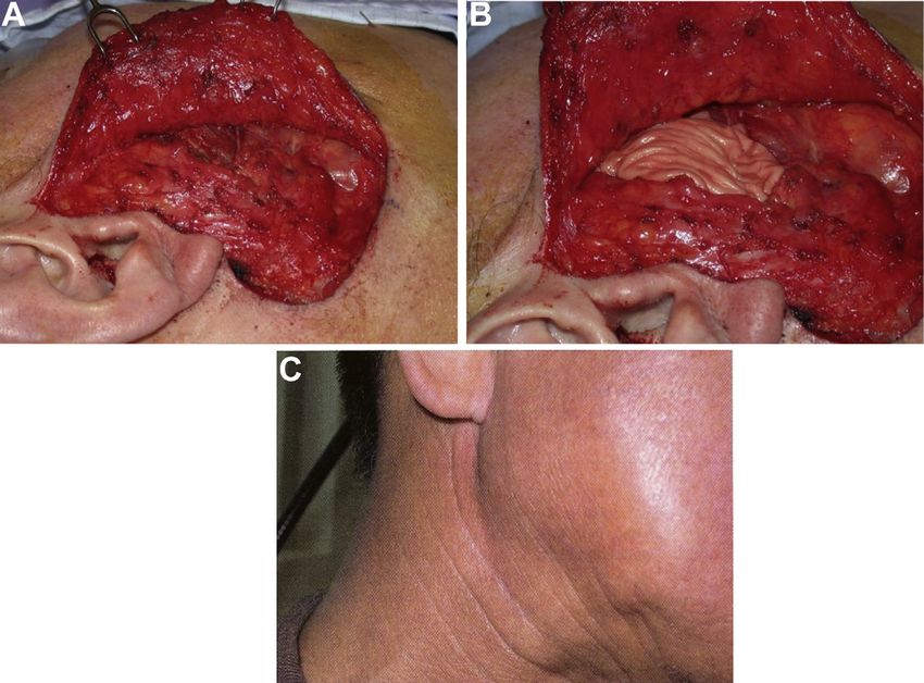
568 Moy et al
to the sympathetic fibers in the skin of the cheek. The manifestations can develop
years after parotidectomy. Although this phenomenon is reported by 38% of patients,
objective testing for Frey syndrome using the minor starch-iodine test shows that up to
96% of patients have this phenomenon after parotidectomy without reconstruction.6
Surgical reconstruction can prevent Frey syndrome by creating a barrier between
the parasympathetic nerves of the parotid bed and the overlying skin. This barrier
can be created with local flaps, fat grafts, acellular dermal matrix, or free tissue
transfer.
This chapter discusses soft tissue reconstruction options for the parotidectomy
defect, including wound healing, Frey syndrome, and tumor surveillance.
SKIN INCISION
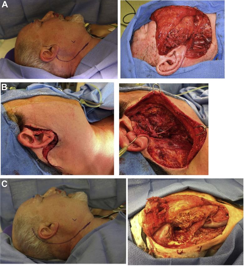
The modified Blair incision (Fig. 1A) is widely used as an approach for parotid surgery.
A reliable incision camouflages well and provides good exposure of the mastoid tip,
Fig. 1. Skin incisions. (A) Modified Blair incision; (B) modified facelift incision; (C) postauric-
ular incision.
Descargado para BINASSS BINASSS (pedidos@binasss.sa.cr) en National Library of Health and Social
Security de ClinicalKey.es por Elsevier en junio 08, 2021. Para uso personal exclusivamente. No se
permiten otros usos sin autorización. Copyright ©2021. Elsevier Inc. Todos los derechos reservados.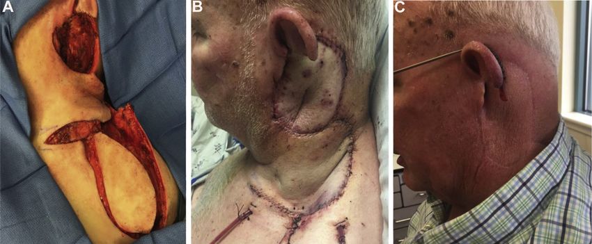
Soft Tissue Reconstruction of Parotidectomy Defect                 569

sternocleidomastoid and posterior digastric belly muscles, and the entire parotid
gland. Alternatively, the modified facelift incision (Fig. 1B) obviates the neck incision
yet provides similar exposure. The modified facelift incision is associated with
improved patient satisfaction without an increase in surgical time or complications.7,8
Caution should be taken in large, anterior tumors, where retrograde dissection of the
facial nerve may be required, as this approach may not provide safe exposure.7 The
contraindications described in the literature are tumors with parapharyngeal space
extension, recurrent tumors, and arteriovenous malformation.9
  When a lateral or subtotal temporal bone resection with or without auriculectomy is
performed, a postauricular incision [Fig. 1C] is made and carried anteroinferiorly to a
neck skin crease.

Reconstructive Options
The volume of removed parotid tissue, the individual patient’s facial anatomy, and the
patient’s desires and values should be considered in choosing a reconstruction tech-
nique. Reconstructive options range from primary closure, fat or acellular implants to
locoregional flaps, and free tissue transfer. Table 1 summarizes each reconstructive
option, with indications, advantages, disadvantages, and surgical tips.
   Primary closure is an excellent option for most superficial parotidectomy defects
with minimal-to-no skin loss. Wide undermining of the soft tissue deep to the superfi-
cial musculoaponeurotic system (SMAS) allows plication and prevents excessive ten-
sion on the skin edges. The scalp does not offer the same laxity as the face and neck
for assisting in primary closure. However, galeal-releasing incisions made at 1-cm in-
tervals can provide some additional reach while maintaining adequate blood supply to
the skin edge. A tension-free repair is especially important in an irradiated field where
there is a propensity for wound healing complications.
   Acellular dermal implants, such as AlloDerm (LifeCell Corporation, Branchburg, New
Jersey) and DermaMatrix (Synthes Corporation, Westchester, Pennsylvania), are off-
the-shelf and ready-to-use sheets derived from human cadaveric skin (Fig. 2).
Because of their availability, simplicity, and lack of donor site morbidity, acellular
dermal implants are a favored reconstructive option for limited parotidectomy defects,
despite concerns for these materials increasing the risk of seroma and sialocele.10 A
meta-analysis of 5 clinical controlled studies showed acellular dermal implants signif-
icantly reduced the rate of Frey syndrome and salivary leaks without an increase in
wound complications.11 Use of a single sheet allows for the best healing potential,
whereas using more than one sheet can increase local wound complications, limiting
the degree of contour improvement that can be achieved. Although contour improve-
ment is underreported, a randomized controlled trial of 36 patients undergoing paro-
tidectomy compared acellular dermis with free fat grafting. This study showed free fat
reconstruction resulted in better aesthetic outcomes, and lower cost and complication
rates,12 suggesting a limitation in the degree of augmentation attainable with Allo-
Derm. AlloDerm has been more widely used for parotidectomy reconstruction than
DermaMatrix. Limited studies directly comparing these materials are available.10
   Fat grafting is a popular reconstructive option for parotidectomy defects, given its
simplicity, limited donor morbidity, and generally satisfactory outcomes. Fat grafting
improves facial symmetry and reduces the incidence of Frey syndrome with better pa-
tient satisfaction scores.4,13–15 This technique has been used in tumors ranging in size
from less than 1 to 7 cm and with volumes of up to 70 cm.3,4,14,16,17 Fat grafts are har-
vested either as a free graft or as a dermal graft from the abdomen or thigh. Free fat
grafts are typically harvested from the periumbilical fat, through a well-hidden incision
in the lower half of the umbilicus.4 Harvesting a single generous piece of abdominal fat

        Descargado para BINASSS BINASSS (pedidos@binasss.sa.cr) en National Library of Health and Social
          Security de ClinicalKey.es por Elsevier en junio 08, 2021. Para uso personal exclusivamente. No se
         permiten otros usos sin autorización. Copyright ©2021. Elsevier Inc. Todos los derechos reservados.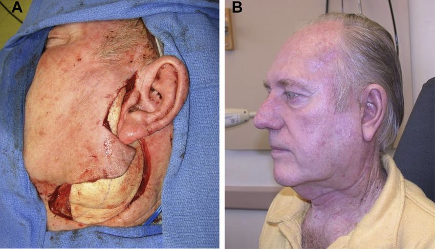
570
Moy et al
Table 1
Descargado para BINASSS BINASSS (pedidos@binasss.sa.cr) en National Library of Health and Social
Pros and cons of reconstructive option
permiten otros usos sin autorización. Copyright ©2021. Elsevier Inc. Todos los derechos reservados.
Security de ClinicalKey.es por Elsevier en junio 08, 2021. Para uso personal exclusivamente. No se
Indicated Defect Advantages Disadvantages Surgical Tips
Primary closure Superficial parotidectomies, No donor morbidity, short OR Only for small defects, Wide undermining, SMAS
tail of parotid time wound healing concerns plicating, and galeal
for postradiated wounds releasing incisions can help
create a tension-free skin
closure
Acellular dermal implant Superficial parotidectomies, No donor morbidity, short OR Cost Limit to 1 sheet of implant to
tail of parotid time prevent complications
Fat graft Superficial parotidectomies, Low-donor site morbidity, Risk of fat necrosis, infection, Harvest as a single-large
tail of parotid, deep lobe similar consistency to and fat reabsorption piece; overcorrect 10%–
parotid tissue, limited 30% in anticipation of
additional operative time atrophy, consider a dermal
fat graft to reduce atrophy
Local and regional grafts Deep/total parotidectomy, Excellent skin match Reach can be limited, wound Inferiorly based flaps require
skin defects healing concerns in strategically placed tacking
postirradiated fields sutures to prevent flap
ptosis and dehiscence
Cervicofacial rotational flap Skin defects Excellent skin match with low Lacks tridimensional volume Can extend onto chest to
donor site morbidity restoration gain additional reach
Temporalis muscle/ Superior defects without skin Short OR time Donor site defect, limited
temporoparietal fascia flap involvement reach inferiorly, relies on
intact superficial temporal
vessels, risk to frontal
branch of the facial nerve
Sternomastoid myofacial flap Tail of parotid, mastoid Minimal morbidity, similar Limited reach, added risk to Use caution with concurrent
defects intraoperative exposure, CN XI neck dissection to prevent
short OR time loss of blood supply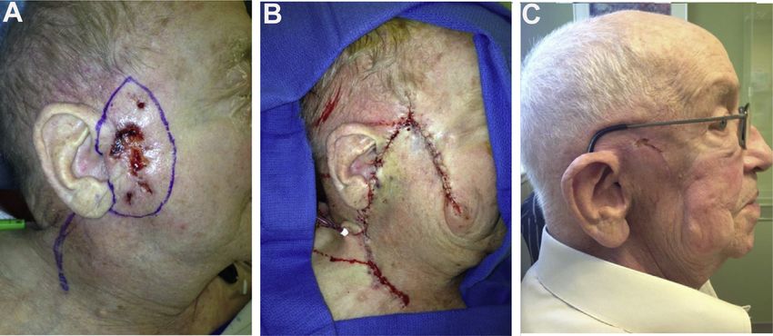
Pectoralis myofacial or Deep/total parotidectomy, Reliable flap with good bulk Donor site morbidity Place superiorly placed
myocutaneous flap skin defects tacking sutures to prevent
ptosis, can add length by
Descargado para BINASSS BINASSS (pedidos@binasss.sa.cr) en National Library of Health and Social
dissecting muscle away
permiten otros usos sin autorización. Copyright ©2021. Elsevier Inc. Todos los derechos reservados.
Security de ClinicalKey.es por Elsevier en junio 08, 2021. Para uso personal exclusivamente. No se
from pedicle as muscle
heads to humerus. Most
common with
auriculectomies
Latissimus dorsi myofascial or Deep/total parotidectomy, Large, thin muscle that can Poor skin match, added OR Place superiorly placed
myocutaneous flap skin defects be contoured in defect, time tacking sutures to prevent
low donor site morbidity ptosis. Most commonly
used with auriculectomies
Keystone island flap Deep/total parotidectomy, Excellent skin match with low Lacks tridimensional volume Can be harvested
skin defects donor site morbidity restoration posterosuperiorly based on
Soft Tissue Reconstruction of Parotidectomy Defect
occipital and posterior
auricular perforators,
anterioinferiorly based on
the facial or submental
perforators, or inferiorly
based on the transverse
cervical or superficial
cervical arteries
Submental island flap Deep/total parotidectomy, Excellent skin match with low Requires intact facial vessels, Harvest with underlying
skin defects donor site morbidity limited bulk muscles for added strength
and bulk
Supraclavicular island flap Deep/total parotidectomy, Excellent skin match with low Can be folded on itself to Most common complication
skin defects donor site morbidity provide adequate bulk is wound dehiscence,
therefore placating deep
dermal sutures to ensure a
tension-free skin closure is
imperative
(continued on next page)
571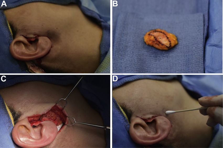
572
                                                                                                        Table 1
                                                                                                        (continued )

                                                                                                                                                                                                                                                                      Moy et al
                                                                                                                                       Indicated Defect             Advantages                       Disadvantages                   Surgical Tips
Descargado para BINASSS BINASSS (pedidos@binasss.sa.cr) en National Library of Health and Social
 permiten otros usos sin autorización. Copyright ©2021. Elsevier Inc. Todos los derechos reservados.
  Security de ClinicalKey.es por Elsevier en junio 08, 2021. Para uso personal exclusivamente. No se

                                                                                                        Free tissue transfer           Total parotidectomy, skin    Highly vascularized to           Increased OR time, donor site   Can be deepithelalized and
                                                                                                                                         defects, chemo/radiation     withstand chemotherapy           morbidity                       buried for bulk
                                                                                                                                                                      and radiation
                                                                                                        Radial forearm                 Skin defect, with minimal    Long pedicle length, reliable,   Lacks bulk, donor site          Can be deepithelalized and
                                                                                                                                         volume loss                  no atrophy                       morbidity, requires skin        buried for bulk or by
                                                                                                                                                                                                       graft                           including upper forearm
                                                                                                                                                                                                                                       subcutaneous fat
                                                                                                        Anterolateral thigh            Total parotidectomy, skin    Excellent bulk that can be       Short pedicle length, can be    Ability to include fascia lata
                                                                                                                                         defects, chemo/radiation     modified to accommodate          too bulky in overweight/       for static facial
                                                                                                                                                                      defect and cover vital           obese patients                 reanimation and/or vastus
                                                                                                                                                                      structures, relatively low                                      lateralis with its motor
                                                                                                                                                                      morbidity                                                       nerve to be grafted for
                                                                                                                                                                                                                                      dynamic facial reanimation

                                                                                                       Abbreviations: CN, cranial nerve; OR, operating room.
Soft Tissue Reconstruction of Parotidectomy Defect 573
Fig. 2. Dermal fat grafting. (A) Retromandibular concavity following parotidectomy; (B)
dermal fat graft, with deepithelialized dermis outlined in black ink; (C) inset of fat graft;
(D) improvement of contour defect following dermal fat graft.
is preferred over multiple small grafts, as it minimizes graft trauma and devasculariza-
tion, which may lead to increased graft necrosis and loss. Dermal fat grafts (Fig. 3)
theoretically support vascularization of the fat through the subdermal plexus. Dermal
fat grafts are harvested from the lower abdomen with a Pfannenstiel incision below the
bikini line.13,18,19 One advantage of a dermal fat graft is that the subdermal tissue can
Fig. 3. MRI postparotidectomy with dermal fat graft. Right parotid gland has normal
appearance. The left side is post–total parotidectomy with mastoidectomy and dermal fat
grafting for recurrent pleomorphic adenoma. (A) Axial T1-weighted without contrast; (B)
axial T1-weighted with fat suppression. Residual disease is easily delineated from surround-
ing fat graft (arrow).
Descargado para BINASSS BINASSS (pedidos@binasss.sa.cr) en National Library of Health and Social
Security de ClinicalKey.es por Elsevier en junio 08, 2021. Para uso personal exclusivamente. No se
permiten otros usos sin autorización. Copyright ©2021. Elsevier Inc. Todos los derechos reservados.574 Moy et al
readily be sutured to allow for better positioning of the graft in the defect. High patient
and surgeon satisfaction with the reconstructive contour and the donor site have been
reported for both techniques of fat grafting16,17,19,20; however, studies directly
comparing free fat grafts with dermal fat grafts are not available.
Local wound complications after fat graft reconstruction of the parotidectomy are
rare. Hematomas, seromas, and wound infections can occur at both the donor and
the reconstructive sites.4,16,17 Fat necrosis can lead to infection, which may improve
with antibiotics and local wound care. In more severe cases, fat necrosis may require
graft removal. Although rare, epithelial cysts can develop in dermal fat grafts if the
epidermis is not carefully removed.19 The primary downside of fat grafting is variable
reabsorption over time. Most surgeons recommend overcorrecting volume loss by
10% to 30%.4,12,17,18,21 Although the fat graft stabilizes after 6 months, it can be
debulked if overcorrection persists.16 Some investigators speculate using a SMAS
flap can improve the viability of the fat graft and decrease resorption17; however,
studies that objectively measure graft survival over time are lacking. A limited case se-
ries reviewed 5 cases of postparotidectomy/fat graft MRI showing stable graft volume
1 to 3 years after implantation.22
Historically, there has been concern that reconstructive techniques can obstruct the
ability to assess tumor recurrence.14 However, advanced imaging techniques,
computed tomography, MRI, and PET imaging can reliably delineate between normal
reconstructive and parotid tissues from tumor recurrence (Fig. 4).4,22 Although some
investigators advocate waiting 6 months to 2 years,19 several have published suc-
cessful use of fat grafting in parotid malignancy reconstruction.4,14,17
Fig. 4. Alloderm reconstruction. (A) Anterior parotid defect; (B) alloderm implant into
defect, (C) postoperative outcome with lack of facial concavity.
Descargado para BINASSS BINASSS (pedidos@binasss.sa.cr) en National Library of Health and Social
Security de ClinicalKey.es por Elsevier en junio 08, 2021. Para uso personal exclusivamente. No se
permiten otros usos sin autorización. Copyright ©2021. Elsevier Inc. Todos los derechos reservados.Soft Tissue Reconstruction of Parotidectomy Defect 575
Local and Regional Flaps
Local and regional flaps, including myofascial, myocutaneous, and fasciocutaneous
flaps, are particularly useful in small to moderate defects. In cases in which skin resto-
ration is necessary, they provide excellent skin color match with lower distant
morbidity. They are well vascularized, allowing longer random pattern paddles with
smaller pedicles than would be allowed elsewhere in the body. However, they do
require incision planning with the ablative surgeons and often lack adequate bulk to
provide good facial contour in larger resections. Because of decreased vasculariza-
tion and fibrosis, rotational flaps are not ideal for moderate to large defects in postir-
radiated patients.
The SMAS can be used as a local muscle flap to assist in volume replacement and
reduction of Frey syndrome incidence. When the SMAS is mobilized, advanced, and
plicated posteriorly into the defect, it can be used for volume restoration. However,
facial asymmetry may result from significant unilateral plication, necessitating contra-
lateral SMAS rhytidectomy.23 Because of its thin nature and limited volume, the SMAS
may not be available in all cases, especially in superficial tumors where resection of
this layer is required for adequate margins.
The cervicofacial rotation flap can cover large skin defects with excellent skin color
match (Fig. 5). Although it lacks tridimensionality, a concurrent muscle flap, such as
temporalis, sternocleidomastoid (SCM), or pectoralis can provide this bulk. The reli-
ability of this flap is compromised by prior radiation, smoking, and sacrifice of the
facial artery during resection or neck dissection. Because of its caudally based
vascular supply, there is reduced survival and increased dehiscence above the
zygoma. However, extension of the incision onto the chest improves reach and
vascularity.
By extending the incision into the temporal region, the temporalis muscle and/or
temporoparietal fascia (TPF) flap can be used as a rotational flap to provide soft tissue
bulk to the parotid defect. Many argue against the use of the temporalis muscle flap for
contour, as it results in temporal “hollowing,” thus creating one defect to fix another. In
addition, the temporalis muscle flap has limited reach beyond the mastoid.24
Conversely, the TPF flap is thin, with excellent pliability that can provide coverage
of the entire parotid bed, including the retromandibular area.25 Although it has excel-
lent vascularity, damage to the superficial temporal vessels during parotidectomy can
Fig. 5. Cervicofacial rotation flap. (A) Cutaneous carcinoma with planned incision drawn; (B)
postablation with cervicofacial rotation flap; (C) six-month follow-up.
Descargado para BINASSS BINASSS (pedidos@binasss.sa.cr) en National Library of Health and Social
Security de ClinicalKey.es por Elsevier en junio 08, 2021. Para uso personal exclusivamente. No se
permiten otros usos sin autorización. Copyright ©2021. Elsevier Inc. Todos los derechos reservados.576 Moy et al
compromise the flap viability. Although these flaps have been shown to reduce the
incidence of Frey syndrome to 8% from 43%, they have added risk to the frontal
branch of the facial nerve, alopecia, hematoma, and increased operative time with
less than optimal cosmetic reconstruction.25
The SCM, pectoralis, and latissimus muscle flaps can also be used as local rota-
tional flaps to provide soft tissue restoration for the parotid defect. The SCM flap is
harvested through the parotidectomy incision with improved bulk compared with
the SMAS flap.26 Several studies have failed to show improved facial contour after
SCM flap,26,27 whereas other studies have shown some degree of improvement.28,29
The SCM flap can be harvested superiorly or inferiorly. A superiorly based flap is har-
vested from the superior third of the SCM, freed from the inferior attachment and
rotated 180 around its anterior margin and sutured to the remnant parotid fascia or
periosteum of the zygoma. The inferiorly based SCM flap is incised superiorly and
anteriorly, then rotated into inferior and midparotid defects. The SCM flap has the po-
tential to create a depression in the donor site and expose the spinal accessory nerve
to damage.28 An additional consideration in patients undergoing concurrent neck
dissection is disrupting the tenuous blood supply to the SCM.
The pectoralis and latissimus flaps can be harvested as myocutaneous flaps to pro-
vide both bulk and cutaneous reconstruction if needed. These muscle flaps are usually
reserved for parotidectomy defects with auriculectomy. The pectoralis flap carries sig-
nificant donor site morbidity with poor contouring capability. The latissimus can be
contoured more easily with the laxity of the posterior back skin allowing nearly all
wounds to be closed primarily with minimal morbidity. When using inferiorly based
rotational flaps, superior tacking sutures are important to prevent descent and
dehiscence.
There has been great success in the use of various island flaps for the reconstruc-
tion of the parotidectomy defect. These include the keystone, submental, and supra-
clavicular island flaps (Fig. 6). Although they have excellent skin color match and low
donor site morbidity, they lack 3-dimensional bulk for large contour defects.30–32 Care
must be taken in patients who have received preoperative radiation, concurrent neck
dissection, or anticipate postoperative radiation.32
Free Tissue Transfer
In patients with large volume composite defects, recurrent tumor, dural defects, and
prior irradiated fields or when postoperative radiation is anticipated, free tissue trans-
fer is the most appropriate reconstructive option. Free flaps offer flexibility in size, bulk,
reliability, and resilience. There are many donor site options for free flaps. The choice
depends on the cutaneous tissue loss, contour defect, or exposed vital structures. The
use of a deepithelialized buried free flap has been used with great success, with good
facial contour, and vascularized soft tissue coverage of vital structures.33
The radial forearm free flap (RFFF) is a reliable flap with a long vascular pedicle that
offers significant surface area coverage. It is limited by its modest bulk, depending on
the habitus of the patient; thus, it is best suited for smaller defects with auricular pres-
ervation. However, additional bulk can be achieved if the upper forearm subcutane-
ous tissue is concurrently harvested. With deepithelialization, the flap can be
placed within the parotid bed as a buried flap for contouring if there is no significant
skin defect. The thin quality of the flap and lack of considerable change over time (ie
muscle atrophy) obviates flap revision. However, closure of the donor site requires an
additional skin graft, contributing to the morbidity in this flap. High-volume composite
defects are best reconstructed with flaps containing muscle or a thick, soft tissue
component.
Descargado para BINASSS BINASSS (pedidos@binasss.sa.cr) en National Library of Health and Social
Security de ClinicalKey.es por Elsevier en junio 08, 2021. Para uso personal exclusivamente. No se
permiten otros usos sin autorización. Copyright ©2021. Elsevier Inc. Todos los derechos reservados.Soft Tissue Reconstruction of Parotidectomy Defect 577
Fig. 6. Supraclavicular island flap. (A) Defect with supraclavicular island flap incised; (B) immediate postoperative reconstruction; (C) six-month
follow-up.
Descargado para BINASSS BINASSS (pedidos@binasss.sa.cr) en National Library of Health and Social
Security de ClinicalKey.es por Elsevier en junio 08, 2021. Para uso personal exclusivamente. No se
permiten otros usos sin autorización. Copyright ©2021. Elsevier Inc. Todos los derechos reservados.578 Moy et al
Fig. 7. Deepithelialized buried anterolateral thigh free flap. (A) Flap placed within parotid
defect before deepithelialization; (B) six-month follow-up.
The anterolateral thigh (ALT) free flap is the ideal reconstructive option for large vol-
ume composite defects, especially with significant cutaneous loss and or lateral skull
base resections with or without dural defects. The pedicle is based off the lateral
circumflex femoral artery and can be up to 12 cm long. The skin paddle is limited to
8 to 12 cm in width to accommodate primary closure of the thigh, whereas the bulk
of this flap largely depends on the gender and habitus of the patient. However, there
are many options to choose from in the ALT flap in order to obtain ideal bulk, contour,
and skin reconstruction. It can be harvested as a myofascial flap, to provide bulk, or a
myocutaneous or fasciocutaneous flap to provide bulk and skin coverage. The ALT
flap can be further deepithealialized for placement as a buried flap, if little to no skin
defect is created (Fig. 7). Although this flap carries excellent resilience through post-
operative radiation,33 it has drawbacks such as the need for frequent debulking pro-
cedures, increased donor site seromas, and wound dehiscence when compared with
the RFFF.34,35
SUMMARY
Soft tissue reconstruction of the parotidectomy defect can vary from primary closure
to acellular dermal implants or fat grafts to local, regional, or free flaps. Many studies
have shown improved facial contour, reduced incidence of Frey syndrome, and
improved patient satisfaction with soft tissue reconstruction. Each reconstructive op-
tion carries specific indications, advantages, and risk. Patient anatomy and desires
should be considered when selecting a reconstruction option.
CLINICS CARE POINTS
Most parotidectomy defects can be successfully reconstructed with acellular matrix, fat
grafts, or locoregional flaps with excellent patient satisfaction, reduced Frey syndrome,
and minimal donor site morbidity.
Descargado para BINASSS BINASSS (pedidos@binasss.sa.cr) en National Library of Health and Social
Security de ClinicalKey.es por Elsevier en junio 08, 2021. Para uso personal exclusivamente. No se
permiten otros usos sin autorización. Copyright ©2021. Elsevier Inc. Todos los derechos reservados.Soft Tissue Reconstruction of Parotidectomy Defect 579
Fat grafts have a propensity to resorb, especially after radiation. Overcorrection, avoiding
piece-meal harvesting, and/or using dermal fat or a local flap concurrently may reduce this
phenomenon.
Ensure to place superiorly placed tacking sutures on inferiorly based rotational flaps to
prevent descent and dehiscence.
Free tissue transfer is a superior option in large composite resections and when adjuvant
radiation is expected.
DISCLOSURE
The authors have no disclosures.
REFERENCES
1. Nitzan D, Kronenberg J, Horowitz Z, et al. Quality of life following parotidectomy
for malignant and benign disease. Plast Reconstr Surg 2004;114(5):1060–7.
2. Ciuman RR, Oels W, Jaussi R, et al. Outcome, general, and symptom-specific quality
of life after various types of parotid resection. Laryngoscope 2012;122(6):1254–61.
3. Dey JK, Ishii M, Boahene KD, et al. Impact of facial defect reconstruction on
attractiveness and negative facial perception. Laryngoscope 2015;125(6):
1316–21.
4. Conger BT, Gourin CG. Free abdominal fat transfer for reconstruction of the total
parotidectomy defect. Laryngoscope 2008;118(7):1186–90.
5. Dey JK, Ishii LE, Byrne PJ, et al. The social penalty of facial lesions: new evi-
dence supporting high-quality reconstruction. JAMA Facial Plast Surg 2015;
17(2):90–6.
6. Linder TE, Huber A, Schmid S. Frey’s syndrome after parotidectomy: a retrospec-
tive and prospective analysis. Laryngoscope 1997;107(11 Pt 1):1496–501.
7. Grover N, D’Souza A. Facelift approach for parotidectomy: an evolving aesthetic
technique. Otolaryngol Head Neck Surg 2013;148(4):548–56.
8. Bianchi B, Ferri A, Ferrari S, et al. Improving esthetic results in benign parotid sur-
gery: statistical evaluation of facelift approach, sternocleidomastoid flap, and su-
perficial musculoaponeurotic system flap application. J Oral Maxillofac Surg
2011;69(4):1235–41.
9. Terris DJ, Tuffo KM, Fee WE Jr. Modified facelift incision for parotidectomy.
J Laryngol Otol 1994;108(7):574–8.
10. Athavale SM, Phillips S, Mangus B, et al. Complications of alloderm and derma-
matrix for parotidectomy reconstruction. Head Neck 2012;34(1):88–93.
11. Zeng XT, Tang XJ, Wang XJ, et al. AlloDerm implants for prevention of Frey syn-
drome after parotidectomy: a systematic review and meta-analysis. Mol Med Rep
2012;5(4):974–80.
12. Wang S, Li L, Chen J, et al. Effects of free fat grafting on the prevention of Frey’s
syndrome and facial depression after parotidectomy: a prospective randomized
trial. Laryngoscope 2016;126(4):815–9.
13. Harada T, Inoue T, Harashina T, et al. Dermis-fat graft after parotidectomy to pre-
vent Frey’s syndrome and the concave deformity. Ann Plast Surg 1993;31(5):
450–2.
14. Curry JM, Fisher KW, Heffelfinger RN, et al. Superficial musculoaponeurotic sys-
tem elevation and fat graft reconstruction after superficial parotidectomy. Laryn-
goscope 2008;118(2):210–5.
Descargado para BINASSS BINASSS (pedidos@binasss.sa.cr) en National Library of Health and Social
Security de ClinicalKey.es por Elsevier en junio 08, 2021. Para uso personal exclusivamente. No se
permiten otros usos sin autorización. Copyright ©2021. Elsevier Inc. Todos los derechos reservados.580 Moy et al
15. Curry JM, King N, Reiter D, et al. Meta-analysis of surgical techniques for
preventing parotidectomy sequelae. Arch Facial Plast Surg 2009;11(5):
327–31.
16. Loyo M, Gourin CG. Free abdominal fat transfer for partial and total parotidec-
tomy defect reconstruction. Laryngoscope 2016;126(12):2694–8.
17. Ambro BT, Goodstein LA, Morales RE, et al. Evaluation of superficial musculoa-
poneurotic system flap and fat graft outcomes for benign and malignant parotid
disease. Otolaryngol Head Neck Surg 2013;148(6):949–54.
18. Chan LS, Barakate MS, Havas TE. Free fat grafting in superficial parotid surgery
to prevent Frey’s syndrome and improve aesthetic outcome. J Laryngol Otol
2014;128(Suppl 1):S44–9.
19. Davis RE, Guida RA, Cook TA. Autologous free dermal fat graft. Reconstruction
of facial contour defects. Arch Otolaryngol Head Neck Surg 1995;121(1):
95–100.
20. Honeybrook A, Athavale SM, Rangarajan SV, et al. Free dermal fat graft recon-
struction of the head and neck: an alternate reconstructive option. Am J Otolar-
yngol 2017;38(3):291–6.
21. Militsakh ON, Sanderson JA, Lin D, et al. Rehabilitation of a parotidectomy pa-
tient–a systematic approach. Head Neck 2013;35(9):1349–61.
22. Lee YJ, Fischbein NJ, Megwalu U, et al. Radiographic surveillance of abdominal
free fat graft in complex parotid pleomorphic adenomas: a case series. Heliyon
2020;6(5):e03894.
23. Cesteleyn L, Helman J, King S, et al. Temporoparietal fascia flaps and superficial
musculoaponeurotic system plication in parotid surgery reduces Frey’s syn-
drome. J Oral Maxillofac Surg 2002;60(11):1284–97, discussion 1297-1288.
24. Chen J, Lin F, Liu Z, et al. Pedicled temporalis muscle flap stuffing after a lateral
temporal bone resection for treating mastoid osteoradionecrosis. Otolaryngol
Head Neck Surg 2017;156(4):622–6.
25. Movassaghi K, Lewis M, Shahzad F, et al. Optimizing the aesthetic result of pa-
rotidectomy with a facelift incision and temporoparietal fascia flap. Plast Reconstr
Surg Glob Open 2019;7(2):e2067.
26. Asal K, Koybasioglu A, Inal E, et al. Sternocleidomastoid muscle flap reconstruc-
tion during parotidectomy to prevent Frey’s syndrome and facial contour defor-
mity. Ear Nose Throat J 2005;84(3):173–6.
27. Gooden EA, Gullane PJ, Irish J, et al. Role of the sternocleidomastoid muscle flap
preventing Frey’s syndrome and maintaining facial contour following superficial
parotidectomy. J Otolaryngol 2001;30(2):98–101.
28. Kerawala CJ, McAloney N, Stassen LF. Prospective randomised trial of the ben-
efits of a sternocleidomastoid flap after superficial parotidectomy. Br J Oral Max-
illofac Surg 2002;40(6):468–72.
29. Fee WE Jr, Tran LE. Functional outcome after total parotidectomy reconstruction.
Laryngoscope 2004;114(2):223–6.
30. Bayon R, Davis AB. Submental flap for soft tissue reconstruction following radical
parotidectomy. Otolaryngol Head Neck Surg 2019;160(6):1130–2.
31. Emerick KS, Herr MW, Lin DT, et al. Supraclavicular artery island flap for recon-
struction of complex parotidectomy, lateral skull base, and total auriculectomy
defects. JAMA Otolaryngol Head Neck Surg 2014;140(9):861–6.
32. Behan FC, Lo CH, Sizeland A, et al. Keystone island flap reconstruction of parotid
defects. Plast Reconstr Surg 2012;130(1):36e–41e.
Descargado para BINASSS BINASSS (pedidos@binasss.sa.cr) en National Library of Health and Social
Security de ClinicalKey.es por Elsevier en junio 08, 2021. Para uso personal exclusivamente. No se
permiten otros usos sin autorización. Copyright ©2021. Elsevier Inc. Todos los derechos reservados.Soft Tissue Reconstruction of Parotidectomy Defect 581
33. Cannady SB, Seth R, Fritz MA, et al. Total parotidectomy defect reconstruc-
tion using the buried free flap. Otolaryngol Head Neck Surg 2010;143(5):
637–43.
34. Thompson NJ, Roche JP, Schularick NM, et al. Reconstruction outcomes
following lateral skull base resection. Otol Neurotol 2017;38(2):264–71.
35. Cigna E, Minni A, Barbaro M, et al. An experience on primary thinning and sec-
ondary debulking of anterolateral thigh flap in head and neck reconstruction. Eur
Rev Med Pharmacol Sci 2012;16(8):1095–101.
Descargado para BINASSS BINASSS (pedidos@binasss.sa.cr) en National Library of Health and Social
Security de ClinicalKey.es por Elsevier en junio 08, 2021. Para uso personal exclusivamente. No se
permiten otros usos sin autorización. Copyright ©2021. Elsevier Inc. Todos los derechos reservados.You can also read