StainGAN: Stain Style Transfer for Digital Histological Images
←
→
Page content transcription
If your browser does not render page correctly, please read the page content below
StainGAN: Stain Style Transfer for Digital
Histological Images
M Tarek Shaban1 , Christoph Baur1 , Nassir Navab1,2 , and Shadi Albarqouni1
1
Computer Aided Medical Procedures (CAMP), Technische Universität München,
Munich, Germany
2
Whiting School of Engineering, Johns Hopkins University, Baltimore, USA
arXiv:1804.01601v1 [cs.CV] 4 Apr 2018
first.last@tum.de
Abstract. Digitized Histological diagnosis is in increasing demand. How-
ever, color variations due to various factors are imposing obstacles to the
diagnosis process. The problem of stain color variations is a well-defined
problem with many proposed solutions. Most of these solutions are highly
dependent on a reference template slide. We propose a deep-learning so-
lution inspired by CycleGANs that is trained end-to-end, eliminating the
need for an expert to pick a representative reference slide. Our approach
showed superior results quantitatively and qualitatively against the state
of the art methods (10% improvement visually using SSIM). We further
validated our method on a clinical use-case, namely Breast Cancer tumor
classification, showing 12% increase in AUC. Code will be made publicly
available.
Keywords: Histology Images, Stain Normalization, Generative Adver-
sarial Networks, Deep Learning.
1 Introduction
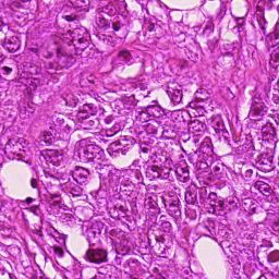
Histology relies on the study of microscopic images to diagnose disease based
on the cell structures and arrangements. Staining is a crucial part of the tissue
preparation process. The addition of staining components (mainly Hematoxylin
and Eosin) transforms the naturally transparent tissue elements to become more
distinguishable ( hematoxylin dyes the nuclei a dark purple color and the eosin
dyes other structures a pink color). However, results from the staining process are
inconsistent and prone to variability; due to differences in raw materials, staining
protocols across different pathology labs, inter-patient variabilities, and slide
scanner variations as shown in 1. These variations not only cause inconsistencies
within pathologists [7] but it also hinders the performance of Computer-Aided
Diagnosis (CAD) systems [5].
Stain normalization algorithms have been introduced to overcome this well-
defined problem of stain color variations. These methods can be broadly classified
into three classes, Color matching based methods that try to match the
color spectrum of the image to that of the reference template image. Reinhard
et al.[11] align the color-channels to match that of the reference image in the
2                               M.T. Shaban, C. Baur, N. Navab, S. Albarqouni

lab color model, However, this can lead to improper color mapping, as the same
transformation is applied across the images and does not take into account the
independent contribution of stain dyes to the final color.
    Further, there are Stain-separation methods where normalization is done
on each staining channel independently. For instance, Macenko et al. [10] find
the stain vectors by transforming the RGB to the Optical Density (OD) space.
On the other hand, the method proposed by Khan et al. [9] estimate the stain
matrix based on a color-based classifier that assigns every pixel to the appropri-
ate stain component. According to Babak et al [2], these methods do not take
the spatial features of the tissue structure into account, which leads to improper
staining. Nevertheless, most of the methods in this class rely on an expertly
picked reference template image, which has a major effect on the outcome of
the methods as we show later. The third class of methods is the Pure learning
based approaches, that handle the problem of stain normalization as a style-
transfer problem, BenTaieb et al.[3] handle the problem using auxiliary classifier
GAN with an auxiliary task on top (classifier or segmentation). Our method
(StainGAN) is also GAN based but as opposed to BenTaieb does not require to
be trained for a specific task in order to achieve stain style transfer.
    We propose a method based on Generative adversarial networks (GANs) that
not only eliminates the need for the reference image but also achieves high visual
similarity to the target domain, making it easier to get rid of the stain varia-
tions thus improving the diagnosis process for both the pathologist and CAD
systems. StainGAN is based on the Unpaired Image-to-Image Translation us-
ing Cycle-Consistent Adversarial Networks (CycleGAN)[16]. Cycle-consistency
allows the images to be mapped to different color-model but preserves the same
tissue structure. We evaluate our approach against the state of the art methods
quantitatively and indirectly through a breast tumor classification. Our contribu-
tions include 1) Using Style-transfer for the classic stain normalization problem
and achieving better results quantitatively than the state of the art methods.
2) Removing the need for a manually picked reference template, as our model
learn the whole distribution. 3) Providing a benchmark for comprehensive com-
parison between the proposed StainGAN method and most of the state of the
art methods.

2   Methodology

Our framework, as depicted in Fig. 2, employs the CycleGAN concept [16] to
transfer the H&E Stain Appearance between different scanners, i.e from Hama-
matsu (H) to Aperio (A) Scanner, without the need of paired data from both do-
mains. The model consists of two generator and discriminator pairs, the first pair
(GH and DH ), tries to map images from domain A to domain H GH : XA → XH .
While the Generator GH tries to generate images that match domain H, the dis-
criminator DH tries to verify if images come from the real domain H or the fake
generated ones. The other pair (GA and DA ), undergoes the same process in the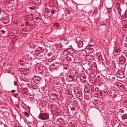
StainGAN: Stain Style Transfer for Digital Histological Images         3

      (a) Source           (b) Target         (c) Reinhard        (d) Macenko

      (e) Khan           (f) Vahadane        (g) StainGAN

Fig. 1: Stain Normalization of various methods, the goal is to match the Target
image.

reverse direction, GA : XH → XA , as

          X̂H = GH (XA ; θH ), X̂A = GA (X̂H ; θA ), s.t. d(XA , X̂A ) ≤ ,         (1)

and
          X̂A = GA (XH ; θA ), X̂H = GH (X̂A ; θH ), s.t. d(XH , X̂H ) ≤ ,         (2)
where d(·, ·) is a distance metric between the given image and the reconstructed
one, so-called a Cycle-Consistency constraint, and both θA and θH are the model
parameters.
    To achieve this, the models are trained to meet the following objective func-
tion,
                               L = LAdv + λLCycle ,                           (3)
where LAdv is the adversarial loss, LCycle is the cycle-consistency loss, and λ is
a regularization parameter.
    The adversarial loss tries to match the distribution of the generated images
to that of the target domain (Forward Cycle), and match the distribution of
the generated target domain back to the source domain (Backward Cycle) as
LAdv = LA          H              A
          GAN + LGAN , where LGAN is given as

   LA
    GAN (GA , DA , XH , XA ) = EXA ∼pdata (XA ) [log DA (XA )]
                                                                                    (4)
                                + EXH ∼pdata (XH ) [log(1 − DA (GA (XH ; θA ))].

    The Cycle-consistency loss ensures that generated images preserve similar
structure as in the source domain. This loss goes in both directions forward and
4                                    M.T. Shaban, C. Baur, N. Navab, S. Albarqouni

Fig. 2: Our StainGAN framework: Images are mapped to domain H and then
back to domain A to ensure structure perseverance. Same process is done in
reverse direction from domain H to domain A.

backward cycles to assure stability.

    LCycle (GA , GH , XA , XH ) = EXA ∼pdata (XA ) [kGA (GH (XA ; θH ); θA )) − XA k1 ]
                                + EXH ∼pdata (XH ) [kGH (GA (XH ; θA ); θH )) − XH k1 ],
                                                                                     (5)

where k · k1 is the `1 -norm.

Network Architectures. In our model, ResNet [6] and 70 × 70 PatchGAN [8]
are employed as network architecture for the generators and the discriminators,
respectively.

3      Experiments and Results

To have a fair comprehensive comparison, we evaluate our model against the
state of the art methods of Reinhard[11] Macenko[10], Khan[9], and Vahadane[13]
as follows; i) Quantitatve comparison between the visual appearances of stained
images, and the effect of varying reference slides, ii) Quantitative comparison
of the Hematoxylin and Eosin stain vectors separation, and iii) Use-case ex-
periment, stain normalization as a preprocessing step in the pipeline of tumor
classifier.

3.1      Stain Transfer

The goal is to be able to map the patches from scanner A(Aperio) to that of
Scanner H(Hamamatsu), the dataset includes same tissue sections scanned with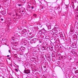
StainGAN: Stain Style Transfer for Digital Histological Images       5

both scanners, Slides from scanner A are normalized to match scanner H, then
compared with the real slides of Scanner H (ground truth). Results are evaluated
using various similarity matrices. Additionally, Stain Vectors are extracted using
Ruifrok’s method [12] and compared to that of the ground truth. Visual results
of the of the various methods are illustrated in 1. It is clear that our results are
more close visually to the target images.

Dataset. The dataset is publicly available as part of the MITOS-ATYPIA
14 challenge 3 . Dataset consists of 284 frames at X20 magnification which are
stained with standard Hematoxylin and Eosin (H&E) dyes. Same tissue section
has been scanned by two slide scanners: Aperio Scanscope XT and Hamamatsu
Nanozoomer 2.0-HT. Slides from both scanners resized to have equal dimensions
of (1539×1376). Rigid registration was performed to eliminate any misalignment.
For the training set, we extract 10,000 random patches from the first 184 full
slide of both scanners. For evaluation, 500 same view section patches of 256×256
were generated from each of the two scanners from the last 100 full slides (unseen
in the training set), patches from the Scanner H are used as the ground truth.

Implementation. For the state of the art methods, a refrence slide was picked
carefully. Our method does not require a reference slide as it learns the distribu-
tion of the whole data, model was trained using 10, 000 random unpaired patches
from both scanners for 26 epochs with the regularization parameter set to λ=
10, learning rate is set to 0.0002, Adam optimizer with a batch size of 4 is used.
Hardware of GeForce GTX TITAN X 12GB and Pytorch framework were used.

Evaluation Metrics. Results are compared to the ground truth using four
similarity measures: Structural Similarity index (SSIM)[14], Feature Similarity
Index for Image Quality Assessment (FSIM) [15], Peak Signal-to-Noise Ratio
(PSNR) and Pearson correlation coefficient similarity[1]. On the other hand,
Euclidian norm distance as reported in [2] was used to evaluate stain vectors
against the ground truth.

Results. As reported in Table 1, our results significantly outperform the state-
of-the-art methods in all similarity metrics (p < 0.01). Further, it has shown a
better stain seperability compared to the ground-truth stain vectors as shown
in Table. 2.

Slide Reference Sensitivity. We ran the same experiment as in the previous
one, but with three different reference images shown in changes of the SSIM
score with respect to the reference images, results are reported in Fig.3, which
shows that the conventional methods are sensitive to the reference image.

3
    https://mitos-atypia-14.grand-challenge.org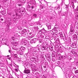
6                                   M.T. Shaban, C. Baur, N. Navab, S. Albarqouni

        Table 1: Stain Transfer Comparison: Mean ± Standard Deviation
Methods               SSIM            FSSIM        Pearson Correlation       PSNR
Reinhard [11]      0.58 ± 0.10      0.67 ± 0.05        0.51 ± 0.21         13.4 ± 1.61
Macenko [10]       0.56 ± 0.12      0.67 ± 0.05        0.45 ± 0.22         14.0 ± 1.68
Khan [9]           0.67 ± 0.11      0.71 ± 0.05        0.54 ± 0.20         16.3 ± 2.11
Vahadane [13]      0.65 ± 0.13      0.71 ± 0.06        0.53 ± 0.22         14.2 ± 2.13
StainGAN          0.71 ± 0.11      0.73 ± 0.06        0.56 ± 0.22         17.1 ± 2.50

Table 2: Stain Vectors Comparison: Mean ± Standard Deviation (the lower the
better) and Processing time taken to normalize the 500 images
              Methods            Staining Separation    Processing time
                                SH            SE          Time (sec)
              Reinhard [11] 20.7 ± 4.85   18.0 ± 3.79        9.80
              Macenko [10] 21.05 ± 4.11   12.2 ± 3.14        63.63
              Khan [9]      21.2 ± 3.65   12.0 ± 2.73      2196.25
              Vahadane [13] 20.0 ± 4.57   12.9 ± 3.45       582.94
              StainGAN      18.0± 4.84    10.0± 3.32         74.55

3.2    Use-Case Application

Stain-normalization is a mandatory preprocessing step in most CAD systems and
has proven to increase performance[5]. The aim of this experiment is to show the
performance of stain normalization in the context of breast cancer classification,
model is trained with patches from the lab 1, testing is done with patches from
lab 2 (different staining appearances). We normalize the test-set to match lab 1
and compare results accordingly.

Dataset. Camelyon16 challenge 4 consisting of 400 whole-slide images collected
in two different labs in Radboud University Medical Center (lab 1) and Univer-
sity Medical Center Utrecht (lab 2). Otsu thresholding was used to remove the
background, Afterwards, 40, 000 256 × 256 patches were generated on the x40
magnification level, 30, 000 were used for training and 10, 000 used for validation
from lab 1 and 10, 000 patches were generated for testing from lab 2.

Implementation. The classifier network is composed of three convolutional
layers with a ReLU activation, followed by a max-pooling layer turning the given
image into a logit vector. Classifier has been trained for 30 epochs with RMSprop
optimizer and binary cross-entropy loss. For the stain normalization, represen-
tative reference slide form lab 2 was picked for the conventional methods, Our
method was trained using the same parameters from the previous experiment
on training set: 68000 patches from both labs.
4
    https://camelyon16.grand-challenge.org/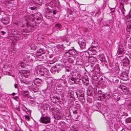
StainGAN: Stain Style Transfer for Digital Histological Images       7

                (a)                                      (b)

Fig. 3: (a) ROC curves of the test-set pre-processed using different stain normal-
ization methods. (b) Box plot shows the variation of the SSIM metric due to the
improper selection of reference slide.

Evaluation Metrics. To evaluate the classifier performance, we report the
Area Under the Curve (AUC) of the Receiver Operating Characteristics (ROC).

Results. The ROC curves and AUC of different classifiers, trained on different
stain normalization methods, are presented in Fig.3. Our proposed StainGAN
method shows a relative improvement of (80%, 36%, 22%, 15%, and 3%) on AUC
over the Un-normalized, Reinhard, Macenko, Khan, and Vahadane, respectively.

4   Discussion and Conclusion

In the paper, we presented StainGAN as a novel method for the stain normaliza-
tion task. Our experiments revealed that our method significantly outperforms
the state of the art. The visual appearance of different methods can be seen in
Fig.1. It clearly shows that images normalized with StainGAN are very similar
to the ground truth. Further, our StainGAN method has been validated in a
clinical use-case, namely Tumor Classification, as a pre-processing step showing
a superior performance. Moreover, the processing time of our method is on par
with Macenko as reported in Table. 2. We believe that end-to-end learning based
approaches are ought to overtake the classic stain normalization methods. Yet,
there is still more room for improvement, for example, we can think of Unified
representation, similar to [4], that can map many to many stain style domains.

Acknowledgment

This work was partially funded by the German Research Foundation (DFG, SFB
824), and Bavarian Research Foundation (BFS, IPN2).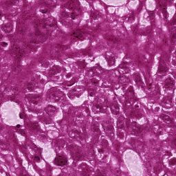
8                                  M.T. Shaban, C. Baur, N. Navab, S. Albarqouni

References
 1. Ahlgren, P., Jarneving, B., Rousseau, R.: Requirements for a cocitation similarity
    measure, with special reference to pearson’s correlation coefficient. Journal of the
    Association for Information Science and Technology 54(6), 550–560 (2003)
 2. Bejnordi, B.E., Litjens, G., Timofeeva, N., Otte-Höller, I., Homeyer, A., Karssemei-
    jer, N., van der Laak, J.A.: Stain specific standardization of whole-slide histopatho-
    logical images. IEEE transactions on medical imaging 35(2), 404–415 (2016)
 3. BenTaieb, A., Hamarneh, G.: Adversarial stain transfer for histopathology image
    analysis. IEEE Transactions on Medical Imaging (2017)
 4. Choi, Y., Choi, M., Kim, M., Ha, J.W., Kim, S., Choo, J.: Stargan: Unified gen-
    erative adversarial networks for multi-domain image-to-image translation. arXiv
    preprint arXiv:1711.09020 (2017)
 5. Ciompi, F., Geessink, O., Bejnordi, B.E., de Souza, G.S., Baidoshvili, A., Litjens,
    G., van Ginneken, B., Nagtegaal, I., van der Laak, J.: The importance of stain
    normalization in colorectal tissue classification with convolutional networks. arXiv
    preprint arXiv:1702.05931 (2017)
 6. He, K., Zhang, X., Ren, S., Sun, J.: Deep residual learning for image recognition. In:
    Proceedings of the IEEE conference on computer vision and pattern recognition.
    pp. 770–778 (2016)
 7. Ismail, S.M., Colclough, A.B., Dinnen, J.S., Eakins, D., Evans, D., Gradwell,
    E., O’sullivan, J.P., Summerell, J.M., Newcombe, R.G.: Observer variation in
    histopathological diagnosis and grading of cervical intraepithelial neoplasia. Bmj
    298(6675), 707–710 (1989)
 8. Isola, P., Zhu, J.Y., Zhou, T., Efros, A.A.: Image-to-image translation with condi-
    tional adversarial networks. arXiv preprint (2017)
 9. Khan, A.M., Rajpoot, N., Treanor, D., Magee, D.: A nonlinear mapping approach
    to stain normalization in digital histopathology images using image-specific color
    deconvolution. IEEE Transactions on Biomedical Engineering 61(6), 1729–1738
    (2014)
10. Macenko, M., Niethammer, M., Marron, J., Borland, D., Woosley, J.T., Guan, X.,
    Schmitt, C., Thomas, N.E.: A method for normalizing histology slides for quanti-
    tative analysis. In: Biomedical Imaging: From Nano to Macro, 2009. ISBI’09. IEEE
    International Symposium on. pp. 1107–1110. IEEE (2009)
11. Reinhard, E., Adhikhmin, M., Gooch, B., Shirley, P.: Color transfer between im-
    ages. IEEE Computer graphics and applications 21(5), 34–41 (2001)
12. Ruifrok, A.C., Johnston, D.A., et al.: Quantification of histochemical staining by
    color deconvolution. Analytical and quantitative cytology and histology 23(4), 291–
    299 (2001)
13. Vahadane, A., Peng, T., Sethi, A., Albarqouni, S., Wang, L., Baust, M., Steiger, K.,
    Schlitter, A.M., Esposito, I., Navab, N.: Structure-preserving color normalization
    and sparse stain separation for histological images. IEEE transactions on medical
    imaging 35(8), 1962–1971 (2016)
14. Wang, Z., Bovik, A.C., Sheikh, H.R., Simoncelli, E.P.: Image quality assessment:
    from error visibility to structural similarity. IEEE transactions on image processing
    13(4), 600–612 (2004)
15. Zhang, L., Zhang, L., Mou, X., Zhang, D.: Fsim: A feature similarity index for
    image quality assessment. IEEE transactions on Image Processing 20(8), 2378–
    2386 (2011)
16. Zhu, J.Y., Park, T., Isola, P., Efros, A.A.: Unpaired image-to-image translation us-
    ing cycle-consistent adversarial networks. arXiv preprint arXiv:1703.10593 (2017)

You can also read