The Development of a Free Radiological Anatomy Software Teaching Tool - SciELO
←
→
Page content transcription
If your browser does not render page correctly, please read the page content below
Int. J. Morphol.,
37(1):205-211, 2019.

                        The Development of a Free Radiological
                            Anatomy Software Teaching Tool

                             Desarrollo de un Software Libre de Anatomia Radiológica
                                        como una Herramienta de Enseñanza

     Marcus Oliveira1; Paulo Geambastiani2, 3 ; Guillermo Lopez1; Mateus Cambui1; Carlos Ubeda4 & Sibusiso Mdletshe5

OLIVEIRA, M.; GEAMBASTIANI, P.; LOPEZ, G.; CAMBUI, M.; UBEDA, C. & MDLETSHE, S. The development of a free
radiological anatomy software teaching tool. Int. J. Morphol., 37(1):205-211, 2019.

         SUMMARY: The purpose of this research was to develop a free radiological anatomy software for radiologic anatomy education
to assist students and professionals in health science. The study was divided into two phases: image acquisition and software development.
The first phase was to obtain plain radiographic images and computed tomographic (CT) scans of an anthropomorphic phantom of head and
neck. In addition, plain radiographic images of an anthropomorphic phantom of the chest were obtained. The second phase was the development
of the anatomy software as an ImageJ macro. The software was developed through the insertion of the radiologic anatomy landmarks into
the images that were obtained and application of multiple choice questions. The software was then tested for usability by getting the
professors to answer the multiple choice questions. The software presented radiologic anatomy from 1) Head projections: Waters view,
Towne view, Caldwell view, Lateral view, Submentovertex, PA view; 2) Thoracic Spine projections: AP and Lateral View and 3) Chest: PA
view, Lateral and Oblique. Tomographic imaging presented one hundred radiologic landmarks of head. In total, there were 354 questions.
A final report containing the score of correct answers, as well as the user ID, Date and Time of the test were showed. The test were available
in three languages (Spanish, English and Portuguese). A user-friendly and inexpensive software was developed and presented. Students and
professionals from several countries are able to practice, repeatedly, the recognition of radiologic anatomical landmarks.

        KEY WORDS: Anatomy; Radiology education; Education technology; Learning.

INTRODUCTION

        Radiography plays an important role in the health                radiography education. Radiography education in the
care services and interacts in a multidisciplinary and                   European community is organized in different ways ranging
interdisciplinary way with various professions (including                from no formal education program to university graduate
nursing and other medical professions). Obtaining skills to              and postgraduate courses. However, there is a great concern
understand radiography becomes an instrumental                           in standardizing the educational level, as well as in
competence, necessary and indispensable to the professional              accrediting training for radiography professionals (Prentakis
of radiography (Challen, 2010). Maintaining workforce                    et al., 2016).
capacity, whilst reacting to the latest clinical demands on
radiographer training, is a key responsibility of radiography            E-learning has been increased as teaching method since 2000,
educators (England et al., 2017).                                        and it has been suggested as an accessible high-quality
                                                                         education method (White & Cheung). In addition, it has been
       The European Federation of Radiographer Societies                 overcoming time and geographic limitations. Most
(EFRS), educational wing, strongly recommends the                        universities and education professionals are supporting this
dissemination and publication of materials and knowledge,                paradigm shift in education, including radiography
including the promotion and development of all levels of                 education.

1
  Department of Heath Technology and Biology, Federal Institute of Bahia, Salvador, Bahia, Brazil.
2
  Radiology Technologist, Cardio Pulmonar Hospital, Salvador, Bahia, Brazil.
3
  EBSERCH, Hospital Universitário Prof. Edgar Santos, Salvador, Bahia, Brazil.
4
  Medical Technology Department, Health Sciences Faculty, Universidad de Tarapacá, Arica, Chile.
5
  HOD: Medical Imaging and Radiation Sciences Department (MIRS),Teaching Advancement at University (TAU) Fellow, Faculty of Health Sciences,
  University of Johannesburg, Johannesburg, South Africa.

                                                                                                                                        205
OLIVEIRA, M.; GEAMBASTIANI, P.; LOPEZ, G.; CAMBUI, M.; UBEDA, C. & MDLETSHE, S. The development of a free radiological anatomy software teaching tool.
Int. J. Morphol., 37(1):205-211, 2019.
Over the last few years there has been a shift in MATERIAL AND METHOD
radiography education with a move to align to the
technological advancements and health education trends e.g.
the use of simulated learning. Among key shifts is the use of This study was conducted at the Federal Institute do
technology for teaching students within the radiography Bahia, Brazil, as a collaborative project between the research
curriculum which is critical because technology can reduce group of radiology technology and Hospital in Bahia, Brazil,
error rates while decreasing administration time and to design a tool for radiologic anatomy education to assist
increasing quality standards. Simultaneously, Anatomy radiographers/radiologic technologist students and
education is at the forefront of utilizing technological professionals.
advancements to increasingly develop learning
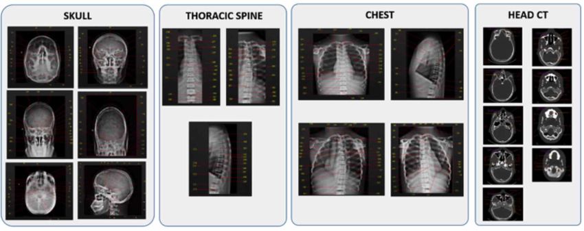
environments. The technology integration into anatomy This study was divided into two phases: image
education has enhanced the student education improvement acquisition and software development (Fig. 1).
(Clunie et al., 2018).
Phase 1: Image Acquisition. The images of an adult
Manufacturers of medical imaging devices also anthropomorphic phantom of head and neck (Radiation
provide courses and credits based on this technology (ISRRT, Support Devices, model RS-230) were obtained in Multix B
2004; American Registry of Radiologic Technologists, 2018) Siemens x-ray unit and a Siemens Somatom Spirit CT
Usually, it assists continued education and maintaining equipment. In addition, an anthropomorphic phantom of chest
professional skills as required by radiologic associations. (Radiation Support Devices, model RS-111) was also imaged
According to challenges and effort to find a proper learning using the same x-ray equipment. A computed radiography (CR)
method, although there are limitations for e-learning was used to obtain the digital radiographic images which was
implementation, it may be considered as an alternative achieved by using a reader and two cassettes (35 x 43 cm, 24
strategy for traditional classes (White & Cheung). x 30 cm). Furthermore, 13 radiographic projections were
performed (Table I). These radiographic projections were used
According to Pinto et al. (2011) the training of owing to the phantom characteristics and limitations. However,
students using suitable approaches to identify radiological in this study, the most frequent radiographic projections used
anatomy accurately is important. This training may reduce in hospital or clinics were included. The tomographic images
the diagnostic errors that are often related to unrecognized were reconstructed in axial plane and bone window. The scan
or unreported abnormalities which may be associated with protocol used is shown in Table II. Figure 2 demonstrates how
high morbidity. Therefore, the aim of this study was to the phantoms were set up for image acquisition.
develop a free radiological anatomy software for radiologic
anatomy education to assist students and professionals in
health science. Fig 1. Study design.
Fig 2. Radiographic projections a) Lateral view of Chest, b) Towne view, c) Submentovertex view.
206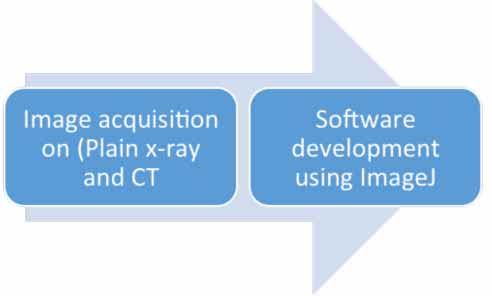
Table I. Radiographic projection position and acquisition method..
SKULL THORACIC SPINE CHEST
PA WATERS CALDWE TOWNE LATERAL SMV AP Lateral Oblique PA LATERAL Oblique
LL
a d
OML MML Forehead OML Head in a IOML MSP Posterior The body MSP Coronal Phantom
perpendic perpendicul against perpendicul true lateral parallel to aligned to half of rotated 20° aligned plane rot ated 45°
ular to IR; ar to plane grid ar to IR; position; Grid; CR and th orax from true with CR perpendicul with left
of Grid; surface; midline of aligned to lateral; and with ar and anter ior
table. CR and midline of sagittal breast
MSPb MSP OML MSP to CR MSP MSP m idline of Spinal grid with plane is against IR
perpendic perpendicul perpendic and to parallel to perpendicul grid column equal parallel to for LAOh
ular to ar t o ular to IR; midline of IR; ar t o the aligned to margins IR. and 45°
midl ine of m idline of the table. midline of CR and between with right
Part e
grid grid MSP IPL the grid midl ine of lateral anter ior
Position
perpendic perpendicul grid thorax and shoulder
ular to ar to IR; sides of against IR
midl ine of IR; for RAO i
the grid. IOMLf
perpendicul Top of IR
ar t o front above 4 to
edge of IR. 5 c m of
chest
apex.
Perpendic Perpendicul 15° 30° caudad Center to a Perpendicul Perpendicul Perpendicul Perpendicul Perpendic Perpendicul Perpendicul
ular to IRc ar t o IR to caudad, to OML; point 5 cm ar to IOML; ar to T7 ( 8 ar t o long ar to T7 ( 8 ular to T7 ar to T7 ( 8 ar to T7 ( 8
and exit at and center superior to to 10 cm axis of to 10 cm (8 to 10 to 10 cm to 10 cm
center ed acanthion. to exit at Center at EAMg Center below th oracic below cm below below below
to exit at nasi on MSP 6.5 cm midway jugular spi ne; jugular jugular jugular jugular
C entral glabella. the glabella between the notch) notch) notch) notch) notch)
R ay to pass gonions. Perpendicul
Int. J. Morphol., 37(1):205-211, 2019.
(CR) through the ar t o T7 ( 8
foramen to 10 cm
magnum at below
the level of ju gular
the base of notch)
the occiput.
a b c dM eI f g h I
Orbitomeatal line; Mediosagittal pl ane; Image receptor; entomeatal Line; nterpupillary Line; infrarbitomeatal; external auditory meatus; left anterior oblique; Right anterior oblique
Table II. Multi-Slice computed tomography scan parameters.
Tube
Product time- Rotation Detector S lice FOV Increment
potential Pitch Reconstruction
current (mAs) Time (s) Collimation (mm) (mm) (mm)
OLIVEIRA, M.; GEAMBASTIANI, P.; LOPEZ, G.; CAMBUI, M.; UBEDA, C. & MDLETSHE, S. The development of a free radiological anatomy software teaching tool.
(kV)
130 240 1.0 32 x 0.6 0,55 Axial Bone 5.0 240 5
207OLIVEIRA, M.; GEAMBASTIANI, P.; LOPEZ, G.; CAMBUI, M.; UBEDA, C. & MDLETSHE, S. The development of a free radiological anatomy software teaching tool.
Int. J. Morphol., 37(1):205-211, 2019.
Phase 2: Software development. The software was The usability of the software was tested by getting a
developed using ImageJ which is a free software accessible group of professors to answer the multiple choice questions.
via the internet (National Institutes of Health, USA). This is After the user’s language selection, the field of identification
an inexpensive method, as it does not require a user license. have to be fitted, image modality and anatomy (spine, head
Besides, it allows the development of macros, which assist or chest) selected and, then the radiological projection chosen
to perform tasks automatically. (Fig. 4). The image and questions were shown. In the end of
evaluation, a reported was presented containing date and
After the image acquisition, DICOM (Digital Imaging time of evaluation, User name and score. The software
and Communications in Medicine, 2019) files were indicated where the user incorrectly identified the anatomy
converted to TIFF. This was followed by the insertion of the (Fig. 5).
arrows and numbers indicating the anatomical structures.
This was done by a professor in radiology. Thereafter, a
template was created relating the structure name according DISCUSSION
to arrow indication. The data was revised by three
experienced professors (Professor 1:20 years, Professor 2:10
years, Professor 3: 10 years) of anatomy who have experience The integration of multimedia and interactivity into
in radiographic and tomographic images. In this digital electronic environment has allowed valuable support for
environment, radiological anatomy reference points were radiography teaching and continuing education (Pinto et al.,
shown and multiple choice questions were applied. These 2008).
questions were presented for anatomical structure
recognition testing by users. Besides, four alternatives were Educational strategies have to be applied for
shown as answers, however just one was correct. The soft- improvement of the learning process. Currently, lecture courses
ware was developed in three languages (Portuguese, Spanish do not provide enough contact time for deeper learning
and English). activities. This results in limitation of students’ learning per-
formance. Furthermore, students become passive recipients
of large amounts of information, leaving them with limited
RESULTS mental capacity to be involved with classes (Cook, 2014).
According to Xiberta & Boada (2016) Microsoft
The software presented radiologic anatomy from 13 PowerPoint is used in more than 80 % of their anatomy and
radiographic views of the head, neck and chest. On the other radiology classes. E-learning platform has been used to
hand, CT images presented more than one hundred anatomic overcome the limitation of the traditional educational
landmarks of the head (Fig. 3). In total 354 radiologic methods. Moreira et al. (2015) developed an e-learning
anatomy references and questions were obtained and course on breast imaging for radiographers. They concluded
performed, respectively. that it was effective and highlighted the need for continuing
Fig 3. Radiographic and CT imaging.
208OLIVEIRA, M.; GEAMBASTIANI, P.; LOPEZ, G.; CAMBUI, M.; UBEDA, C. & MDLETSHE, S. The development of a free radiological anatomy software teaching tool.
Int. J. Morphol., 37(1):205-211, 2019.
Fig 4. Software flowchart.
Fig 5. Screenshot of software and test result.
education. According to Cook, the e-learning method led to courses are not presented in practical classes. Moreover, the
a reduction of delayed self-study and consequent amassed content creation is high time consuming (Roe et al., 2010;
information before exams. Other platforms were developed Xiberta & Boada).
to assist in radiological subjects in an on-line environment
Eg. MyPacs (Weinberger et al., 2002), COMPARE In this study, a free radiological anatomy software as
(Grunewald et al., 2003), KICLA (Rowe et al., 2014) and a teaching tool was presented. ImageJ is an open-source soft-
RadStax (Colucci et al., 2015). There is a limitation with ware and works independently of the operating system.
radiology education platform because usually e-learning Taking into account that Portuguese, English and Spanish
209OLIVEIRA, M.; GEAMBASTIANI, P.; LOPEZ, G.; CAMBUI, M.; UBEDA, C. & MDLETSHE, S. The development of a free radiological anatomy software teaching tool.
Int. J. Morphol., 37(1):205-211, 2019.
are widespread languages spoken around the world, the use dividió en dos fases: adquisición de imágenes y desarrollo de
of this software could assist teachers and students at no cost. software. La primera fase consistió en obtener imágenes
According to Zafar et al. (2014), sustainable educational radiográficas simples y tomografías computarizadas (TC) de
models generate positive implications supporting the idea un fantasma antropomórfico de cabeza y cuello. Además, se
of lifelong learning, emphasizing that combined forms of obtuvieron imágenes radiográficas simples de un fantasma
learning are even more effective. antropomórfico del tórax. La segunda fase fue el desarrollo
del software de anatomía como una macro ImageJ. El soft-
E-Learning efficiency is related to reliability, ware se desarrolló a través de la inserción de los puntos de
functionality, user friendliness of technological tool for the referencia de la anatomía radiológica en las imágenes que se
accomplishment of a purpose (Pójanowicz et al., 2014). The obtuvieron y la aplicación de preguntas de opción múltiple.
radiologic anatomy software may be considered an extremely Luego, se probó la usabilidad del software haciendo que los
accessible tool. Moreover, this software will assist to redu- profesores respondieran las preguntas de opción múltiple. El
ce the practice of working intensively, to absorb a large software presentó la anatomía radiológica de 1) Proyecciones
volume of informational material in a short amountsof time de la cabeza: vista de aguas, vista de Towne, vista de Caldwell,
by students. The students can practice, exhaustively, the vista lateral, Submentovertex, vista de PA; 2) proyecciones
recognition of radiological anatomy landmarks, everywhere de la columna torácica: vista AP y lateral y 3) Cofre: vista de
and independently of internet. It could also assist with PA, lateral y oblicua. Las imágenes tomográficas presenta-
continuing education for professionals. ron cien puntos de referencia radiológica de la cabeza. En
total, hubo 354 preguntas. Se mostró un informe final con la
A user-friendly and inexpensive software was puntuación de las respuestas correctas, así como la identifi-
presented. Radiographers, students and professionals from cación del usuario, la fecha y la hora de la prueba. Las prue-
several countries are able to repeatedly practice, the bas estaban disponibles en tres idiomas (español, inglés y
recognition of radiologic anatomical landmarks. This soft- portugués). Se desarrolló y presentó un software fácil de usar
ware can be applied as a feasible technological tool for y de bajo costo. Estudiantes y profesionales de varios países
enhancing learning environment. pueden practicar, repetidamente, el reconocimiento de pun-
tos de referencia anatómicos radiológicos.
ACKNOWLEDGEMENTS PALABRAS CLAVE: Anatomía; Radiología edu-
cacional; Tecnología educacional; Enseñanza.
The authors thank the Federal Institute of Bahia LAFIR
and GTecRad (Grupo de Pesquisa em tecnologia em
REFERENCES
Radiologia), Brazil; Universidad de Tarapacá, Arica, Chile;
Faculty of Health Sciences, University of Johannesburg,
Johannesburg, South Africa for their support in conducting American Registry of Radiologic Technologists (ARRT). St. Paul, The
this study. American Registry of Radiologic Technologists, 2018. Available from:
https://www.arrt.org/docs/default-source/Governing-Documents/
The readers can obtain the software contacting the continuing-education-requirements.pdf
Challen, V. Radiography Education in Europe, Vision of HENRE (Higher
first author and developer by email: Education Network for Radiography Education). Vienna, European
marcusradiology@gmail.com Association of Nuclear Medicine, 2010. Available from: https://
www.eanm.org/content-eanm/uploads/CTE-Archive/2010/Lunch/
lunch_1.pdf
Clunie, L.; Morris, N. P.; Joynes, V. C. T. & Pickering, J. D. How
comprehensive are research studies investigating the efficacy of
OLIVEIRA, M.; GEAMBASTIANI, P.; LOPEZ, G.; technology-enhanced learning resources in anatomy education? A
CAMBUI, M.; UBEDA, C. & MDLETSHE, S. Desarro- systematic review. Anat. Sci. Educ., 11(3):303-19, 2018.
llo de un software libre de anatomia radiológica como una Colucci, P. G.; Kostandy, P.; Shrauner, W. R.; Arleo, E.; Fuortes, M.; Griffin,
herramienta de enseñanza. Int. J. Morphol., 37(1):205-211, A. S.; Huang, Y. H.; Juluru, K. & Tsiouris, A. J. Development and
utilization of a web-based application as a robust radiology teaching
2019. tool (radstax) for medical student anatomy teaching. Acad. Radiol.,
22(2):247-55, 2015.
RESUMEN: El propósito de esta investigación fue Cook, D. A. The value of online learning and MRI: finding a niche for
desarrollar un software gratuito de anatomía radiológica para expensive technologies. Med. Teach., 36(11):965-72, 2014.
Digital Imaging and Communications in Medicine (DICOM). Arlington,
la educación de anatomía radiológica para ayudar a estu- National Electrical Manufacturers Association, 2019. Available from:
diantes y profesionales de ciencias de la salud. El estudio se https://www.dicomstandard.org/
210OLIVEIRA, M.; GEAMBASTIANI, P.; LOPEZ, G.; CAMBUI, M.; UBEDA, C. & MDLETSHE, S. The development of a free radiological anatomy software teaching tool.
Int. J. Morphol., 37(1):205-211, 2019.
England, A.; Geers-van Gemeren, S.; Henner, A.; Kukkes, T.; Pronk-Larive, Corresponding author:
D.; Rainford, L. & McNulty, J. P. Clinical radiography education across Marcus Vinicius Linhares de Oliveira
Europe. Radiography (Lond.), 23 Suppl. 1:S7-15, 2017. Department of Health Technology and Biology
Grunewald, M.; Heckemann, R. A.; Gebhard, H.; Lell, M. & Bautz, W. A.
Federal Institute of Bahia Emídio dos Santos – s/n –
COMPARE radiology: creating an interactive Web-based training
program for radiology with multimedia authoring software. Acad.
Salvador
Radiol., 10(5):543-53, 2003. Bahia
Moreira, I. C.; Ventura, S. R.; Ramos, I. & Rodrigues, P. P. Development BRAZIL
and assessment of an e-learning course on breast imaging for
radiographers: a stratified randomized controlled trial. J. Med. Internet
Res., 17(1):e3, 2015. E-mail: marcusradiology@gmail.com
Pinto, A.; Acampora, C.; Pinto, F.; Kourdioukova, E.; Romano, L. &
Verstraete, K. Learning from diagnostic errors: a good way to improve
education in radiology. Eur. J. Radiol., 78(3):372-6, 2011.
Pinto, A.; Selvaggi, S.; Sicignano, G.; Vollono, E.; Iervolino, L.; Amato,
Received: 09-08-2018
F.; Molinari, A. & Grassi, R. E-learning tools for education: regulatory Accepted: 17-10-2018
aspects, current applications in radiology and future prospects. Radiol.
Med., 113(1):144-57, 2008.
Pójanowicz, W.; Roszak, M.; Koodziejczak, B. & Bre˛borowicz, A. An
Analysis Of The Effectiveness and Quality of E-Learning in Medical
Education. In: Smyrnova-Trybulska, E. (Ed.). E-learning and
Intercultural Competences Development in Different Countries.
Katowice, Studio NOA, University of Silesia, 2014. pp.177-96.
Prentakis, A. G.; Stefanoyiannis, A. P.; Georgiadis, K.; Coleman, L.; Foley,
S. J.; Herlig, D.; Kollas, P.; Kowalik, A.; Tomczak, J. & Chatziioannou,
S. N. Education, training, and professional issues of radiographers in
six European countries: a comparative review. J. Eur. CME, 5(1):31092,
2016
Roe, D.; Carley, S. & Sherratt, C. Potential and limitations of e-learning in
emergency medicine. Emerg. Med. J., 27(2):100-4, 2010.
Rowe, S. P.; Siddiqui, A. & Bonekamp, D. The key image and case log
application. Acad. Radiol., 21(7):916-30, 2014.
Weinberger, E.; Jakobovits, R. & Halsted, M. MyPACS.net: a Web-based
teaching file authoring tool. A. J. R. Am. J. Roentgenol., 179(3):579-
82, 2002.
White, P. & Cheung, A. K. Y. E-learning in an undergraduate radiography
programme: Example of an interactive website. Radiography,
12(3):244-52, 2006.
Xiberta, P. & Boada, I. A new e-learning platform for radiology education
(RadEd). Comput. Methods Programs Biomed., 126:63-75, 2016.
Zafar, S.; Safdar, S. & Zafar, A. N. Evaluation of use of e-Learning in
undergraduate radiology education: a review. Eur. J. Radiol.,
83(12):2277-87, 2014.
211You can also read