THE IMPACT OF SARS-COV-2 MULTIPLE SPIKE PROTEIN MUTATIONS ON COVID-19 OUTCOMES
←
→
Page content transcription
If your browser does not render page correctly, please read the page content below
The Impact of SARS-CoV-2 Multiple Spike Protein
Mutations on COVID-19 Outcomes
Gunadi ( drgunadi@ugm.ac.id )
Universitas Gadjah Mada/Dr. Sardjito Hospital
Mohamad Saifudin Hakim
Universitas Gadjah Mada
Hendra Wibawa
Disease Investigation Center, Wates, Yogyakarta, Ministry of Agriculture
Marcellus
Universitas Gadjah Mada
Ika Trisnawati
Universitas Gadjah Mada/Dr. Sardjito Hospital
Endah Supriyati
Universitas Gadjah Mada
Afiahayati
Universitas Gadjah Mada
Riat El Khair
Universitas Gadjah Mada/Dr. Sardjito Hospital
Kristy Iskandar
Universitas Gadjah Mada/ UGM Academic Hospital
Siswanto
Universitas Gadjah Mada/ UGM Academic Hospital
Irene
Balai Besar Teknik Kesehatan Lingkungan dan Pengendalian Penyakit
Nungki Anggorowati
Universitas Gadjah Mada/Dr. Sardjito Hospital
Edwin Widyanto Daniwijaya
Universitas Gadjah Mada/ UGM Academic Hospital
Dwi Aris Agung Nugrahaningsih
Universitas Gadjah Mada
Yunika Puspadewi
Universitas Gadjah Mada/Dr. Sardjito Hospital
Susan Simanjaya
Universitas Gadjah Mada
Dyah Ayu Puspitarani
Page 1/21Universitas Gadjah Mada
Hana Fauzyyah Hanifin
Universitas Gadjah Mada
Alvina Alexandra Setiawan
Universitas Gadjah Mada
Irene Tania
Universitas Gadjah Mada
Cita Shafira Amalia
Universitas Gadjah Mada
I Putu Aditio Artayasa
Universitas Gadjah Mada
Haries Rachman
Universitas Gadjah Mada
Herdiyanto Mulyawan
Disease Investigation Center, Wates, Yogyakarta, Ministry of Agriculture
Nur Rahmi Ananda
Universitas Gadjah Mada/Dr. Sardjito Hospital
Eggi Arguni
Universitas Gadjah Mada/Dr. Sardjito Hospital
Titik Nuryastuti
Universitas Gadjah Mada
Tri Wibawa
Universitas Gadjah Mada
Research Article
Keywords: COVID-19 outcomes, comorbidity, genomic surveillance, multiple spike protein mutations,
phylogenetic analysis, SARS-CoV-2 variants, whole genome sequencing
Posted Date: July 26th, 2021
DOI: https://doi.org/10.21203/rs.3.rs-700897/v1
License: This work is licensed under a Creative Commons Attribution 4.0 International License.
Read Full License
Page 2/21Abstract
Background: Recent studies focusing on the association of SARS-CoV-2 variants of concern (VOC) on
COVID-19 outcomes have been reported. However, studies of the impact of multiple mutations within the
spike (S) protein of SARS-CoV-2 on COVID-19 illness are limited. This study determined the association
between multiple mutations within the S protein, prognosis factors, and the disease outcomes of SARS-
CoV-2 infection.
Methods: We included 51 COVID-19 patients from Yogyakarta and Central Java, Indonesia. Whole
genome sequences of SARS-CoV-2 were determined by the Illumina MiSeq next-generation sequencer,
followed by the phylogenetic analysis of 170 full-genomes of SARS-CoV-2 from different regions. We
analyzed characteristics of COVID-19 patients and multiple mutations in association with different
outcomes.
Results: Among 51 patients, the clinical manifestations of COVID-19 were as follows: without any
symptoms (13.7%), mild (47%), moderate (19.6%), severe (4%), critical (2%), and died (13.7%). The age of
hospitalized patients (53.4 ± 18 years) was higher than non-hospitalized patients (34.6 ± 19) (p=0.001). A
significant association between diabetes, hypertension, and anticoagulant and the hospitalization of
patients was noted with p-value of 0.039 (OR=4.47 [95% CI=1.07-18.58]), 0.001 (OR=17 [95% CI=2-144]),
and 0.02 (OR=27.97 [95% CI=1.54-507.13]), respectively; whereas a strong association between patients’
age, diabetes, anticoagulant, and steroid with the mortality of patients was revealed with p-value of 0.016
(OR=8.44 [95% CI=1.5-47.49]), 0.019 (OR=8.5 [95% CI=1.43-50.66]), 0.001 (46.8 [95% CI=4.63-472.77]),
and 0.009 (OR=15.75 [95% CI=2-123.86]), respectively. All viruses contained the D614G variant, except
one case. Accordingly, the samples were classified as the following clade: L (2%), GH (84.3%), GR (11.7%),
and O (2%). Besides the D614G, the most common variants in the S protein were L5F (18.8%), V213A
(18.8%), and S689R (8.3%). There was no significant association between multiple S protein variants with
either hospitalization or mortality of COVID-19 (p=0.11 and 0.69, respectively). Multivariate analysis
showed that hypertension and anticoagulant were the strong factors affecting the hospitalization and
mortality of patients with COVID-19 with a p-value of 0.009 (OR=17.06 [95% CI=2.02-144.36]) and 0.001
(OR=46.8 (95% CI=4.63-472.77), respectively. Interestingly, the multiple S protein variants almost reached
a significant level affecting the hospitalization of patients (p=0.07). Phylogenetic analysis showed that
although most of the viruses from this study belonged to clade GH, none were detected as the variant of
concern (VOC) and the variant of interest (VOI) of SARS-CoV-2.
Conclusions: Here, we show for the first time the association between SARS-CoV-2 mutations within the S
protein besides the VOC with the COVID-19 outcomes. Our findings suggest that multiple mutations in the
S protein might affect the severity of COVID-19. Our study further suggests the importance of genomic
surveillance to monitor SARS-CoV-2 variants, particularly those that might influence the outcomes of
COVID-19 patients.
Introduction
Page 3/21After one year of the COVID-19 pandemic, SARS-CoV-2 has infected approximately 185 million people
and causes 4 million deaths worldwide [1,2]. Indonesia has documented 2,379,397 COVID-19 cases and
62,908 deaths on July 7, 2021, and become the highest cases country in the South-East Asian region [3].
The outcome of SARS-CoV-2 infection is determined by multiple factors, including the viral and host
genetics and age and comorbidities [4,5]. It is hypothesized that the host genetic factors might influence
the outcome of SARS-CoV-2 infection. Three genes encoding the angiotensin-converting enzyme 2
(ACE2), the human leukocyte antigen (HLA), toll-like receptor, and complement pathway are suggested to
be the primary determinant of COVID-19 outcomes [6]. For viral genetic factors, a previous study indicated
that variations within the ORF1ab (4715L) and S protein (614G) had a significant positive correlation with
fatality rates of COVID-19 [7]. The viral mutation may affect the presentation to MHC-I and MHC-II and
consequently determine the magnitude of cellular immune responses [8]. The emergence of variants of
concern (VOC) has attracted public health authorities to assess its impact on clinical presentation and
severity. Indeed, the currently known VOCs (alpha, beta, gamma and delta) have been associated with a
possible increased risk of hospitalization and disease severity [9].
SARS-CoV-2 has continuously and rapidly spread worldwide, providing a high opportunity for mutation
events, especially on the S protein. However, the studies of the impact of multiple mutations within the
spike (S) protein of SARS-CoV-2 on COVID-19 illness are limited. We determined the association between
the SARS-CoV-2 mutations within the S protein identified from non-VOC isolates and the COVID-19
outcomes.
Material And Methods
RNA extraction and whole genome sequencing
RNA was extracted from all samples of COVID-19 patients from Yogyakarta and Central Java provinces
using QiAMP Viral RNA mini kit (Qiagen, Hilden, Germany), followed by real-time polymerase chain
reaction (RT-PCR) using Real-Q 2019-nCoV Detection Kit (BioSewoom, Seoul, South Korea) with
LightCycler® 480 Instrument II (Roche Diagnostics, Mannheim, Germany).
The double-stranded cDNA was synthesized using Maxima H Minus Double-Stranded cDNA Synthesis
(Thermo Fisher Scientific, MA, United States), followed by purification of cDNA using a GeneJET PCR
Purification Kit (Thermo Fisher Scientific, MA, United States) and library preparations using the Nextera
DNA Flex for Enrichment using Respiratory Virus Oligos Panel. Next-generation sequencing (NGS) was
performed to sequence the whole-genome of SARS-CoV-2 using the Illumina MiSeq instrument (Illumina,
San Diego, CA, United States) with Illumina MiSeq reagents v3 150 cycles (2 x 75 cycles).
The assembly of our sample genomes was mapped into the reference genome from Wuhan, China
(hCoV-19/Wuhan/Hu-1/2019, GenBank accession number: NC_045512.2) using Burrow-Wheeler Aligner
(BWA) algorithm embedded in UGENE v. 1.30 [10]. Single nucleotide polymorphisms (SNPs) were
identified using the number of high confidence base calls (consensus sequence variations of the
Page 4/21assembly) that disagree with the reference bases for the genome position of interest, followed by exporting all SNPs a vcf file and visualizing them in MS Excel. Phylogenetic study For the phylogenetic study, we utilized a dataset of 170 available SARS-CoV-2 genomes from our region and other countries that were retrieved from GISAID (Acknowledgment Table is provided in Supplementary Table 1), followed by multiple nucleotide sequence alignment using the MAFFT program (https://mafft.cbrc.jp/alignment/server/). Neighbour Joining statistical method with 1,000 bootstrap replications was used to construct a phylogenetic tree from 29.563 nt length of the open reading frame (ORF) of SARS-CoV-2 virus genome [11,12]. The Kimura 2-parameter method and the gamma distribution with estimated shape parameter (α) for the dataset were utilized to compute the evolutionary distances and model the rate variation among sites, respectively [13]. DAMBE version 7 [14] was used to calculate the estimation of the α gamma distribution, while MEGA version 10 (MEGA X) [15] was utilized for all other analyses. Patients and prognostic factors This study was a retrospective study. We included all patients with COVID-19 from Yogyakarta and Central Java provinces, Indonesia, who sent their samples for whole genome sequencing into our institution from June to October 2020. The exclusion criteria were incomplete medical records. Various clinical manifestations of COVID-19 have been noted, including asymptomatic until pneumonia with varying degrees. The degree of pneumonia of COVID-19 was classified according to the WHO classifications: 1) mild, without evidence of hypoxia or pneumonia; 2) moderate, pneumonia but not severe; 3) severe, pneumonia plus one of the following signs: respiratory rate > 30 breaths/minute (or based on age for children), severe respiratory distress, or SpO₂
Results
Association between prognostic factors and hospitalization of patients with COVID-19.

Among 51 patients, the clinical manifestations of COVID-19 were as follows: without any symptoms
(13.7%), mild (47%), moderate (19.6%), severe (4%), critical (2%), and died (13.7%). The age of
hospitalized patients (53.4 ± 18 years) was higher than non-hospitalized patients (34.6 ± 19) (p = 0.001)
(Table 1).

                                                 Page 6/21
Table 1
Association between prognostic factors and hospitalization of patients with COVID-19.
Characteristics All (n Hospitalized Non-hospitalized (n = 22) (N, p- OR (95%
= 51) (n = 29) %; mean ± SD) value CI)
(N, %; mean ±
SD)
RT-PCR Ct 20.3 ± 4.2 18.9 ± 3.9 0.26
value
Age (years) 53.4 ± 18 34.6 ± 19 0.001* 3.81 (0.72–
20.16)
▪ ≥ 65 10 8 (27.6) 2 (9.1) 0.12
▪ < 65 41 21 (72.4) 20 (90.9)
Sex
▪ Male 30 19 (65.5) 11 (50) 0.27 1.9 (0.61–
5.9)
▪ Female 21 10 (34.5) 11 (50)
Comorbidity
▪ Obesity 3 3 (10.3) 0 0.25 5.94 (0.29-
121.31)
▪ Diabetes 15 12 (41.4) 3 (13.6) 0.039*
4.47 (1.07–
▪ Hypertension 14 13 (44.8) 1 (4.5) 0.001* 18.58)
▪ 9 8 (27.6) 1 (4.5) 0.06 17 (2-144)
Cardiovascular
disease 2 2 (6.9) 0 0.37 8 (0.92–
69.72)
▪ Chronic
kidney disease 4.09 (0.19–
89.65)
Smoking 4 1 (3.4) 3 (13.6) 0.21 0.23 (0.02–
2.34)
Therapy
▪ ACEI/ARB 4 4 (13.8) 0 0.17 7.94 (0.33–
66.14)
▪ 11 11 (37.9) 0 0.02*
Anticoagulant 27.97
5 5 (17.2) 0 0.12 (1.54-
▪ Steroid 507.13)
10.1 (0.53-
193.23)
*, significant (p < 0.05); ACEI, angiotensin-converting enzyme inhibitors; ARB angiotensin receptor
blocker; CI, confidence interval; OR, odds ratio
Page 7/21Association between prognostic factors and mortality of
patients with COVID-19
A significant association between diabetes, hypertension, and anticoagulant and the hospitalization of
patients was found with p-value of 0.039 (OR = 4.47 [95% CI = 1.07–18.58]), 0.001 (OR = 17 [95% CI = 2-
144]), and 0.02 (OR = 27.97 [95% CI = 1.54-507.13]), respectively (Table 1).A strong association between
patients’ age, diabetes, anticoagulant, and steroid and the mortality of patients was revealed with p-value
of 0.016 (OR = 8.44 [95% CI = 1.5-47.49]), 0.019 (OR = 8.5 [95% CI = 1.43–50.66]), 0.001 (46.8 [95% CI =
4.63-472.77]), and 0.009 (OR = 15.75 [95% CI = 2-123.86]), respectively (Table 2).
Page 8/21Table 2
Association between prognostic factors and mortality of patients with COVID-19.
Characteristics All (n = Died (n = 7) Survived (n = 44) p- OR (95% CI)
51) value
(N, %; mean ± (N, %; mean ±
SD) SD)
RT-PCR Ct value 18.7 ± 5.0 19.9 ± 3.9 0.57
Age (years) 66.8 ± 14 41.8 ± 19 0.002* 8.44 (1.5-
47.49)
▪ ≥ 65 10 4 (57.1) 6 (13.6) 0.016*
▪ < 65 41 3 (42.9) 38 (86.4)
Sex 0.69 1.9 (0.33–
10.88)
▪ Male 30 5 (71.4) 25 (56.8)
▪ Female 21 2 (28.6) 19 (43.2)
Comorbidity
▪ Obesity 3 1 (14.3) 2 (4.5) 0.34 3.5 (0.27–
44.75)
▪ Diabetes 15 5 (71.4) 10 (22.7) 0.019*
8.5 (1.43–
▪ Hypertension 14 4 (57.1) 10 (22.7) 0.07 50.66)
▪ Cardiovascular 9 3 (42.9) 6 (13.6) 0.08 4.53 (0.87–
disease 23.72)
2 1 (14.3) 1 (2.3) 0.18
▪ Chronic kidney 4.75 (0.84–
disease 26.71)
7.17 (0.39-
130.31)
Smoking 4 0 4 (9.1) 0.74 0.6 (0.03–
12.34)
Therapy
▪ ACEI/ARB 4 2 (28.6) 2 (4.5) 0.05 8.4 (0.96–
73.43)
▪ Anticoagulant 11 6 (85.7) 5 (11.4) 0.001*
46.8 (4.63-
▪ Steroid 5 3 (42.9) 2 (4.5) 0.009* 472.77)
15.75 (2-
123.86)
*, significant (p < 0.05); ACEI, angiotensin-converting enzyme inhibitors; ARB angiotensin receptor
blocker; CI, confidence interval; OR, odds ratio
Molecular and phylogenetic analysis
Page 9/21All viruses contained the D614G variant, except one isolate. Accordingly, the samples were classified as
the following clade: L (2%), GH (84.3%), GR (11.7%), and O (2%). Besides the D614G mutation, the most
common mutation in the S protein was L5F (18.8%), V213A (18.8%), and S689R (8.3%) (Table 3).
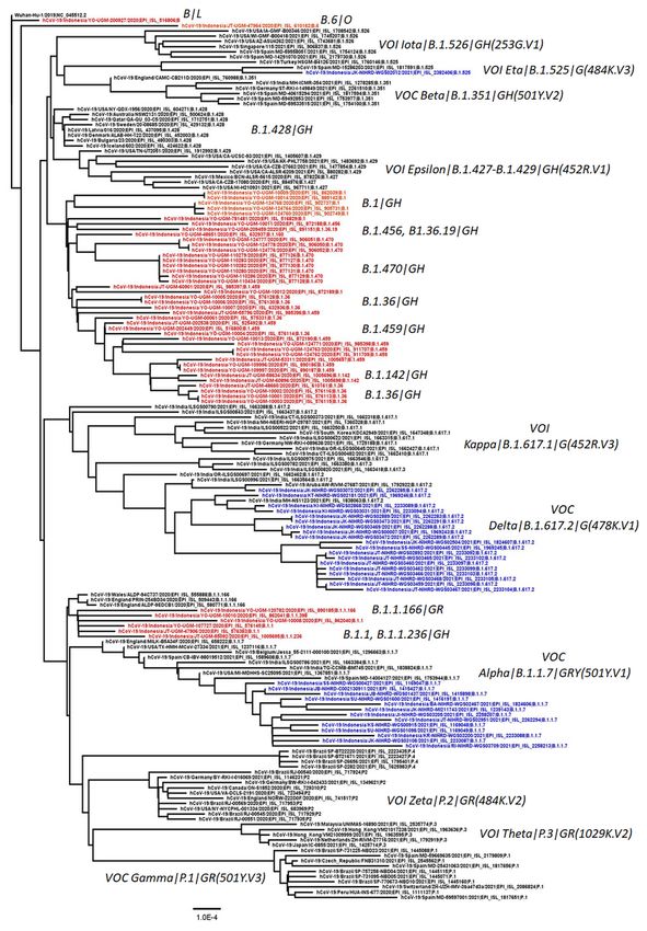
Phylogenetic analysis showed that although most virus samples belonged to the clade GH and followed
by the clade GR, none of these was detected as the variant of concern (VOC) and the variant of interest
(VOI) of SARS-CoV-2 (Fig. 1). While two viruses of clade L and O formed separate clusters to the GH and
GR clades.
Association between multiple S protein variants with
COVID-19 patients’ outcomes and prognostic factors
There was no significant association between multiple spike protein with either hospitalization or
mortality of COVID-19 patients (p = 0.11 and 0.69, respectively) (Table 4). Moreover, none of the
prognostic factors was associated with multiple spike protein mutations (p > 0.05) (Table 4).
Page 10/21Table 4
Association between multiple spike protein mutations with outcomes of patients with COVID-19 and
prognostic factors
Variables S protein mutation OR (95% CI) p-
value
Multiple (N, %; mean None/Single (N, %; mean
± SD) ± SD)
Hospitalized 0.36 (0.11–1.25) 0.11
▪ Yes 16 (48.5) 13 (72.2)
▪ No 17 (51.5) 5 (27.8)
Survival 1.43 (0.25–8.23) 0.69
▪ Died 5 (15.2) 2 (11.1)
▪ Live 28 (84.8) 16 (88.9)
RT-PCR Ct value 19.2 ± 3.7 20.6 ± 4.7 0.26
Age (years) 1.35 (0.30-6.0) 0.70
▪ ≥ 65 7 (21.2) 3 (16.7)
▪ < 65 26 (78.8) 15 (83.3)
Sex 19 (57.6) 11 (61.1) 0.86 (0.27–2.79) 0.81
▪ Male 14 (42.4) 7 (38.9)
▪ Female
Comorbidity
▪ Obesity 2 (6.1) 1 (5.6) 1.10 (0.09-13.0) 0.94
▪ Diabetes 11 (33.3) 4 (22.2) 1.75 (0.46–6.59) 0.41
▪ Hypertension 6 (18.2) 8 (44.4) 0.28 (0.08-1.0) 0.05
▪ 6 (18.2) 3 (16.7) 1.11 (0.24–5.09) 0.89
Cardiovascular
disease 2 (6.1) 0 2.94 (0.14– 0.49
64.55)
▪ Chronic kidney
disease
Smoking 2 (6.1) 2 (11.1) 0.51 (0.07–4.01) 0.53
Page 11/21Variables S protein mutation OR (95% CI) p-
value
Therapy
▪ ACEI/ARB 3 (9.1) 1 (5.6) 1.7 (0.16–17.65) 0.66
▪ Anticoagulant 7 (21.2) 4 (22.2) 0.94 (0.23–3.78) 0.93
▪ Steroid 4 (12.1) 1 (5.6) 2.34 (0.24– 0.46
22.73)
*, significant; ACEI, angiotensin-converting enzyme inhibitors; ARB, angiotensin receptor blocker; CI,
confidence interval; OR, odds ratio
Multivariate analysis
Multivariate analysis showed that hypertension and anticoagulant were the substantial factors affecting
the hospitalization and mortality of patients with COVID-19 with a p-value of 0.009 (OR = 17.06 [95% CI =
2.02-144.36]) and 0.001 (OR = 46.8 (95% CI = 4.63-472.77), respectively. Interestingly, the multiple S
protein mutations almost reached a significant level affecting the hospitalization of patients (p = 0.07)
with the OR of 4.64 (95% CI = 0.87–24.68) (Table 5).
Page 12/21Table 5
Multivariate analysis of the association between prognostic factors and outcomes of patients with
COVID-19
Prognostic Factor Hospitalized Mortality
OR (95% CI) p-value OR (95% CI) p-value
Multiple S protein mutations 4.64 (0.87–24.68) 0.07 0.91 (0.04–22.85) 0.96
Age (≥65 years) 0.10 (0.004–3.07) 0.19 4.56 (0.01-2267.77) 0.63
Sex (male) 2.5 (0.5–12.6) 0.24 3.45 (0.01-941.16) 0.67
Comorbidity
▪ Obesity - 1 0.05 (0.0001–23.23) 0.33
▪ Diabetes 2.74 (0.33–22.76) 0.35 14.27 (0.16-1286.04) 0.25
▪ Hypertension 17.06 (2.02-144.36) 0.009 3.44 (0.03-386.31) 0.61
▪ Cardiovascular disease 5.52 (0.18-168.92) 0.33 1.31 (0.01-340.36) 0.92
▪ Chronic kidney disease - 1 3.68 (0.04-353.38) 0.58
Smoking 6.72 (0.23-197.53) 0.27 - 1
Therapy
▪ ACEI/ARB - 1 13.69 (0.02-11919.51) 0.45
▪ Anticoagulant - 1 46.8 (4.63-472.77) 0.001
▪ Steroid - 1 43.96 (0.05-41926.76) 0.28
*, significant (p < 0.05); ACEI, angiotensin-converting enzyme inhibitors; ARB, angiotensin receptor
blocker; CI, confidence interval; OR, odds ratio
Discussion
Our study is able to show the effect of hypertension and the use of anticoagulants on the severity of
COVID-19 patients from the Indonesian population. The patients with hypertension have a ~ 17-fold
higher risk of hospitalization than those without hypertension, in line with previous reports [17,19]. The
effect of hypertension on COVID-19 severity is controversial [20]. The pathogenesis of hypertension
affecting the COVID-19 severity is complex [20]. The impact of hypertension on the severity of COVID-19
is significant when accompanied by cardiovascular diseases, including myocardial injury [21]. However,
our study did not show an association between the use of angiotensin-converting enzyme
inhibitors/angiotensin receptor blockers (ACEI/IRB) and COVID-19 severity. Similar to hypertension, the
effect of ACEI/IRB on COVID-19 severity is still inconclusive [20]. The S protein of SARS-CoV-2 binds to
the ACE2 receptor to enter the human cells, suggesting that the use of ACEI/IRB might worsen the
Page 13/21prognosis of COVID-19 [22]. However, current reports showed that ACEI/IRB was not associated with the
poorer outcomes of COVID-19 [23,24].
Our study also demonstrated the association between the use of anticoagulants and COVID-19 mortality
with an increased risk of approximately ~ 47-fold. SARS-CoV-2 often induces a pro-coagulative state due
to several mechanisms, including endothelial dysfunction, cytokine storm, and complement
hyperactivation [25]. While a recent study showed that the use of anticoagulants decreased the mortality
of patients with COVID-19, it was not the case with our findings. These differences probably because we
grouped the hospitalized and non-hospitalized into one group, classified into anticoagulant versus non-
anticoagulant groups. Of note, we have only a limited sample size. These limitations should be
considered during the interpretation of our findings. Further study with larger sample size is necessary to
clarify and confirm our study.
Most previous reports focused on the impact of VOC on the COVID-19 outcomes, including B.1.1.7
(alpha), B.1.351 (beta), P1 (gamma), and the most recent VOC, B.1.617.2 (delta) [26–29, 30]. Indonesia
has reported the identification of alpha, beta, and delta variants since January 2021 [31]. In this present
study, we have not found the VOC, and VOI strains in our samples of the sample collection occurred
during the period from June to October 2020 or before the first detection of VOC (B.1.1.7 lineage) in
Indonesia in January 2021. Currently, the delta variant is identified as the most frequent VOC [31].
However, the actual frequency of the circulating VOCs in Indonesia might be biased due to our limited
whole-genome sequencing capacities.
Interestingly, we revealed that patients with multiple S protein mutations might have a ~ 5-fold higher
possibility of being hospitalized than those with none or a single S protein mutation. However, the
association between mutation and clinical outcome of COVID-19 is inconclusive. A study in Uruguay
found that mutation in structural and non-structural protein was not associated with COVID-19 fatalities
[32]. Another recent study analyzed the association between viral genomic variants and COVID-19
outcomes. They showed that 17 variants had a two-fold higher risk of severe COVID-19, while 67 variants
were associated with less severe COVID-19 [33]. This is in line with another study from France and the US,
suggesting that different viral variants may result in different infection severity and risk of hospitalization
[34,35]. Since SARS-CoV-2 is an RNA virus, its dynamic evolution is expected to influence its biological
characteristic [36], including its virulence and pathogenicity [33]. Interestingly, as an RNA virus, the critical
aspect of the SARS-CoV-2 life cycle is not implied by the protein sequence [37]. Indeed, one study showed
the importance of synonymous substitutions on the selection of SARS-CoV-2 [38].
All our samples, except one, contained the D614G variant. It has already been established that the D614G
mutation was not associated with the COVID-19 illness [39,40]. Indeed, almost all viruses circulating
globally consist of the D614G mutation [31]. We also observed other S protein mutations in our samples,
including L5F, V213A, and S68SR. None of the mutations lies on the receptor-binding domain (RBD) of
the S protein.
Page 14/21Interestingly, a previous report showed that one variant in non-RBD S protein, V1176F, might lead to RBD-
ACE2 binding changes and was associated with a high mortality rate of COVID-19 [41]. Moreover, a recent
study revealed that variants within the different proteins of SARS-CoV-2 have associated with different
patients' outcomes [42]. Further in vitro study is essential to clarify whether multiple non-RBD S protein
variants associate with the COVID-19 severity in our patients.
There are several limitations of our study. First, we have only a limited sample size that may result in bias
in our analysis. Second, the S protein continuously evolves and may result in new mutation(s) that
significantly affect virulence and disease pathogenesis. Third, we only analyzed mutations located within
the S protein. Mutations in other structural and non-structural proteins may significantly influence the
COVID-19; however, they are not investigated in our study.
Conclusions
Here, we show for the first time the association between the SARS-CoV-2 mutations within the S protein
besides the VOC and the COVID-19 outcomes, revealing that multiple S protein mutations might affect the
severity of COVID-19, in addition to hypertension and the use of anticoagulants. Our study further
suggests the importance of genomic surveillance to monitor the SARS-CoV-2 variants, particularly those
that might influence the outcomes of COVID-19 patients.
Abbreviations
CI, confidence interval; OR, odds ratio; S protein, spike protein; SNPs, single nucleotide polymorphisms;
VOC, variant of concern; VOI, variant of interest
Declarations
Ethics approval and consent to participate
This study was approved by the Medical and Health Research Ethics Committee, Faculty of Medicine,
Public Health and Nursing, Universitas Gadjah Mada/Dr Sardjito Hospital, Yogyakarta, Indonesia
(KE/FK/0563/EC/2020) and written informed consent was obtained. The research has been performed
following the Declaration of Helsinki.
Consent to publish
All participants or guardians signed written informed consent for participating in this study.
Availability of data and material
All data generated or analyzed during this study are included in the submission. The sequence and
metadata are shared through GISAID (www.gisaid.org).
Page 15/21Competing interests
The authors declared no potential conflicts of interest concerning the research, authorship, and/or
publication of this article.
Funding
The Ministry of Education, Culture, Research and Technology, Indonesia,funded our study. The funders
had no role in study design, data collection and analysis, decision to publish, or manuscript preparation.
Authors’ contributions
G, KI, and NA conceived the study. G drafted the manuscript, and MSH, HW, and TW critically revised the
manuscript for important intellectual content. M, SS, DAP, HFH, AAS, ITa, CSA, IPAA, HR, and HM
performed the library preparation and NGS. G, MSH, M, IT, REK, I, A, S, EWD, ES, DAAN, YP, NRA, EA, TN
and TW collected the data; and G, MSH, HW, M, and A analyzed the data. All authors have read and
approved the manuscript and agreed to be accountable for all aspects of the work in ensuring that
questions related to the accuracy or integrity of any part of the work are appropriately investigated and
resolved.
Acknowledgements
We thank the Collaborator Members of the Yogyakarta-Central Java COVID-19 study group: Eko Budiono,
Heni Retnowulan, Sumardi, Bambang Sigit Riyanto, Munawar Gani, Satria Maulana, Ira Puspitawati,
Osman Sianipar (Faculty of Medicine, Public Health and Nursing, Universitas Gadjah Mada [FK-KMK
UGM]/RSUP Dr. Sardjito), Bagoes Poermadjaja (Balai Besar Besar Veteriner Wates, Yogyakarta), Indaryati
and Havid Setyawan (Balai Besar Teknik Kesehatan Lingkungan dan Pengendalian Penyakit,
Yogyakarta), Ludhang Pradipta Rizki and Sri Fatmawati (FK-KMK UGM), Safitriani and Muhammad
Taufiq Soekarno (PT. Pandu Biosains). We gratefully acknowledge the authors, the originating and
submitting laboratories for their sequence and metadata shared through GISAID. We also thank to Besar
Besar Veteriner Wates (Disease Investigation Center Wates) where the virus samples were sequenced
using the NGS Illumina MiSeq instrument. All submitters of data may be contacted directly via
www.gisaid.org. The Acknowledgments Table for GISAID is reported as Supplementary Table 1.
References
1. World Health Organization. https://www.who.int/news/item/29-06-2020-covidtimeline Accessed on
July 2, 2021.
2. Phelan AL, Katz R, Gostin LO. The novel coronavirus originating in Wuhan, China: challenges for
global health governance. JAMA. 2020 323, 709–710, doi:10.1001/jama.2020.1097
3. World Health Organization. https://covid19.who.int/table Accessed on July 2, 2021.
Page 16/214. Brodin P. Immune determinants of COVID-19 disease presentation and severity. Nat Med.
2021;27(1):28-33. doi:10.1038/s41591-020-01202-8
5. Awortwe C, Cascorbi I. Meta-analysis on outcome-worsening comorbidities of COVID-19 and related
potential drug-drug interactions. Pharmacol Res. 2020;161:105250. doi:10.1016/j.phrs.2020.105250
6. Debnath M, Banerjee M, Berk M. Genetic gateways to COVID‐19 infection: Implications for risk,
severity, and outcomes. The FASEB Journal. 2020;34(7):8787-8795. doi:10.1096/fj.202001115r
7. Toyoshima Y, Nemoto K, Matsumoto S, Nakamura Y, Kiyotani K. SARS-CoV-2 genomic variations
associated with mortality rate of COVID-19. J Hum Genet. 2020;65(12):1075-1082.
doi:10.1038/s10038-020-0808-9
8. de Sousa E, Ligeiro D, Lérias J et al. Mortality in COVID-19 disease patients: Correlating the
association of major histocompatibility complex (MHC) with severe acute respiratory syndrome 2
(SARS-CoV-2) variants. International Journal of Infectious Diseases. 2020;98:454-459.
doi:10.1016/j.ijid.2020.07.016
9. World Health Organization. https://www.who.int/publications/m/item/weekly-epidemiological-
update-on-covid-19---8-june-2021 Accessed on July 2, 2021.
10. About UGENE - Unipro UGENE Online User Manual v. 1.30 - WIKI [Internet]. Ugene.net. 2020 [cited 22
December 2020]. Available from: https://ugene.net/wiki/display/UUOUM30/About+UGENE.
11. Saitou N. and Nei M. The neighbor-joining method: A new method for reconstructing phylogenetic
trees. Molecular Biology and Evolution. 1897;4:406-425.
12. Felsenstein J. Confidence limits on phylogenies: An approach using the bootstrap. Evolution.
1985;39:783-791.
13. Kimura M. A simple method for estimating evolutionary rate of base substitutions through
comparative studies of nucleotide sequences. Journal of Molecular Evolution. 1980;16:111-120.
14. Xia X. DAMBE7: New and improved tools for data analysis in molecular biology and evolution. Mol
Biol Evol. 2018;35:1550–1552.
15. Kumar S. Stecher G, Li M, Knyaz C, Tamura K. MEGA X: Molecular Evolutionary Genetics Analysis
across computing platforms. Mol Biol Evol. 2018;35:1547-1549
16. Gunadi, Wibawa H, Marcellus, Hakim MS, Daniwijaya EW, Rizki LP, et al. Fulllength genome
characterization and phylogenetic analysis of SARS-CoV-2 virus strains from Yogyakarta and Central
Java, Indonesia. PeerJ. 2020;8:e10575 doi: 10.7717/peerj.10575
17. Beeching NJ, Fletcher TE, Fowler R. BMJ best practice. Coronavirus Disease 2019 (COVID-19).
https://bestpractice.bmj.com/topics/en-us/3000168/prognosis Accessed on May 23, 2020.
18. Gunadi, Wibawa H, Hakim MS, Marcellus, Trisnawati I, Khair RE, et al. Molecular epidemiology of
SARS-CoV-2 isolated from COVID-19 family clusters. BMC Med Genomics. 2021;14(1):144. doi:
10.1186/s12920-021-00990-3.
19. Alrashed AA, Khan TM, Alhusseini NK, Asdaq SMB, Enani M, Alosaimi B, et al. Severity of COVID-19
infection in ACEI/ARB users in specialty hospitals: A retrospective cohort study. J Infect Public
Page 17/21Health. 2021;14(6):726-733.
20. Clark CE, McDonagh STJ, McManus RJ, Martin U. COVID-19 and hypertension: risks and
management. A scientific statement on behalf of the British and Irish Hypertension Society. J Hum
Hypertens. 2021;35(4):304-307.
21. Guo T, Fan Y, Chen M, Wu X, Zhang L, He T, et al. Cardiovascular implications of fatal outcomes of
patients with coronavirus disease 2019 (COVID-19). JAMA Cardiol. 2020;5:811–18.
22. Sanders JM, Monogue ML, Jodlowski TZ, Cutrell JB. Pharmacologic treatments for coronavirus
disease 2019 (COVID-19): a review. JAMA. 2020;323:1824–36.
23. Mancia G, Rea F, Ludergnani M, Apolone G, Corrao G. Renin–angiotensin–aldosterone system
blockers and the risk of covid-19. N Engl J Med. 2020;382:2431–40.
24. Reynolds HR, Adhikari S, Pulgarin C, Troxel AB, Iturrate E, Johnson SB, et al. Renin–angiotensin–
aldosterone system inhibitors and risk of covid-19. N Engl J Med. 2020;382:2441–8.
25. Carfora V, Spiniello G, Ricciolino R, Di Mauro M, Migliaccio MG, Mottola FF, et al. Anticoagulant
treatment in COVID-19: a narrative review. J Thromb Thrombolysis. 2021;51(3):642-648.
26. Singh J, Samal J, Kumar V, Sharma J, Agrawal U, Ehtesham NZ, Sundar D, Rahman SA, Hira S,
Hasnain SE. Structure-Function Analyses of New SARS-CoV-2 Variants B.1.1.7, B.1.351 and
B.1.1.28.1: Clinical, Diagnostic, Therapeutic and Public Health Implications. Viruses. 2021;13(3):439.
27. Esper FP, Cheng YW, Adhikari TM, Tu ZJ, Li D, Li EA, Farkas DH, Procop GW, Ko JS, Chan TA, Jehi L,
Rubin BP, Li J. Genomic Epidemiology of SARS-CoV-2 Infection During the Initial Pandemic Wave and
Association With Disease Severity. JAMA Netw Open. 2021;4(4):e217746.
28. Horby P, Huntley C, Davies N, et al. NERVTAG paper on COVID-19 variant of concern B.1.1.7. London:
Department of Health and Social Care, Scientific Advisory Group for Emergencies, January 2021
(https://www.gov.uk/government/publications/nervtag-paper-on-covid-19-variant-of-concern-b117.
opens in new tab)
29. Abdool Karim SS, de Oliveira T. New SARS-CoV-2 Variants - Clinical, Public Health, and Vaccine
Implications. N Engl J Med. 2021;384(19):1866-1868.
30. World Health Organization. https://www.who.int/docs/default-source/coronaviruse/situation-
reports/20210511_Weekly_Epi_Update_39.pdf Accessed on May 23, 2021.
31. 2021. Pandemic coronavirus causing COVID-19 [Online]. Available at https://platform.
gisaid.org/epi3/cfrontend#8dc5e (accessed 24 May 2021).
32. Elizondo V, Harkins GW, Mabvakure B, et al. SARS-CoV-2 genomic characterization and clinical
manifestation of the COVID-19 outbreak in Uruguay. Emerg Microbes Infect. 2021;10(1):51-65.
doi:10.1080/22221751.2020.1863747
33. Voss JD, Skarzynski M, McAuley EM, Maier EJ, Gibbons T, Fries AC, Chapleau RR. Variants in SARS-
CoV-2 Associated with Mild or Severe Outcome. medRxiv 2020.12.01.20242149; doi:
10.1101/2020.12.01.20242149
Page 18/2134. Dao TL, Hoang VT, Nguyen NN, et al. Clinical outcomes in COVID-19 patients infected with different
SARS-CoV-2 variants in Marseille, France [published online ahead of print, 2021 May 24]. Clin
Microbiol Infect. 2021;S1198-743X(21)00270-6. doi:10.1016/j.cmi.2021.05.029
35. Nakamichi K, Shen JZ, Lee CS, et al. Hospitalization and mortality associated with SARS-CoV-2 viral
clades in COVID-19. Sci Rep. 2021;11(1):4802. Published 2021 Feb 26. doi:10.1038/s41598-021-
82850-9
36. Armengaud J, Delaunay-Moisan A, Thuret JY, van Anken E, Acosta-Alvear D, Aragón T, et al. The
importance of naturally attenuated SARS-CoV-2in the fight against COVID-19. Environ Microbiol.
2020;22(6):1997-2000.
37. Berrio A, Gartner V, Wray GA. Positive selection within the genomes of SARS-CoV-2 and other
Coronaviruses independent of impact on protein function. PeerJ. 2020;8:e10234.
38. Velazquez-Salinas L, Zarate S, Eberl S, Gladue DP, Novella I, Borca MV. Positive Selection of ORF1ab,
ORF3a, and ORF8 Genes Drives the Early Evolutionary Trends of SARS-CoV-2 During the 2020 COVID-
19 Pandemic. Front Microbiol. 2020;11:550674.
39. Korber B, Fischer WM, Gnanakaran S, Yoon H, Theiler J, Abfalterer W, et al. Tracking Changes in
SARS-CoV-2 Spike: Evidence that D614G Increases Infectivity of the COVID-19 Virus. Cell.
2020;182(4):812-827.e19.
40. Volz E, Hill V, McCrone JT, Price A, Jorgensen D, O'Toole Á, et al. Evaluating the Effects of SARS-CoV-
2 Spike Mutation D614G on Transmissibility and Pathogenicity. Cell. 2021;184(1):64-75.e11.
41. Farkas C, Mella A, Haigh JJ. Large-scale population analysis of SARS-CoV2 whole genome
sequences reveals host-mediated viral evolution with emergence of mutations in the viral Spike
protein associated with elevated mortality rates. medRxiv 2020.10.23.20218511; doi:
https://doi.org/10.1101/2020.10.23.20218511.
42. Nagy Á, Pongor S, Győrffy B. Different mutations in SARS-CoV-2 associate with severe and mild
outcome. Int J Antimicrob Agents. 2021;57(2):106272.
Table 3
Table 3 can be found in the Supplemental Files
Figures
Page 19/21Figure 1
The evolutionary history was inferred using the Neighbor-Joining method [11]. The optimal tree is shown.
The percentage of replicate trees in which the associated taxa clustered together in the bootstrap test
(1000 replicates) are shown next to the branches [12]. The tree is drawn to scale, with branch lengths in
the same units as those of the evolutionary distances used to infer the phylogenetic tree. The
evolutionary distances were computed using the Kimura 2-parameter method [13] and are in the units of
Page 20/21the number of base substitutions per site. This analysis involved 170 nucleotide sequences. All
ambiguous positions were removed for each sequence pair (pairwise deletion option). There was a total
of 29,563 positions in the final dataset. Evolutionary analyses were conducted in MEGA10.
Supplementary Files
This is a list of supplementary files associated with this preprint. Click to download.
Table3Gunadi.xlsx
SuppTable1Gunadi.xlsx
Page 21/21You can also read