The radiologist's handbook for future excellence 2020: Four technologies to amplify success
←
→
Page content transcription
If your browser does not render page correctly, please read the page content below

The radiologist’s handbook
for future excellence 2020:
Four technologies to amplify success
THE RADIOLOGIST’S HANDBOOK FOR FUTURE EXCELLENCE 2020   2

Introduction
New technology and ways of thinking are reshaping the imaging landscape, enabling
radiologists equipped with the right technology and skills to compete not only on
price, but on the value and level of service they provide. Radiologists who leverage
leading-edge technology to facilitate their diagnoses will have an advantage over
those who do not—both when it comes to delivering higher-quality services faster
and preventing themselves from becoming burned out. Improving productivity
is about much more than just retrieving image pixels faster. A more holistic
approach is needed as well as the use of smart tools on all levels of the workflow.

This handbook provides exclusive guidance on key technologies within four areas
that, if handled well, will truly enhance radiologists’ skills, scope and ability to cope
with future demands and spend their time more wisely. The technology areas we will
detail are:

  »»Workflow orchestration
  »»Artificial intelligence
  »»Multiparametric MRI
  »»Cross-discipline collaboration with pathology

After each section, we provide a checklist with the most significant functionalities and
prerequisites needed for you to stay ahead of the curve. Let’s get started.

                                                   ARTIFICIAL
                                                 INTELLIGENCE

                                    FOUR TECHNOLOGIES
                                    TO AMPLIFY SUCCESS
                     WORKFLOW                                                MULTIPARAMETRIC
                   ORCHESTRATION                                                   MRI

                                              CROSS-DISCIPLINE
                                        COLLABORATION WITH PATHOLOGY
THE RADIOLOGIST’S HANDBOOK FOR FUTURE EXCELLENCE 2020   3

An unmanageable situation for radiology
—a need for tools to manage workloads
Radiologists today face increased pressure as a result of
steadily growing volumes and increasingly complex image
reviews. The impending shortage of radiologists and ongo-
ing prevalence of burnout will further strain healthcare over
the next few years and onward. A survey (15,000 respon-
                                                                                          45%
dents in October 2018) in the Medscape National Physician
Burnout, Depression & Suicide Report 20191 found that an
incredible 45% of US radiologists have reported burn-
out symptoms during the last year. Radiology came 12th
                                                                                    American radiologists reporting
place (out of 29) among all specialties1.                                        burnout symptoms during the last year.

   It is projected that by 2025, a national shortage of radiologists in the US will leave tens of
   thousands of positions unfilled. AuntMinnie.com details the magnitude in an article from
   20182, looking overseas to the UK where this already is a hard reality:

                 “… 99% of radiology departments reported that they were unable to meet their
                     prescribed reading thresholds. For patients, more than 230,000 have had
                                             to wait a month or more for their imaging results.”
                                                                                         Lafleur, 2018

From another angle, radiology also faces increased financial pressure, with commod-
itization forcing radiology service providers to compete on price rather than the value
they provide. This adds to the pressure on radiologists to cope with higher volumes.
Studies report that, in some cases, radiologists must interpret an average of one
image every three to four seconds throughout an eight-hour workday to meet
their workload demands3. When the pressure on radiologists increases, we see alarm-
ing figures of the number of physicians experiencing burnout symptoms.

    For the last four years, commoditization of radiology has been named the biggest threat to
    radiology in AuntMinnie.com’s yearly “Minnies awards”4. However, physician burnout is likely
    to close in as the leading threat.
THE RADIOLOGIST’S HANDBOOK FOR FUTURE EXCELLENCE 2020   4

New technology can help radiologists to increase their productivity, improve their
work/life balance, and move towards value-based imaging. Precedents and signs are
beginning to emerge indicating that payers and referrers are looking for—and are
prepared to pay more for—high-quality radiology services.

Adopting the right technology will not only enable radiologists to spend their time
more wisely, but also shift focus to the additional clinical value that can be provided
by modern technologies.

    One example illustrating that competition in radiology services has moved from price to
    quality is the fact that the American retail corporation Walmart signed a contract with
    a health analytics company in 2019 to help their employees find facilities that provide
    the most accurate imaging services5.

Make use of the right new technology
The commoditization of radiology has morphed over the years due to different tech-
nological advancements. From teleradiology, the genesis of commoditization, through
to today’s fears that artificial intelligence (AI) technology will make radiologists redun-
dant, the practice of radiology as we know it is being challenged4. But time has proven
that adopting the right technologies leads to an advantage over practices that do not
and will successfully help overcome the current challenges. The task of filtering out
new advantageous technologies to invest in and add to the diagnostic toolbox, how-
ever, is not an easy one. In addition, you must ensure that those new technologies
can be accessed through your enterprise imaging system.

Accelerated technology adoption—don’t end up at a dead end
Access to new technology is often dependent on a new software release. A prerequi-
site that should not be overlooked is your vendor’s capacity and willingness to up-
grade your systems.

Some enterprise imaging vendors choose to keep all customers on the latest ver-
sions, while others only seem to deliver new functionality to new customers. This
is often reflected in customer satisfaction. According to a 2018 report from KLAS
Research6, there is a strong correlation between customer satisfaction and a vendor’s
capability to innovate and being able to deliver new technology. To evaluate both
parameters, KLAS Research uses a metric called technology score, defined as “a com-
bination of ratings for the following metrics: delivery of new technology, product has
needed functionality, and supports integration goals”.
THE RADIOLOGIST’S HANDBOOK FOR FUTURE EXCELLENCE 2020   5

To be equipped with new key technologies, the first step is to make sure you have
a platform that will be upgraded on a reasonably frequent basis. In addition to
scrutinizing the vendor’s technology score in the KLAS report, here are a few ques-
tions to ask about their upgrade track record:

 »»How long are their upgrade cycles?
 »»How often do they release new versions?
 »»Are upgrades included in the service agreement? (This is central, otherwise your
   own organization’s financial priorities might become an obstacle.)
 »»Do they have a successful track record of developing new technology?

Once you have ensured the system you are on, or are considering, will be kept on a
reasonably new release, the next step will be to filter out the right new technol-
ogies to use. In the following sections, we will outline the fundamentals in identify-
ing, deploying and adopting key technologies. We want to help radiology to not only
survive but thrive in 2020 and onwards.

        If you want to see and experience some of the new technology mentioned
        in this handbook in action, we invite you to a live demonstration in
        Sectra’s booth #6113 at RSNA 2019, December 1–5. Make your demo
        reservation at medical.sectra.com/rsna.
THE RADIOLOGIST’S HANDBOOK FOR FUTURE EXCELLENCE 2020   6

Four technology areas
for radiology longevity

The following sections address functionality within each of the four tech-
nology areas that are pivotal for future radiology success—both those
already available and those to keep on your radar to ensure they are part
of your imaging vendor’s long-term plans.

                                        ARTIFICIAL
                                      INTELLIGENCE

                       FOUR TECHNOLOGIES
                       TO AMPLIFY SUCCESS
     WORKFLOW                                                              MULTIPARAMETRIC
   ORCHESTRATION                                                                 MRI

                                  CROSS-DISCIPLINE
                            COLLABORATION WITH PATHOLOGY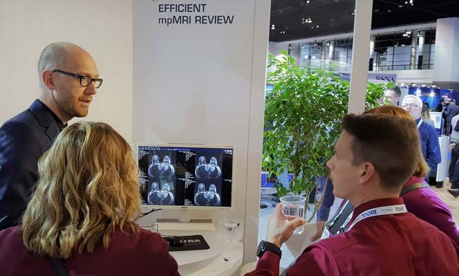
Workflow orchestration
           As more and more healthcare providers consolidate, and radiology
becomes more sub-specialized, the need for efficiently managed workflows has
never been greater. To prevent physician overload and burnout, and to adhere to
service-level agreements (SLAs), providers need to ensure that the right radiologist
reviews the right exam in the right order. You need to be equipped with tools that
can ensure an efficient workflow and maximize the probability that studies will be
read within agreed upon SLAs, while evenly and fairly distributing the workload with
respect to complexity.

The value of efficient workflow management is substantial. According to industry
leaders interviewed in a 2018 article7 published in Diagnostic Imaging, approximately
$15–20 billion of radiology’s expenditures are unnecessary and avoidable, caused
by shortages in the workflow. Either the right test is conducted at the wrong time, or
patients receive an unnecessary repeat exam.

Workflow management from an enterprise imaging perspective
It is fundamental that workflow orchestration tools are tightly integrated with, or pref-
erably a part of, the enterprise imaging system. Patients are moved between depart-
ments where different data, images and requests are generated, and radiology is only
one component in the optimization equation. Alongside proper integration, authenti-
cation and contextual synchronization with other systems such as the PACS, EMR and
viewers, a ‘multi-ology’ enterprise imaging system is a must for realizing proper
workflow management.

Information regarding future referrals and follow-ups is generated in various depart-
ments. Integrations with surrounding systems provide the enterprise imaging system
with the necessary information to create a centrally controlled and unified workload
distribution. In the future, access to information from various systems will become
even more important as these systems become smarter at predicting and estimating
workloads.

An enterprise imaging-centric approach towards workflow orchestration also con-
tributes with a unified user interface. A single, easy-to-administer user interface will
minimize interruptions to the already pressured workflow for radiologists and tech-
nologists. Launching a separate system should be avoided at all costs.

Specific worklist tools
Many vendors offer good support for semi-automated workflow management. This
is often based on adjustable worklists that can be configured to distribute the right
exam to the right radiologist based on a set of criteria. These include worklists for
acute cases and specific sub-specialties, as well as peer review and teaching and
resident workflows. Adjustable worklists need to be tailored to suit the needs of each
department.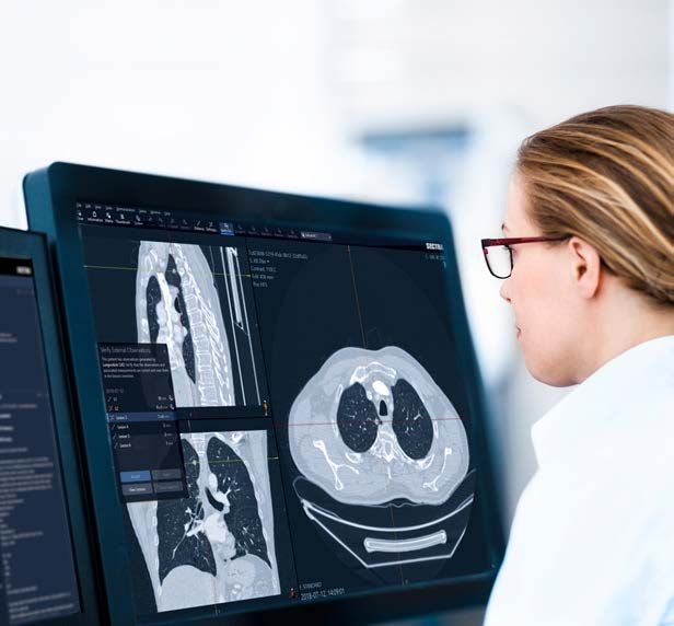
THE RADIOLOGIST’S HANDBOOK FOR FUTURE EXCELLENCE 2020   8

Communication tools
In addition, communication tools such as built-in chat func-
tionality and automatic notifications are key to facilitating
workflow orchestration. One example that highlights the
importance of efficient communication tools was found in a
study conducted at Massachusetts General Hospital, where
they implemented an automatic alert-based intervention that
reduced the number of delayed cases by 78 percent8.
                                                                                         78%
Real-time and two-way communication was ranked as one of
the most essential workflow-enhancing functionalities in an
article7 published in Diagnostic Imaging in 2018. Insights from                 Reduction in delayed cases according
                                                                                    to a study8 due to efficient
several leading vendors of workflow orchestration solutions                            communication tools.
were summarized as follows:

            “Real-time communication is one of the most effective ways to transfer and
            manage data and side-step problems. It helps eliminate patient care delays
                            and can prevent both clinical and medical record errors.”
                                                                              Palmer, 2018

The interviews also reveal that instantaneous connection accelerates
the workflow and can free up time for other responsibilities7.

Workflow overview, alerts and analytics
To make the right decisions, it is important to gain access to
an overview of the current workload and performance for
individual radiologists as well as for groups, departments and
connected sites. The system needs to provide a real-time,
customizable dashboard that gives an overview of work and
allocations, identifies bottlenecks, and reallocates exams                            ~67%
based on clinical status or SLAs.

The importance of having a system that can alert radiology
managers to problems that need to be resolved was demon-
                                                                                  Reduction of examination-to-PACS
strated in a study from 20179 that tracked exams from image
                                                                                     delays due to efficient alert
acquisition to the PACS. Using a system that tracked and                          functionality was fund in a study9.
alerted managers in real time based on outliers, the study
confirmed that active alerts produced a threefold
reduction in examination-to-PACS delays. Their conclusion was:

            “By providing supervisors with information about exactly where delays emerge in
            their workflow and alerting the correct staff to take action, applications like ours
                  create more robust radiology workflow with predictable, timely outcomes.”
                                                                               Pianykh et al., 2017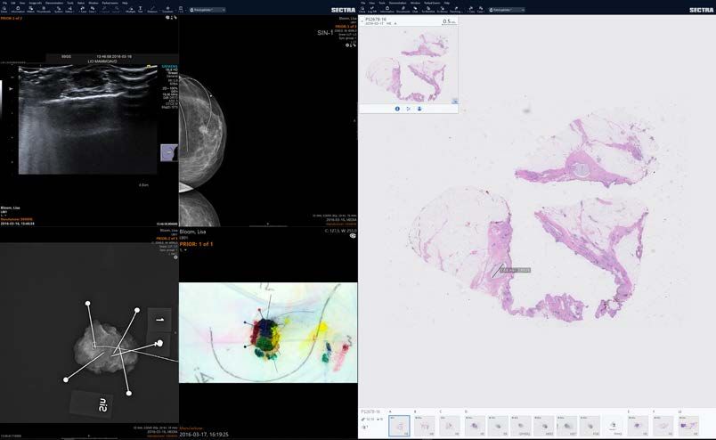
THE RADIOLOGIST’S HANDBOOK FOR FUTURE EXCELLENCE 2020   9

Another functionality that should not be underestimated is the ability to extract ana-
lytical reports as a basis for decision-making to justify further recruitments or invest-
ments.

The future: Automatic reallocation
Workflow orchestration that relies on AI-assisted workflow management is under
development and some early pilots are under way. “Smart” automatic reallocation of
exams should be based on the workload situation and different events, and be capa-
ble of managing the following tasks:

  »»Automatically plan and organize exams, using pre-defined reading sessions desig-
    nated by department, specialty, time of day, and other factors
  »»Using dynamic exam assignment, automatically escalate and assign studies to the
    most appropriate and available reader based on radiologists’ availability, sub-
    specialties, location, time of day, etc.
  »»Automatically balance workloads across your enterprise by matching exam work-
    load with reading capacity
  »»Prioritize reading queues and set configurable due-in-time thresholds for escalat-
    ing and monitoring SLA exams
  »»Send specific exams for AI-assisted pre-analysis based on the request and type of
    exam. For example, detection of lung noduli, lung embolisms or pneumothorax

AI-based automated workflow systems that will be available in the coming years will
be able to intelligently use and apply data and assist with operations management.
For instance, they will automatically recognize bottlenecks and use data to appropri-
ately route exams to available radiologists to reduce waiting times and streamline
care delivery. As an early adopter, implementing AI-based workflow orchestration
capability can give you a jump start when it comes to increasing productivity in your
overall reading workflow.

The future: Forecasting capabilities and predictive analytics
In addition, forecasting capabilities based on patient visit data generated from the
EMR and surrounding systems will be a key component of tomorrow’s workflow
orchestration engines. Efficiency gains will be realized through better planning for
future recruitments and vacation periods, and by adjusting the exam reallocation.
Now is a great time to scrutinize your enterprise imaging vendor’s capability
to integrate with various EMR vendors.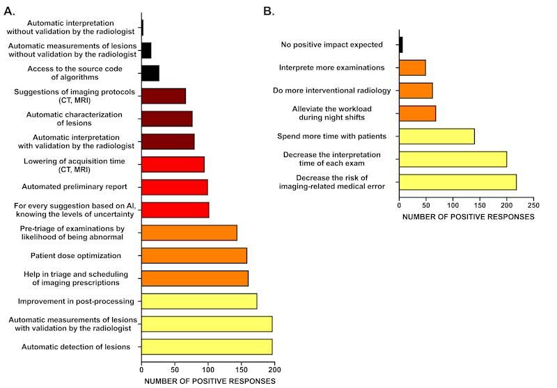
THE RADIOLOGIST’S HANDBOOK FOR FUTURE EXCELLENCE 2020   10

Workflow orchestration—The checklist
;;Workflow orchestration tools should be tightly integrated with,
  or preferably a part of, the enterprise imaging system. Only then
  will the platform provide a single user interface that is efficient to
  administer, and the workflow can be optimized across specialties

;;Workflow functionality should manage the integration, authentica-
  tion and contextual synchronization with various IT systems, such
  as the enterprise imaging system, the EMR, and advanced visualiza-
  tion software

;;Adjustable worklists should be configured to distribute the right
  exam to the right radiologist based on a set of criteria and tailored
  for each department’s needs

;;Real-time and two-way communication tools are a must-have

;;A real-time and customizable overview dashboard is necessary for
  identifying bottlenecks and reallocating exams based on clinical
  status or SLAs. The functionality should also be able to provide
  analytical reports for decision-making

;;The system should be able to generate alerts based on outliers

;;Keep your eyes open for AI-assisted workflow management; being
  a pioneer in a pilot could improve your workflow before the tech-
  nology is generally available

;;Confirm your vendor’s EMR integration capabilities to enable future
  forecasting opportunities
Artificial intelligence
           “PACS should be smarter than a 5th-grader” was a popular article10 pub-
lished on AuntMinnie.com in 2012. Based on Dr. Eliot Siegel’s presentation at the New
York Medical Imaging Informatics Symposium, the article made reference to a popular
American TV game show—“Are You Smarter than a 5th Grader?”—in describing the
modern PACS of the time as a “dumb” system. Seigel’s mission was to highlight the
need for adopting AI in radiology—an idea that today is close to becoming reality.

Much of the functionality that he asked for seven years ago is now available or on
their way to become. Many of these developments—for example, PACS doing
automatic measurements and tracking of tumor growth, auto-populating fields in a
structured report, matching of prior exams, or integrating relevant data from the EMR
that could be useful in a diagnostic review—are due to recent advancements in deep
learning. Many of today’s radiologists are prepared to adopt the AI-based tools11 that
Dr. Siegel called for back in 2012 to increase productivity.

Performance of current AI
At present, we are witnessing narrow, task-specific AI applications that can match and
occasionally surpass the performance of radiologists in specific applications. Radiol-
ogists will potentially benefit from AI, as it will enable them to increase their efficiency
and deliver higher-quality diagnoses3.

Today, most people agree that AI will not replace the radiologists, but that it has the
potential to improve their capabilities, efficiency and accuracy. A good metaphor for
the symbiosis between AI and radiologists was used in an article12 on this topic, com-
paring the relationship between the radiologist and AI with pilots flying a plane with
assistance from a highly advanced navigation system:

               “… the world’s best airplanes with automatic navigation systems still have at least
                 two “human pilots” on board. Anything that deals with human lives, simply can’t
                                              be trusted on with machines or algorithms alone.”
                                                                                       Mohan, 2018

As with the adoption of any new technology, it will take time before regulations, med-
ical guidelines and healthcare—together with vendors—harmonize on how AI should
be used. It will also take time to see which applications from different providers work
well. Predicting the future is difficult but looking at the expectations of the radiology
community as a whole provide at least general guidance on what we will see in clinical
use the coming years.
THE RADIOLOGIST’S HANDBOOK FOR FUTURE EXCELLENCE 2020 12 Expectations for AI Probably the most extensive research into what radiologists expect from AI comes from a French study published in May 201913, in which a large number of radiologists were surveyed. Based on the results, the authors concluded that most radiologists believe AI will: »»Reduce medical errors, »»Reduce the time spent per exam, and »»Allow them to spend more time with patients. Looking deeper into the study13 on what specific tools radiologists expect from AI, three features were mentioned as the most expected (also summarized in Figure 1). »»Automatic detection of lesions »»Automatic measurements of lesions »»Improvements in post-processing Figure 1. A: Ranking of expected technical features of AI-based tools, depending on the number of positive responses. B: Ranking of expected practical impact of AI-based tools on daily practice, depending on the number of positive responses. Chart courtesy of Dr. Thibaut Jacques, Dr. Q. Waymel, et al. In summary, the study concluded that there is great interest in and willingness to adopt AI, but there is also a need to accelerate the introduction of AI. The bright side is that many of the applications mentioned by radiologists in the study are already available.
THE RADIOLOGIST’S HANDBOOK FOR FUTURE EXCELLENCE 2020             13

Examples of useful AI available today
In contrast to the systems available in 2012 when Siegel expressed his frustration
with “dumb” PACS10, AI is no longer just a thing of the future. AI is here, and some
applications are ready to augment radiologists. Applications that radiologists soon will
be able to benefit from include:

 »»AI-assisted workflow and triage
   • External AI apps can communicate with PACS to indicate the presence of
     potential findings and the nature of these findings. This information can be used
     to control the workflow, how exams are to be prioritized and the worklist they
     should be on.

   In a survey from 201711 that asked radiologists whether they                   4%
   would trust an AI system to help them to prioritize cases, 44%                      7%
   either agreed completely or to some extent (see graph). This
   number is probably much higher today.                                  19%

                                                                                              37%

        Statement 2: I would trust an autonomous system to make             33%                           Agree completely
                                                                                                          Partly agree
     the decisions for me in terms of what to read and when based                                         Neither agree nor disagree
                                                                                                          Partly disagree
    upon findings from the automatic processing of the image data.                                        Completely disagree

 »»AI-assisted diagnostic review and reporting
   • External AI apps can communicate information about detected and charac-
     terized findings, which can act as an additional information source to support
     radiologists during review. One example is automatic lesion tracking, where the
     system finds and matches the same tumor on several sets of exams and mea-
     sures its development.
   • Information about detected and characterized findings can be linked to a
     reporting template to create pre-filled structured reports.
   • Matching series highlighting has been proven to save a great amount of time. This
     is a typical feature where previous matching examinations are called attention
     to, that was previously prototyped and requested by many users and is now
     available.

Future development of AI
In the coming years, we will witness a rapid development of AI, with better and
quicker access to a large variety of best-of-breed AI applications. One example that
Dr. Siegel mentioned10, and that many vendors are working on, is smart display proto-
cols that provide suggestions on appropriate hanging protocols based upon previ-
THE RADIOLOGIST’S HANDBOOK FOR FUTURE EXCELLENCE 2020 14 ous preferences. Also, automatic identification of relevant priors is a feature that will save significant amount of time. Such applications will probably be accessed through platforms provided by the enterprise imaging vendors. At the same time, we will see a development towards workflows and tools for easier evaluation, monitoring and validation of third-party AI applications, which will further facilitate adoption. How to facilitate AI adoption As it is still unclear which AI applications and vendors will add the most value, it is important to be equipped with a platform that offers a portfolio of AI applications from different vendors. These platforms—sometimes referred to as “AI application stores”—provided by enterprise imaging vendors, could be a way to spur adoption. Designed correctly, AI platforms could solve several of the main challenges related to adoption. We have identified three fundamental aspects for successful AI platforms: 1. Seamless integration into existing workflow AI applications need to be part of, and be tightly integrated into, existing workflows to enable adoption in everyday radiology. There should be no external launch and the AI should be accessed as easily as any tool in the radiologist’s diagnostic work- space. Most helpful would be a platform that provides tight, validated and tested integrations of AI applications into existing diagnostic workflows. 2. AI made “unremarkable” Every time the AI component catches the user’s attention without contributing any value, it instead degrades the user’s efficiency. Therefore, AI should only provide guidance when necessary and needs to be made “unremarkable” by being very carefully tailored to fit the clinical scenarios where it is to be used. Nuisance issues, such as having to log in to another application or being offered assistance when you don’t need it, can and will prevent adoption. The design and integration offered by the enterprise imaging vendor should ensure that the AI tools accessed through the diagnostic review offer guidance only when it adds value. 3. Rich portfolio access through neutrality Most AI vendors have focused on a small area of interest. But a radiology depart- ment requires access to a rich portfolio of applications within many different areas, including breast, lung, liver and chest—preferably from one contracting party. Han- dling different contracts and business models with various vendors is most likely an arduous task for most radiology departments. The enterprise imaging vendors’ AI platform should allow for a broad set of AI solutions for various tasks, through a single user interface and contract. To summarize, the first step to facilitating AI adoption is investing in a well-integra- ted AI platform that can provide a rich portfolio of “unremarkable” and reliable AI applications in the long term.
THE RADIOLOGIST’S HANDBOOK FOR FUTURE EXCELLENCE 2020   15

Artificial intelligence—The checklist
;;AI application platforms provided by enterprise imaging vendors
  will speed up and facilitate the adoption of AI in radiology

;;An AI application platform should be able to:
   • Offer a tight integration into the existing workflow
   • Offer “unremarkable” AI and only assist when needed
   • Offer a rich portfolio by being vendor neutral
   • Handle contracts with various AI vendors
   • Provide applications where the integration is tested and quality
     assured

;;Examples of applications already available and ready to use:
   • AI-assisted workflow and triage
   • AI-assisted diagnostic review and reporting:
       • Automatic lesion tracking
       • Pre-filled structured reports
       • Matching series highlighting

;;Make sure the vendor’s roadmap contains workflows and tools for
  easier evaluation and validation of AI applications
Multiparametric MRI
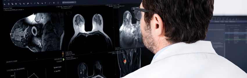
There are a few modality-specific technologies on the rise, such as
dual-energy CT, PET/CT and multiparametric MRI (mpMRI), all of which imposing new
demands on PACS and advanced diagnostic applications for efficient image review.
The need for high-performance applications is particularly significant for mpMRI
review—the use of mpMRI has become a central part of cancer diagnostics and
volumes are steadily growing14, especially for breast and prostate that, together with
lung, are the most common cancer types15. This development has mainly been fueled
by a range of recent studies showing significant positive clinical value by using mpMRI
over other alternatives. However, mpMRI review takes more time than many
other exam types. Radiologists who are equipped with tools to review these studies
more efficiently will perform better than those who are not.
Efficient mpMRI diagnostic tools—a must-have for
managing workloads
Using MRI to derive the PI-RADS® score for prostate in order to predict cancerous
lesions provides a very accurate diagnostic tool with high specificity for high-grade
disease14. Radiologists who can separate “bad” lesions from those that do not require
surgery will not only avoid unnecessary patient suffering, but also focus resources
and speed up the time to surgery for those who need it.
“Through the use of dynamic contrast-enhanced and diffusion weighted series [mpMRI, Ed.],
prostate tumors that were previously not visible have become identifiable and quantifiable.The
ability of MRI to improve staging and identification of clinically significant disease has resulted in
increased utilization for different aspects of prostate cancer care.”
Hutchinson and Lotan, 2017
The need for tools to increase productivity in prostate cancer care is significant,
partly based on its massive increase in cost. The expenditures for the care of
prostate cancer are expected to constitute the largest single portion of projected
cancer care cost increases in the US in the coming years14.
Demand for MRI diagnostics is also growing within breast cancer, indicating a steady
increase in volumes, mainly because of its high sensitivity and superior ability to
detect cancers. A study from 201916 comparing different methods for breast cancer
diagnostics concluded that mpMRI provides far more information than alternative
imaging—namely high-resolution, cross-sectional lesion morphology—as well asTHE RADIOLOGIST’S HANDBOOK FOR FUTURE EXCELLENCE 2020 17
functional information on a variety of tissue perfusion, vessel permeability, tissue
relaxation times, tissue cellularity/proliferation rate, and interstitial pressure. This is
useful information for tissue characterization, and for distinguishing between benign
and malignant lesions16.
Although the use of mpMRI has led to a
paradigm shift in the form of improved
diagnosis and monitoring of prostate and
breast cancer, it has also led to longer
examination reading times for radiolo-
gists16 and changes in the practice of breast
cancer review14. Hence, the benefits gained
from mpMRI imaging must outweigh the
increased costs of longer review times for
practices and departments. Highly effi-
cient tools for mpMRI diagnostics for
breast and prostate are a therefore a
key priority, and an area where enterprise
imaging vendors differ in their offerings.
Moreover, the use of mpMRI on other
organs such as neuro and colon is growing, A picture from the Sectra booth at RSNA 2018, where prototypes of mpMRI functionality
which will make such support important in were shown to attendees. This functionality is now released and will be demonstrated at
this year’s RSNA.
the future.
Ease of use and performance are key
Experiences from early users of Sectra’s mpMRI tools have shown that ease of use is
more important than the actual number of features, and that advanced functionality
for mpMRI must be seamlessly integrated into the diagnostic workflow. Ease of use
also must be complemented by high performance since there will be no tolerance for
switching windows or waiting for image processing. If your enterprise imaging vendor
does not offer in-house developed functionality for mpMRI, you need to verify that
it can smoothly integrate with third-party applications. Either way, it is essential that
your vendor provides tools for mpMRI review that are well integrated into the existing
diagnostic window and can meet your demands in terms of processing time.
It is worth mentioning that the same criteria are valid also for seamlessly integrated
advanced visualization functionality for PET/CT images. A good diagnostic user
interface would not require any separate logins or switching of windows to use such
functionality.THE RADIOLOGIST’S HANDBOOK FOR FUTURE EXCELLENCE 2020 18
Don’t take shortcuts on performance that compromise
on image quality
Another area related to the performance of mpMRI functionality is the frequently dis-
cussed topic of high-speed breast tomosynthesis review. Make sure you are equipped
with an application for tomosynthesis images that provides high performance and
doesn’t compromise on image quality—you need both. According to an article from
201717, for example, the use of lossy image streaming is disallowed by the Mammog-
raphy Quality Standards Act (MQSA) for breast imaging and mammography in the US.
Their review concluded that:
“…compression in medical imaging have led several professional organizations to develop guide-
lines for the use of lossy compression in clinical practice. However, these guidelines are not
always consistent among each other. […] the use of lossy compression of mammography data
is disallowed by the MQSA in the US.”
Liu et al., 2017
Integrated structured reporting
Another application worth mentioning that supports the mpMRI review is structured
reporting. The standardized methods for the acquisition, interpretation and report-
ing of breast and prostate cancer, BI-RADS® and PI-RADS®, could be streamlined by
utilizing functionality for structured reporting. Ideally, it would automatically populate
necessary information into the report, acting as both a time saver and decision sup-
port tool for users. It is a good idea to make sure you are equipped with mpMRI tools
that can automatically populate measurements into structured reports.
Example of functionality for structured reporting where prostate measurements are automatically populated into the template.THE RADIOLOGIST’S HANDBOOK FOR FUTURE EXCELLENCE 2020 19 Multiparametric MRI—The checklist ;;Make sure your enterprise imaging vendor provides tools for prostate and breast mpMRI, either developed in-house or tightly integrated third-party applications ;;Make sure the mpMRI functionality is well integrated into the exist- ing diagnostic window, is easy to use, and can meet your demands in terms of processing time. Functionality for advanced visualization of PET/CT is another area where these requirements are important ;;mpMRI review will likely grow in other sub-specialties outside breast and prostate. Your vendor’s ability to provide efficient tools for neuro and colon review will be valuable ;;Make sure you are equipped with an application for tomosynthesis images that provide high performance and do not compromise on image quality. For example, lossy image streaming is not allowed by MQSA guidelines ;;Functionality should be available to automatically populate struc- tured reports to support BI-RADS and PI-RADS scoring
Cross-discipline collaboration with pathology
           Most radiologists would likely agree that better coordination among the
medical specialties involved in cancer care would mean fewer errors and more effi-
cient use of time. Radiology is becoming increasingly dependent on collaboration with
other specialists and can thus take the lead in managing cross-disciplinary workflows.
As one of the most IT-knowledgeable disciplines, radiology can blaze the trail by
utilizing new digital technology to apply so called integrated diagnostics,
especially with pathologists who are now moving towards digital image review.

Today, information exchange between specialties is often an analog-based and
time-consuming task, mainly due to poor IT support. In a recent positive develop-
ment, many healthcare providers are moving towards enterprise imaging systems
that manage all patient-related multimedia, including images, documents and reports,
in a single platform. Separate departmental PACS are becoming a thing of the past. A
unified, central system provides radiologists with access to information, reports and
images from all specialties, thus saving time and even increasing their confidence in
decision-making.

We have identified three main areas in cross-disciplinary collaboration where the right
functionality can provide substantial time savings:

  »»Information and image access
  »»Identification of discordance
  »»Tumor board preparation, presentation and follow-up

Information and image access
Make sure your enterprise imaging system supports a multimedia workflow for the
most important collaborators with whom you exchange information. For most radiol-
ogists, this currently includes pathology, cardiology and breast imaging, and in the
future will also include genomics.

Leading institutions around the world are taking action in this area and are in the pro-
cess of implementing one enterprise imaging system that supports the workflows of
all of these collaborators. Using one system for all -ologies is probably not a prerequi-
site for efficient image and report access, but it does minimize the number of inte-
grations. The result is a system more likely to run at lower costs, deliver higher
stability, and provide a better user experience.

When looking to digital pathology support for your enterprise imaging system, fast and
intuitive image review is fundamental as this will determine if pathologists can and will
leave the microscope behind in favor of digital review. Additionally, the system should
offer functionality for driving the pathologists’ workflow, with worklists similar to those
THE RADIOLOGIST’S HANDBOOK FOR FUTURE EXCELLENCE 2020 21
used by radiologists for years, tools for image analysis, and proper integrations with
the laboratory information system (LIS). Few enterprise imaging vendors can live up
to pathologists’ requirements today.
The demands are similar for breast imaging, where the system should offer fast diag-
nostic review for both high-volume screening and advanced diagnostics, including
support for tomosynthesis. Moreover, both pathology and breast imaging place high
requirements on tight integration with third-party applications for image analysis and
CAD.
Although the collaboration with cardiology is not as intense as with the other special-
ties mentioned, including cardiology as part of the enterprise imaging system would
represent a strong win not least from an IT perspective. This places particularly high
demands on good integrations with various best-of-breed cardiology applications. For
cardiologists to use the enterprise imaging system, it is essential that it can provide a
single diagnostic user interface without a need for multiple logins or to launch a new
window for each specific application. Having one user interface also minimizes the
need for training, which is often a struggle for cardiologists.
Identify rad-path discordance
Errors and discrepancies in radiology practices are uncomfortably common, with an
estimated day-to-day rate of 3–5% of studies reported. Much higher rates are men-
tioned in many targeted studies18. In some circumstances, diagnoses are proven by
a pathologic examination, and this proof can be used to evaluate prior radiological
diagnoses. The use of radiological-pathological correlation in decision-making, where
possible, can avoid some erroneous assumptions, and can ingrain the practice of
seeking histological proof of diagnoses before accepting them as incontrovertible18.
Implementing a rad-path correlation workflow poses both technical and organizatio-
nal challenges. To support a discrepancy workflow technically, the enterprise imaging
system should automatically be able to alert users to discrepancies between diagnos-
tic findings. While not yet on the market, this functionality would reduce errors in
diagnoses and potentially save time by avoiding unnecessary tumor board discus-
sions and preparation time.
To be on the ready to adopt leading-edge technology such as discordance workflow
functionality, radiology already needs to prepare and ensure it has the right prerequi-
sites in place. Several studies point to readiness, illuminating the need for communi-
cation flows and linking of structured diagnostic results:
“To this end, pathology-radiology integration workflows must ensure the flow of communications
and specimens and link structured diagnostic results from pathologists with those of radiologists.”
Liu et al., 2017THE RADIOLOGIST’S HANDBOOK FOR FUTURE EXCELLENCE 2020 22
Another study19 summarized some of the key elements of a system supporting inte-
grated diagnostic workflows as follows:
»»Structured reporting capabilities: necessary to automate concept extraction from
reports and perform comparisons
»»Supporting both radiology and pathology clinical workflows and the integration of
textual, image and quantitative data generated by both disciplines
»»Being able to measure changes in the efficiency and accuracy of diagnosis, treat-
ment timing and, ultimately, patient outcomes
»»The system should support interoperability in situations where radiology and
pathology are housed in different institutions
Tumor board preparation, presentation and follow-up
A single, joint platform for radiology, pathology and breast imaging will, together with
the right set of tumor board tools, improve productivity in the preparation, presenta-
tion and follow-up of tumor boards. This was experienced in a pilot study conducted
by University Hospitals in Cleveland, Ohio in 2018, where digital pathology images
and reports where integrated into their enterprise imaging system and used in their
breast tumor boards20:
The Rad-Path team agreed that integrated conferences like this could greatly enhance
the clinical care and efficiency system-wide, especially for tumor boards.
Another benefit was found in image-guided biopsy procedures carried out by the
radiologists:
“This is why radiologic and pathologic correlation with every image-guided biopsy is so important
[…] it helps us move toward a world where we really integrate radiologic, pathologic,
genomic, and clinical data into kind of one unifying system. There is huge potential.”
Hannah Gilmore, MD Chief of Anatomic Pathology and Director
of the Breast Pathology Service at University Hospitals Cleveland Medical CenterTHE RADIOLOGIST’S HANDBOOK FOR FUTURE EXCELLENCE 2020 23 Accessing the pathologists’ reports before the meeting helps determine whether new exams are required and if so, avoids unnecessary preparations and meeting time. It is not unusual that radiologists prepare cases for the meeting that require an additional biopsy or examination. Using one system in the presentation facilitates the discussion itself as images and reports can be accessed and reviewed on the fly to allow for a more efficient discussion. After the tumor board, accessing the pathologists’ reports will provide feedback on previous statements, including whether or not the suspected lesion was malignant. All in all, the key to efficiency lies in tools that span from case preparation to presentation to follow-up requirements. Example of an enterprise imaging system giving access to all kinds of medical images and reports necessary in the tumor board discussion.
THE RADIOLOGIST’S HANDBOOK FOR FUTURE EXCELLENCE 2020   24

Cross-disciplinary collaboration—The checklist
;;Ensure that the enterprise imaging system supports various multi-
  media and a workflow for the most important collaborators with
  whom you exchange information
   • Digital pathology: Fast and intuitive image review. Functionality
     for driving the pathologist workflow, tools for image analysis, and
     proper integrations with the LIS
   • Breast imaging: Fast diagnostic workstation for both mammogra-
     phy screening and clinical breast diagnostics, including support
     for tomosynthesis
   • Both pathology and breast imaging should be able to tightly inte-
     grate with third-party applications for image analysis and CAD
   • Integrate best-of-breed cardiology applications without a need
     to launch a new window or perform multiple logins. One user
     interface also minimizes training needs
   • If possible, adopt an enterprise PACS that can handle both
     radiology and pathology in the same user interface to facilitate
     the tumor board scenario

;;Ability to access the pathologists’ reports before, during and after
  the tumor board

;;In the future, when functionality is available, the enterprise imaging
  system should be able to automatically alert physicians to discrep-
  ancies between diagnostic findings, primarily between radiology
  and pathology. Begin today by building the most important prereq-
  uisites for this workflow, i.e.:
   • Structured reporting capabilities for both radiology and
     pathology
   • Support for both radiology and pathology clinical workflows and
     the integration of textual, image and quantitative data generated
     by both disciplines
THE RADIOLOGIST’S HANDBOOK FOR FUTURE EXCELLENCE 2020 25
Handbook summary
To thrive and prevent physician burnout, radiology must evolve. A paramount part of
this change will be to embrace the right technologies to empower radiologists to do
the same amount of work in less time. Improving productivity is about much more
than just retrieving image pixels faster. A more holistic approach is needed as well as
the use of smart tools on all levels of the workflow. The four areas of technology we
have identified—workflow orchestration, AI, mpMRI and cross-disciplinary collaboration—
will hopefully help and guide the radiology specialty to cope with growing volumes,
increase productivity and manage workloads. The checklists under each area offer
specific, short- and long-term advice on which functionality to adopt as part of your
enterprise imaging platform.
A prerequisite to benefit from new technology is to make sure you have a platform
that will be upgraded on a reasonably frequent basis. Scrutinizing the vendor’s “tech-
nology score” in the KLAS Research Performance Report and asking questions about
upgrade cycles will minimize the risk of ending up at a dead end.
When it comes to which key technologies to adopt, the following summary looks at
the ingredients for a system that will amplify your future success.
ARTIFICIAL
INTELLIGENCE
FOUR TECHNOLOGIES
TO AMPLIFY SUCCESS
WORKFLOW MULTIPARAMETRIC
ORCHESTRATION MRI
CROSS-DISCIPLINE
COLLABORATION WITH PATHOLOGYTHE RADIOLOGIST’S HANDBOOK FOR FUTURE EXCELLENCE 2020 26 Workflow orchestration »»Tools should be tightly integrated with, or preferably a part of, the enterprise imaging system »»Adjustable worklists should be configured to distribute the right exam to the right radiologist based on a set of criteria and tailored for each department’s needs »»Real-time and two-way communication tools are a must-have Artificial intelligence »»AI application platforms provided by enterprise imaging vendors will speed up and facilitate the adoption of AI in radiology. These platforms should be able to: • Offer a tight integration into the existing workflow • Offer “unremarkable” AI and only assist when needed • Offer a rich portfolio by being vendor neutral • Handle contracts with various AI vendors • Provide applications where the integration is tested and quality assured Multiparametric MRI »»Make sure your enterprise imaging vendor provides tools for prostate and breast mpMRI »»Make sure the mpMRI functionality is well integrated into the existing diagnostic window, is easy to use, and can meet your demands in terms of processing time »»Make sure you are equipped with an application for tomosynthesis images that provide high performance and do not compromise on image quality. For example, lossy image streaming is not allowed by MQSA guidelines »»Functionality should be available to automatically populate structured reports to support BI-RADS and PI-RADS scoring
THE RADIOLOGIST’S HANDBOOK FOR FUTURE EXCELLENCE 2020   27

Cross-discipline collaboration with pathology
  »»Ensure that the enterprise imaging system supports various multimedia and
    a workflow for the most important collaborators with whom you exchange
    information
   • Digital pathology: Fast and intuitive image review. Functionality for driving the
     pathologist workflow, tools for image analysis, and proper integrations with
     the LIS
   • Breast imaging: Fast diagnostic workstation for both high-volume screening and
     advanced diagnostics, including support for tomosynthesis
   • Integrate best-of-breed cardiology applications without a need to launch a new
     window or perform multiple logins. Having one user interface also minimizes
     training needs
   • If possible, adopt an enterprise PACS that can handle both radiology and
     pathology in the same user interface to facilitate the tumor board scenario

As technology is ever evolving, this handbook will soon be out of date. But no worries,
we will update it as shifts and changes occur with respect to technology, regulations
or medicine. To stay up to date, please subscribe to our knowledge-sharing updates
at medical.sectra.com/subscribe.

       Join us at RSNA
       If you want to see and experience some of the new technology mentioned
       in this handbook in action, we invite you to a live demonstration in Sectra’s
       booth #6113 at RSNA 2019, December 1–5. Make your demo reservation at
       medical.sectra.com/rsna.
THE RADIOLOGIST’S HANDBOOK FOR FUTURE EXCELLENCE 2020 28
Sources and inspiration
Bibliography
1. Medscape National Physician Burnout, Depression & Suicide Report 2019. s.l.:
Medscape, 2019. https://www.medscape.com/slideshow/2019-lifestyle-burn-
out-depression-6011056#3.
2. Lafleur, Laurie. Are you ready for a radiologist shortage? s.l. : AuntMinnie.com,
2018. https://www.auntminnie.com/index.aspx?sec=sup&sub-
=ic&pag=dis&ItemID=120391.
3. Artificial intelligence in radiology. Ahmed Hosny, Chintan Parmar,
John Quackenbush, Lawrence H. Schwartz, Hugo J. W. L. Aerts. 8, s.l. : Nat Review
Cancer, 2018, Vol. 18. https://www.ncbi.nlm.nih.gov/pmc/articles/PMC6268174/.
4. Biggest Threat to Radiology. AuntMinnie.com. [Online] 09 26, 2018. [Cit-
ed: 05 17, 2019.] https://www.auntminnie.com/index.aspx?sec=nws&sub-
=rad&pag=dis&ItemID=121882.
5. Phil Galewitz. To Improve Health, Cut Costs, Walmart Pushes For Better
Medical Imaging For Workers. npr. [Online] 05 17, 2019. [Cited: 05 26, 2019.]
https://www.npr.org/sections/health-shots/2019/05/17/724300217/to-im-
prove-health-cut-costs-walmart-pushes-for-better-medical-imaging-for-work-
er?t=1558874251136.
6. KLAS Research. PACS 2018: Achieving Success from Selection to Go-Live. s.l. :
KLAS Research, 2018. https://klasresearch.com/report/pacs-2018/1329.
7. Whitney J. Palmer. How to streamline radiology workflow. Diagnostic Imaging.
[Online] Medern Medicine Network, 08 27, 2018. [Cited: 05 28, 2019.] https://
www.diagnosticimaging.com/article/how-streamline-radiology-workflow.
8. The Pareto Principle. H. Benjamin Harvey, MD, Susan T. Sotardi, MD. 6, Boston :
Journal of the American College of Radiology, 2018, Vol. 15. https://www.jacr.org/
article/S1546-1440(18)30257-6/fulltext.
9. Improving Radiology Workflow with Automated Examination Tracking and Alerts.
Oleg S. Pianykh, PhD et. al. 7, s.l. : Journal of the American College of Radiology,
2017, Vol. 14. https://www.jacr.org/article/S1546-1440(17)30345-9/fulltext.
10. Siegel. PACS should be smarter than a 5th-grader. AuntMinnie.com. [Online]
AuntMinnie, 09 12, 2012. [Cited: 05 23, 2019.] https://www.auntminnie.com/in-
dex.aspx?sec=sup&sub=pac&pag=dis&ItemID=100540.
11. Sectra. Where radiologists see the added value of machine learning. Linkoping :
Sectra, 2017. https://medical.sectra.com/resources/where-radiologists-see-the-
added-value-of-machine-learning/THE RADIOLOGIST’S HANDBOOK FOR FUTURE EXCELLENCE 2020 29
12. Artificial intelligence in radiology – Are we treating the image or the patient?
Chander Mohan, SM. 2, s.l. : Indian J Radiol Imaging, 2018, Vol. 28. https://www.
ncbi.nlm.nih.gov/pmc/articles/PMC6038229/.
13. Impact of the rise of artificial intelligence in radiology: What do radiologists
think? al., Waymel Q et. Lille, France : Diagnostic and Interventional Imaging,
2019. https://www.ncbi.nlm.nih.gov/pubmed/31072803.
14. Cost consideration in utilization of multiparametric magnetic resonance imag-
ing in prostate cancer. Hutchinson, Ryan and Lotan, Yair. 3, s.l. : Transl Androl
Urol, 2017, Vol. 6. https://www.ncbi.nlm.nih.gov/pmc/articles/PMC5503976/.
15. National Cancer Institute (NIH). Common Cancer Types. s.l. : National Cancer
Institute (NIH), 2019. https://www.cancer.gov/types/common-cancers.
16. Abbreviated Magnetic Resonance Imaging for Breast Cancer Screening:
Concept, Early Results, and Considerations. Eun Sook Ko, MD and Elizabeth A.
Morris, MD. 4, s.l. : Korean J Radiol, 2019, Vol. 20. https://www.ncbi.nlm.nih.gov/
pmc/articles/PMC6424827/.
17. Feng Liu, Miguel Hernandez-Cabronero, Victor Sanchez, Michael W.
Marcellin, Ali Bilgin. The Current Role of Image Compression Standards
in. s.l. : MDPI, 2017. https://pdfs.semanticscholar.org/b80d/9e1d3a8f-
f72e2af1014a2521dd6463a67335.pdf.
18. Error and discrepancy in radiology: inevitable or avoidable? Brady, Adrian P. 1,
s.l. : Insights Imaging, 2017, Vol. 8. https://www.ncbi.nlm.nih.gov/pmc/articles/
PMC5265198/.
19. Integrating pathology and radiology disciplines: an emerging opportunity?
James Sorace, corresponding author, et al. 100, s.l. : BMC Medicine , 2012, Vol.
10. https://www.ncbi.nlm.nih.gov/pmc/articles/PMC3523019/.
20. Tierny, Mary. Digital Pathology Pilot Predicts Prosperity: Pondering
Pathology’s Pivot. s.l. : Health Imaging, 2019. https://www.healthimaging.
com/sponsored/1068/topics/imaging-informatics/digital-pathology-pilot-pre-
dicts-prosperity-pondering.
Sectra AB • info.medical@sectra.com • medical.sectra.com
This is a marketing material and may be changed at any time without prior notice.
Sectra will not be held liable for any errors or misconceptions herein.
DOC-EKAN-BGMBZM-1.0 © 2019 Sectra ABYou can also read