The renal collecting duct carcinoma (CDC) (Bellini duct carcinoma), case report - A. Barakauskienė, L. Neverauskienė, T. Petraitis, J. Strimaitienė
←
→
Page content transcription
If your browser does not render page correctly, please read the page content below
The renal collecting duct
 carcinoma (CDC)
(Bellini duct carcinoma), case
  report
 A. Barakauskienė, L. Neverauskienė,
 T. Petraitis, J. Strimaitienė
The renal collecting duct carcinoma (CDC)
      exhibits special clinicopathological
features, high degree of malignancy and    poor
prognosis

      Synonyms like Bellini duct carcinoma,
medullary renal carcinoma, distal renal
tubular carcinoma and distal nephron
carcinoma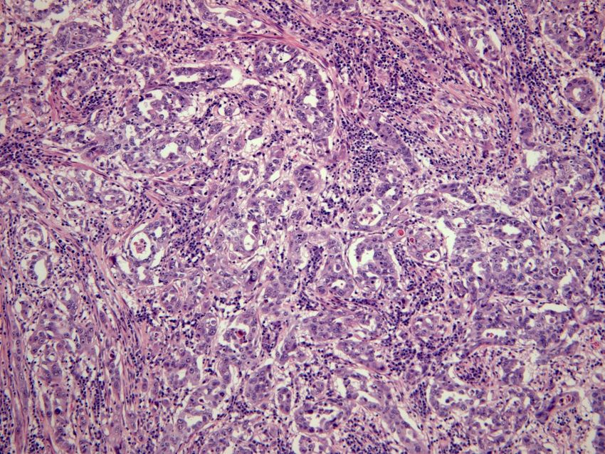
The diagnosis depends on the
histopathological examination

     Early detection and early surgical treatment
     are still the main methods to improve the
prognosis of patients with CDC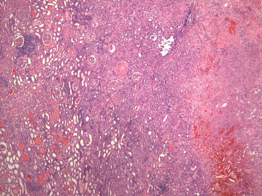
CDC is located in the renal medulla and
originates from the epithelial cells of Bellini
collecting ducts

       Currently, WHO names it as Bellini duct
carcinoma, it’s considered to be an independent
histological type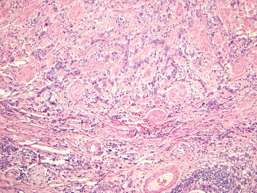
CDC is an unusual variant of renal cell
carcinoma and accounts for about 1-2% of all
renal cell carcinomas

      The average onset age is 58 years and male
      patients account for 72% of the cases

      CDC metastasizes to regional lymph nodes in
      approximately 80% of cases, to the lung or
adrenal gland in 25% and to the liver in 20%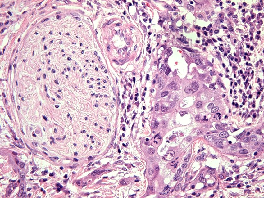
Painless gross hematuria, lumbar abdominal
       pain, waist and abdominal mass, fatigue,
fever, and weight loss

     Metastasis occurs in most of patients before
     treatment, including bone metastasis and
lymph node metastasis

      Average survival time has been reported to be
      22 months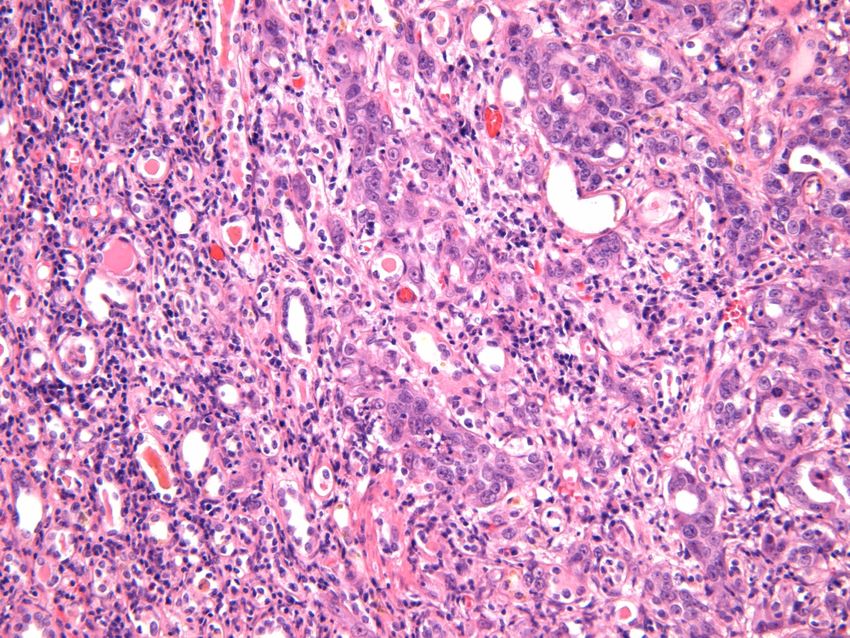
Imaging examinations are the main methods for
CDC diagnosis

The tumors are hypo-vascular with ill-defined
border

Pose invasions to the renal cortex and renal
sinus
CT is able to detect the invasions of tumors into
pelvis and renal cortex

Calcification and hemorrhage can also be seen
in some cases

Mild to moderate uneven delayed enhancement
can be detected in dynamic contrast-enhanced
scan

MRI gives iso-intensity or hyper-intensity
CDC doesn’t have specific imaging features
       that distinguish it from other types of renal cell
       carcinoma such as renal medullary carcinoma,
       sarcomatoid renal cell carcinoma and renal
pelvis carcinoma

      Its diagnosis requires pathological examination
      - the gold standard for diagnosis of CDC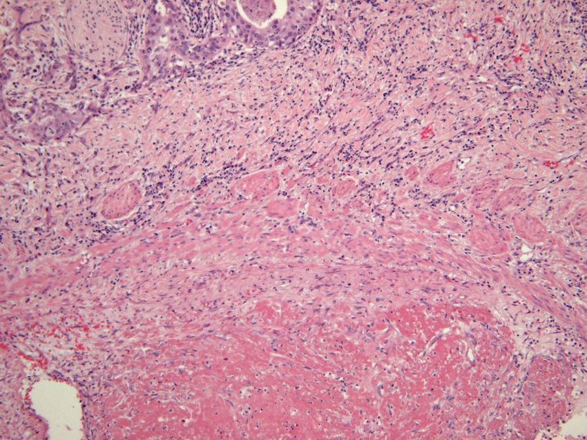
Our case report of CDC
                      (woman 76 years old)

•   2011 Painless microhematuria
•   2012.04 Abdomen pain, CT: renal upper part tumor 6,5 cm, no
    mts
•   Performed nephrectomia et lymphonodectomia per
    lumbotomiam infiltrate into renal cortex: Ca renis pT3aG3
    N0M0 anemia (C64)
•   2012.07 Progresing to liver 4,5 cm mts and lower v. cava 2 cm
    l/m, mutiple pulmonary mts 5 mm 3x1cm recidivating tumour in
    the same location, aortocavalis l/m 1x1 cm l/m.
•   2012.08 Biological Chemotherapy Sunitinibi: 37,5-12,5 mg 4/2
    weeks. After 2 weeks: diarrhea, weakness.
•   2012.04 – 2012.12 (8 month) Ca renis pT3aG3 N0M0 anemia
    (C64)
Our case report of CDC
• Pathology • Pathology
macroscopic data microscopic data
• Renal upper part tumor • Presents with
tubulopapillary
7x6x2 cm: yellow with
architecture
multiple necrosis zones,
• tumor cells form hobnail
infiltrates portal part,
pattern along the
subcapsular fat glandular tube
• infiltrate into renal cortex,
agressive perineural
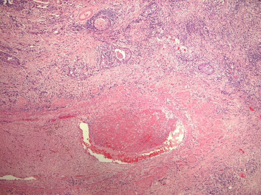
infiltrationTumor cells form tubulopapillary hobnail pattern along the glandular tube
Tumor cells form
tubulopapillary
hobnail pattern
along the
glandular tube
CDC morphology

• Tubulopapillary architecture with the
  hobnail-shaped cells protruding into the
  glandular lumen, and accompanied by
  interstitial fibrosis and dysplasia of
  epithelial cells in collecting ducts adjacent
  to the tumors
Dysplasia of
epithelial
cells in
collecting
ducts
adjacent to
the tumors
Dysplasia of
epithelial cells
in collecting
ducts
Normal
tubul
Neoplastic
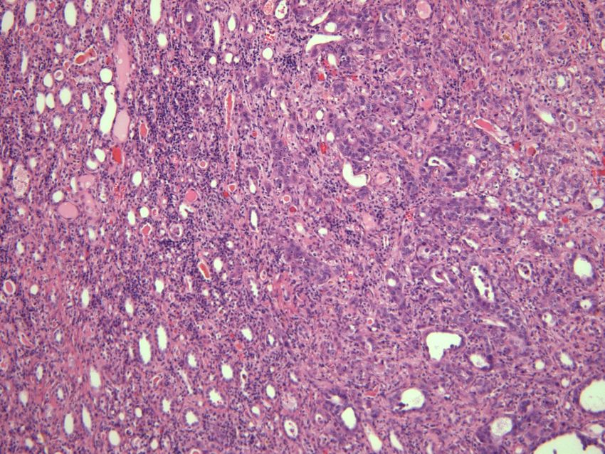
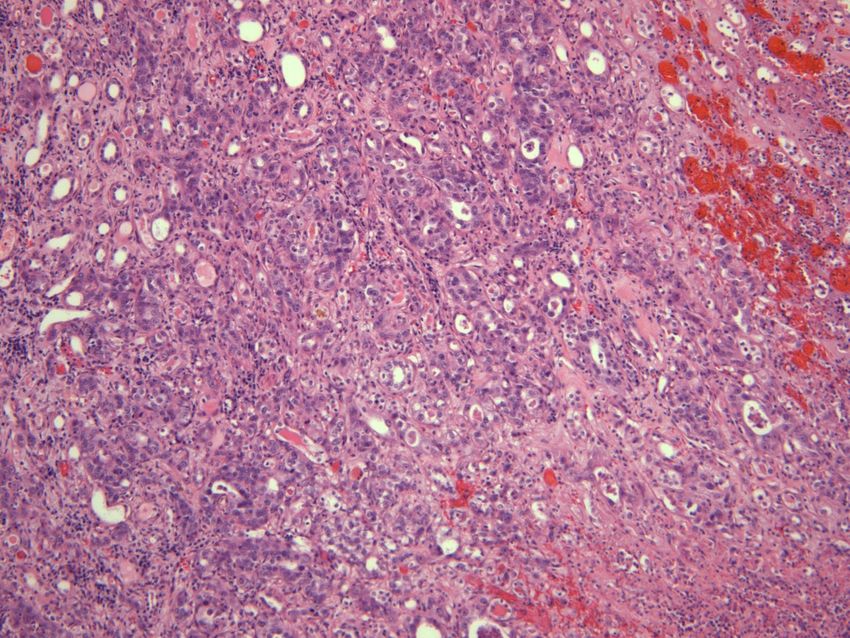
tubular pattern• Poorly differentiated tumor cells show nest-shaped, • rope-like, sarcomatoid • or adenoid cystic morphology, with or without interstitial connective tissue reaction
Interstitial fibrosis
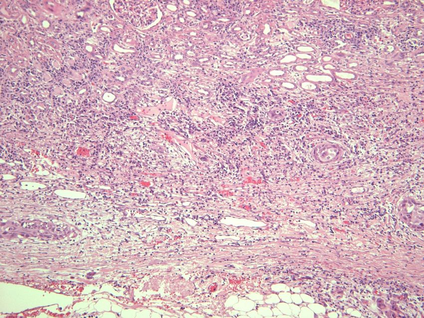
Agressive perineural
                       infiltration

Agressive perineural
infiltration
Agressive perineural
infiltration
Intravascular
trombusInfiltrate into renal cortex, subcapsular fat
Immunofenotype
• Cancer cell have positive expressions of CK
  (AE1/AE3), CK7, CK19, EMA, vimentin,
  CK34BE12, PNA and ulex europeus agglutinin
  (UEA), and negative expression of CD10 and
  CK20
• Combination of CK34BE12 and PNA is able to
  detect 90% of CDC
• Our case: PANCK CK (AE1/AE3) (+++) 90%,
  CK7 (+++) 90%, EMA 80%(+++), vimentin (+)
  10%, CD10 (+) 30%, CK20 (-), TTF1 (-)
PANCK / CK7 +++
100%
EMA +++
100%
CD10 + 30%
VIM ++ 10%

   EMA +++ 100%
Treatment
• Recently, there are a few reports on the
  effectiveness of targeted therapy with
  Sunitinib (multiple tyrosine kinase
  inhibitor) and sorafenib in treatment of
  CDC
• However, there’s a study indicating no
  response of targeted therapy
• Postoperative interferon immunotherapy is
  recommended
Survival time

The postoperative survival time for the
cases of stage IV 5 to 6 months
for the patients of stage III 9 to 12 months
for the case of stage II 18 months
Conclusions

• CDC is an unusual variant of renal cell carcinoma
  and accounts for about 1-2% of all RCC.
• CDC doesn’t have specific imaging features.
• Pathological examination is the gold standard for
  diagnosis of CDC.
• Radical nephrectomy is the major method to treat
  CDC.
• Treatments and corresponding outcomes are
  valuable information for guiding future clinical
  practice, therefore, the efficacy of targeted therapy
  on CDC remains to be demonstrated.
You can also read