Theme 4 Biomedical Engineering and Imaging 2019/20 - MRC DTP
←
→
Page content transcription
If your browser does not render page correctly, please read the page content below
Theme 4
Biomedical Engineering
and Imaging
2019/20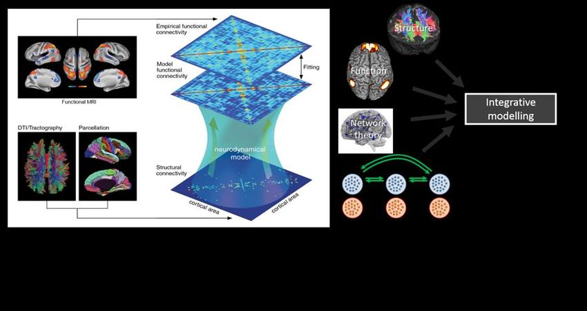
Contents
 1.4 The neonatal virtual brain: computational models of emerging connectivity using
 simultaneous EEG and MRI ........................................................................................................................................ 4
 2.4 Molecular Imaging of Collagen in Cardiac Remodelling .......................................................................... 6
 3.4 Diffusion and structural MR histology: new high-resolution MR imaging methodologies for
 quantitative mapping of cortical organisation during development ......................................................... 9
 4.4 Unravelling the role of pericellular matrix mechanics in regulating stem cell fate in 3D ....... 10
 5.4 Detection of bone marrow cancer using high sensitivity 7T MRI ...................................................... 11
 7.4 ‘RAGE’ as an imaging biomarker in Alzheimer’s disease - Structure-driven development,
 radiosynthesis and evaluation of a positron emission tomography radiotracer towards the
 Receptor for Advanced Glycation Endproducts (RAGE)............................................................................... 13
 8.4 Development of siRNA-based therapeutics for intestinal delivery in inflammatory bowel
 disease using imaging ............................................................................................................................................... 14
 9.4 Development of sideromycin derivatives as fluorescent probes for the imaging of Gram+/-ve
 bacterial infections .................................................................................................................................................... 15
 10.4 Design of novel 18F molecular probes for positron emission tomography brain imaging of
 the cannabinoid-1 receptor .................................................................................................................................... 17
 11.4 Using molecular imaging to identify early cardiotoxicity due to cancer chemotherapy ................... 19

                                                                                                                                                                        2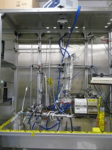
Imaging and Biomedical Engineering

This theme focuses on the link between biomedical and physical sciences – particularly
physics, engineering and computational approaches. Clinical functional and molecular
imaging (MRI, PET, X-MR and PET-MR) is a major strength, along with computational
modelling and biomaterials (particularly in the Dental Institute).

Lead: Professor Phil Blower

When choosing a project from this catalogue in the funding section of the online
application form please enter: MRCDTP2019_Theme4

Deadline for application: Sunday 25th November 2018
Shortlisted candidates will be contacted in early January.

Interviews: 30th & 31st January 2019
The 2019/20 studentships will commence in September 2019.

For further Information or queries relating to the application process please
contact mrc-dtp@kcl.ac.uk

 Projects listed in this catalogue are subject to amendments, candidates
   invited to interview will have the opportunity to discuss projects in
                                further detail.

                                                                                    3
1.4 The neonatal virtual brain: computational models of emerging connectivity using
simultaneous EEG and MRI

Co-Supervisor 1A:             Dr. Dafnis Batalle
School/Division & CAG: IoPPN, Forensic & Neurodevelopmental Sciences
KCL/KHP E-mail:               dafnis.batalle@kcl.ac.uk
KCL/KHP Website:              https://kclpure.kcl.ac.uk/portal/en/persons/dafnis-batalle(594a5a66-51aa-
                              421c-8863-39c245d3d93c)/projects.html

Co-Supervisor 1B:            Dr. Tomoki Arichi
School/Division & CAG: Biomedical Engineering & Imaging Sciences
KCL/KHP Email:               tomoki.arichi@kcl.ac.uk
KCL/KHP Website:             https://kclpure.kcl.ac.uk/portal/en/persons/tomoki-arichi(eb157769-4047-
                             47d1-b47a-95731abf11fe).html

Project description:

State-of-the-art mapping of structural and functional brain connectivity can enable the first use of
computational models of brain connectivity in newborn infants. This emerging field uses mathematical models
of neuronal activity to couple MRI-derived measures of white-matter structure and correlated functional activity
(Figure). As neuronal activity is fast and MRI suffers from low temporal resolution, further model refinement is
possible through incorporating high temporal resolution electrophysiology (EEG). We expect that this will
provide important insights into both the emergence of brain connectivity and the biological alterations which
underpin neurodevelopmental impairments resulting from perinatal injury.

This project will apply computational modelling techniques to a unique neonatal dataset of simultaneously
acquired EEG and MRI. Building on existing models derived from adult data, the student will develop
computational algorithms to link structural connectivity (from diffusion MRI) and functional connectivity (from
fMRI and EEG). This will include exploring different approaches such as Hopf Bifurcation and dynamic mean
field models and the adaptations needed for neonatal data. They will then aim to identify imaging phenotypes
and altered model parameters associated with neurodevelopmental disorders such as autism spectrum
disorder.

                                                                                                             4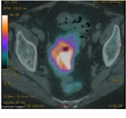
Objectives:

Year 1/3: Training in neuroimaging and modelling. Characterisation of structural/functional connectivity from
MRI and EEG.
Year 2/3: Development of computational models linking structure and function.
Year 3/3: Identify model characteristics as biomarkers of brain pathology.

The successful student will receive training in neuroimaging, computational modelling, and developmental
neuroscience through the supervisory team; the School’s research and educational program; and external
educational courses and conferences.

One representative publication from each co-supervisor:

[1] Batalle D, Hughes EJ, Zhang H, Tournier J-D, Tusor N, Aljabar P, Wali L, Alexander DC, Hajnal JV, Nosarti C,
Edwards AD, Counsell SJ; Early development of structural networks and the impact of prematurity on brain
connectivity; Neuroimage, 2017, 149, pp. 379-392

[2] Arichi T, Whitehead K, Barone G, Pressler R, Padormo F, Edwards AD, Fabrizi L. Localization of spontaneous
bursting neuronal activity in the preterm human brain with simultaneous EEG-fMRI. eLIFE 2017; e27814

                                                                                                             5
2.4 Molecular Imaging of Collagen in Cardiac Remodelling
Co-Supervisor 1A: Prof René M. Botnar
Research Division or CAG: Imaging Sciences and Biomedical Engineering, KCL
E-mail: rene.botnar@kcl.ac.uk
Website: https://kclpure.kcl.ac.uk/portal/rene.botnar.html
Co-Supervisor 1B: Alkystis Phinikaridou (Lecturer)
Research Division or CAG: Imaging Sciences and Biomedical Engineering, KCL
Email: alkystis.1.phinikaridou@kcl.ac.uk
Website: https://kclpure.kcl.ac.uk/portal/alkystis.1.phinikaridou.html
Project description:
Remodeling of collagen plays a crucial role in the pathogenesis of several diseases including heart failure,
atherosclerosis, aortic aneurysm dissection, lung and liver fibrosis. The Aim of this project is to investigate the
role of collagen deposition (fibrosis) on left ventricular (LV) remodeling and cardiac function post myocardial
infarction (MI) and the potential impact of therapies that aim at modulating the fibrotic response. The project
will utilize a commercially available collagen I-binding MR contrast agent and an established murine model of
post-MI remodeling. In parallel, we will develop and evaluate peptides that selectively bind to collagen types I
or III and which allow discrimination between the different types of collagen and thus enhance our
understanding of collagen turnover in disease progression. The probes can be labelled with radioisotopes for
initial screening tool in biological studies using PET/SPECT imaging. The most promising probe can then be
labelled with gadolinium for MRI experiments. All in vivo imaging experiments would be complemented by ex
vivo analysis.
Background:
Cardiovascular diseases (CVD) including coronary heart disease, stroke, heart failure (HF), cardiomyopathy, and
atrial fibrillation affect more that 2.3 million people and account for ~27% of all deaths in the UK. Amongst all
CVD, myocardial infarction (MI) and heart failure are the most frequent with ~175,000 people experiencing
episodes of acute MI per year and more than 500,000 people are living with HF in the UK.
The role of extracellular matrix (ECM) in post-MI remodeling and heart failure:
The structure and composition of the ECM is crucial to maintain cardiac anatomy and function as it mediates
cell-to-cell and cell-to-ECM molecular signaling and interactions. Myocardial fibrosis characterized by increased
ECM deposition is a key compensatory and repair mechanism. There are two forms of myocardial fibrosis; (i)
replacement fibrosis, following cardiomyocyte loss post-MI contributes to maintaining the cardiac
macroanatomy and (ii) reactive fibrosis, triggered by myocardial stress or inflammatory mediators that often
results in left ventricular (LV) stiffening, functional deterioration, and development of heart failure. Collagen is
a major ECM protein involved in post-MI remodeling. Following injury, myofibroblasts and other cell types
synthesize pro-collagens, mostly type 1 and type 3. Pro-collagen proteinases initially convert these precursor
molecules into collagen fibers which subsequently become cross-linked into collagen fibrils by lysyl-oxidase. In
parallel, upregulation of matrix metalloproteinases (MMPs) causes ECM degradation that not only facilitates
adaptive changes (including removal of cellular debris by inflammatory cells and migration of myofibroblasts)
but also contributes to the weakening of the myocardium and ventricular dilatation. Thus, a fine tuned balance
of collagen turnover is crucial for myocardial healing and preservation of cardiac function post-MI.
Imaging of Myocardial Fibrosis:
The size, location and extent of a myocardial infarction (MI) are important criteria for the evaluation of post-MI
remodeling. Multimodality molecular imaging including SPECT, PET, cardiac MRI, and optical approaches is
offering new insights into the pathophysiology of MI and left ventricular remodeling in small-animal models.
6Molecular imaging of collagen during myocardial healing was achieved with a Tc-99m-labelled collagelin tracer.
Because of its excellent spatial and temporal resolution and high soft-tissue contrast, MRI has evolved as the
gold-standard for cardiac imaging that enables evaluation of myocardial function, perfusion. Late-gadolinium
enhancement (LGE) using non-targeted contrast agents has been extensively used to indirectly detect fibrosis
by quantifying ECM volume. Although, LGE can accurately detect replacement fibrosis caused by myocardial
infarction it cannot detect diffuse fibrosis due to the lack of image contrast in normal myocardium. To address
this challenge, quantitative T1 mapping methods have been developed and been shown to accurately measure
the extracellular volume (ECV), which is associated with fibrosis and collagen formation. Although very
promising, T1 mapping does not directly detect or quantify collagen and cannot differentiate between the
different collagen sub-types. A recently developed collagen-binding MRI contrast agent (EP-3600) thus may be
an alternative and provide true molecular information. EP-3600 has been used to detect chronic myocardial scar
and myocardial perfusion defects under steady-state conditions, overcoming many of the limitations associated
with molecular quantification of myocardial remodeling and first-pass perfusion imaging (low spatial resolution,
quantification).
Objectives:
AIM 1- Year 1: In this project we will initially use a commercially available and well characterised gadolinium
binding contrast agent to image cardiac remodelling and fibrosis following MI in a murine model using a clinical
3T MR scanner. This is a well-established murine model of left-anterior descending coronary artery ligation that
it is already established within the Division. The temporal evolution of the biological processes that occur in this
animal model are well known and hence the choice of those particular imaging time points proposed in the
project. Animals will be scanned at days d1-d7-d14 and d21 post-MI with the contrast agent injected before the
MRI scan. Subsequently, tissues will be collected for ex vivo histological and other biochemical analysis. AIM 1
will provide:
a) Information on the role of the size, location and extent of collagen deposition and cardiac function
b) A comparison between the collagen-agent and late-gadolinium enhancement (LGE) using non-
targeted contrast agents for the accurate detection of the remodeling area using ex vivo histology as the gold-
standard
c) A comparison between quantitative T1 mapping methods that measure the extracellular volume (ECV)
and collagen-enhanced T1 mapping,
d) A comparison between first-pass perfusion and perfusion defects detected by the collagen-agent under
steady-state conditions.
e) How treatments that alter the collagen turnover affect collagen deposition and subsequently cardiac
function.
AIM 2 - Year 2: We have identified peptides that could potentially have selectivity towards collagens I and III and
we have performed preliminary in vitro binding essays. The peptides carry a DOTA chelate and thus have the
flexibility to be labelled with lanthanides (europium or gadolinium) or radioisotopes for SPECT and/ or PET. In
aim 2, the student will expand the in vitro binding essays and subsequently use radiolabelled peptides for initial
biodistribution and biological studies in control and diseased using PET and SPECT imaging. The most
promising probe will then be conjugated with gadolinium for in vivo MRI experiments described in Aim 3. This
aim requires input and guidance from a chemist and for this reason Dr Michelle Ma and Prof. Phil Blower with
extensive experience in chemistry have been added to the supervisory team. There is also an ongoing
collaboration with Dr Lacerda at the CNRS (France) who has performed similar work in the past and she agreed
to provide guidance and expertise as needed.
AIM 3 - Year 3: AIM 3 will be followed by in vivo testing of the peptides that were identified to be the best
candidates in AIM 2. The peptides will be then used for in vivo imaging of collagens I and III in the mouse model
of MI to study how the size, location and extent of each collagen sub-type affects cardiac remodelling and
subsequently left ventricular function. The experiments will follow the same design as outlined in AIM1 and they
will include MRI, PET/SPECT imaging.
7Techniques:
Generation, genotyping and maintenance of murine colonies, animal handling, tail injections,
anesthesia, tissue harvesting.
Surgical skills: permanent coronary artery ligation and ischemia-reperfusion.
Peptide chemistry for the co-ordination of lanthanides (e.g., gadolinium, Europium) or radioactive
compounds to peptides.
In vitro and in vivo PET/SPECT imaging experiments.
In vitro and in vivo cardiac MRI protocols including late-gadolinium enhancement, T1 mapping protocols
and 1st pass perfusion.
Ex vivo binding assays of radioactive isotopes or Europium labelled peptides (DELFIA assays).
Ex vivo histology, immunohistochemistry, proteomics, western blotting, ELISA, PCR, FACS, ICP-MS of
extracted tissues or cell lines.
Image processing and analysis software
Statistical analysis and software.
All of the infrastructure, equipment, and animal models are currently available within the Schools of Imaging
Sciences and Cardiovascular at KCL that has a multidisciplinary team of supervisors who have been working
closely together on several projects. We will ensure that the student will be trained and supervised on all of the
required techniques by experienced members of the Division.
Suggested undergraduate backgrounds: Biology or Chemistry.
One representative publication from each co-supervisor:
[1] Increased Vascular Permeability Measured With an Albumin-Binding Magnetic Resonance Contrast Agent Is
a Surrogate Marker of Rupture-Prone Atherosclerotic Plaque.
Phinikaridou A, Andia ME, Lavin B, Smith A, Saha P, Botnar RM.
Circ Cardiovasc Imaging. 2016 Dec;9(12). pii: e004910.
[2] Noninvasive magnetic resonance imaging evaluation of endothelial permeability in murine atherosclerosis
using an albumin-binding contrast agent.
Phinikaridou A, Andia ME, Protti A, Indermuehle A, Shah A, Smith A, Warley A, Botnar RM.
Circulation. 2012 Aug 7;126(6):707-19. doi: 10.1161/CIRCULATIONAHA.112.092098. Epub 2012 Jun 29.
83.4 Diffusion and structural MR histology: new high-resolution MR imaging methodologies for
quantitative mapping of cortical organisation during development
Co-Supervisor 1A: Flavio Dell’Acqua
School/Division & CAG: Academic Psychiatry - Forensic and Neurodevelopmental Sciences
KCL/KHP E-mail: Flavio.dellacqua@kcl.ac.uk
KCL/KHP Website: https://www.scopus.com/authid/detail.uri?authorId=24757840500
https://kclpure.kcl.ac.uk/portal/en/persons/flavio-dell-acqua(3de057d5-4de7-
43d9-a44a-70ff1aca7963).html
Co-Supervisor 1B: Marija-Magdalena Petrinovic
School/Division & CAG: Academic Psychiatry - Forensic and Neurodevelopmental Sciences
KCL/KHP Email: marija-magdalena.petrinovic@kcl.ac.uk
KCL/KHP Website: https://kclpure.kcl.ac.uk/portal/marija-magdalena.petrinovic.html
Project description:
The cortex is one of the most complex regions of the brain, characterized by an extraordinary diversity of cell
types, whose coordinated development and function underlie memory, cognition, language, and
consciousness. The formation of laminar and columnar organisation during development is a fundamental
prerequisite for proper cortical function. Decoding this organisation during development has been at the core
of neuroscientists’ work for decades. While traditional histological techniques provided critical insights about
cortical features they are limited by their invasive nature, they are time consuming and provide only a 2D-type
of information of a small part of the brain. Opposingly, MRI techniques non-invasively provide 3D-whole-brain
coverage, but not the high spatial resolution required to visualise cortical organisation. Aim of this project is to
develop new MRI protocols that will enable the quantitative characterisation of the laminar and columnar
organisation during typical and atypical (e.g. neurodevelopmental disorders) development in rodents.
In this project, the student will have access to the state-of-the-art preclinical imaging facilities at KCL including
the new 9.4T MRI scanner, the Nikon Imaging Centre and wet labs. The student will develop, and validate (e.g.
histology, cell cultures) new MRI structural and diffusion histology methods and will combine them with novel
microstructure diffusion imaging techniques. The goal is to develop new ultra-high resolution structural and
diffusion imaging methods able to probe cortical features at near histological resolution to map cortical
development in rats. Year 1-the student will learn technical skills and begin to acquire MRI and histological data.
Year 2/3-data analysis and methods development. This multidisciplinary project provides an outstanding array
of skills and a potential to rapidly impact upon our understanding of cortical development and organisation.
One representative publication from each co-supervisor:
[1] Catani, M., Dell’Acqua, F., Budisavljevic, S., Howells, H., Thiebaut De Schotten, M., Froudist-Walsh, S.,
Williams, S.C. (2018). Frontal networks in adults with autism spectrum disorder. Brain, 139(2).
[2] Horder, J.,* Petrinovic, M.M*., Mendez, M.A., Bruns, A., Takumi, T., Spooren, W., Barker, G.J., Künnecke,
B., Murphy, D.G. (2018). Glutamate and GABA in autism spectrum disorder-a translational magnetic resonance
spectroscopy study in man and rodent models. Translational Psychiatry. 8(1).
94.4 Unravelling the role of pericellular matrix mechanics in regulating stem cell fate in 3D
Co-Supervisor 1A: Cecile Dreiss
School/Division & CAG: Cancer & Pharmaceutical Sciences
KCL/KHP E-mail: cecile.dreiss@kcl.ac.uk
KCL/KHP Website: https://www.kcl.ac.uk/lsm/research/divisions/ips/about/people/dreiss/index.aspx
Co-Supervisor 1B: Eileen Gentleman
School/Division & CAG: Dental Institute/Centre for Craniofacial and Regenerative Biology
KCL/KHP Email: eileen.gentleman@kcl.ac.uk
KCL/KHP Website: https://kclpure.kcl.ac.uk/portal/eileen.gentleman.html
Project description:
Many tissue engineering (TE) paradigms aim to replace diseased/damaged tissues by directing human
mesenchymal stem cells (hMSC) differentiation with soluble chemicals/growth factors; however, physical
factors such as stiffness also influence differentiation. Using modifiable 3D hydrogels, we recently discovered
that in addition to detecting stiffness, encapsulated hMSC also actively modify their pericellular matrix (PCM)
to direct their fate (Ferreira, Nature Communications), and that PCM-mediated differentiation is driven by
hMSC’s ability to sense the stiffness of their secreted PCM.
To understand how secreted PCM drives lineage specification, this PhD project will build on our established
modifiable hydrogels to examine how hMSC mechanically modify their surroundings. Using multiple particle
tracking microrheology (MPT), which images fluorescent beads in live cultures, this project will measure
pericellular stiffening/softening in situ as differentiation proceeds. We will then determine mechanistically if
PCM mechanics direct differentiation, and if specific secreted proteins mediate this process. This will involve
treating cells with cytoskeletal inhibitors, incorporating non-cell-mediated controlled softening/stiffening into
hydrogels, and using targeted RNAi, protein secretion inhibitors, and tethering of specific proteins to the
hydrogel.
This interdisciplinary project requires a motivated student with a background in either the biological or physical
sciences who is willing to cross the boundaries of stem cell biology, mechanics and biomaterials synthesis.
Skills training:
hMSC culture, live cell imaging, microrheology, hydrogel synthesis, peptide design/synthesis, molecular
biology techniques
Objectives:
Year 1: MPT on hMSC in hydrogels
Year 2: Design/synthesise softening/stiffening/protein-tethered hydrogels
Year 3: Mechanistic molecular understanding of PCM-driven fate.
One representative publication from each co-supervisor:
[1] Ferreira SA, Motwani MS, Faull PA, Seymour AJ, Yu TTL, Enayati M, Taheem DK, Kania EM, Oommen OP,
Ahmed T, Loaiza S, Parzych K, Dazzi F, Auner HW, Varghese OP, Festy F, Grigoriadis AE, Snijders AP, Bozec L,
Gentleman E (2018) “Bi-directional cell-pericellular matrix interactions direct stem cell fate.” In Press, Nature
Communications
[2] Serres-Gomez M, González-Gaitano G, Kaldybekov DB, Mansfield ED, Khutoryanskiy VV, Isasi JR, Dreiss CA
(2018) “Supramolecular hybrid structures and gels from host-guest interactions between -cyclodextrin and
PEGylated organosilica nanoparticles”, Langmuir, DOI: 10.1021/acs.langmuir.8b01744
105.4 Detection of bone marrow cancer using high sensitivity 7T MRI
Co-Supervisor 1A: Dr Shaihan Malik
School/Division & CAG: BMEIS
KCL/KHP E-mail: Shaihan.malik@kcl.ac.uk
KCL/KHP Website: https://kclpure.kcl.ac.uk/portal/shaihan.malik.html
Co-Supervisor 1B: Professor Vicky Goh
School/Division & CAG: BMEIS, Imaging CAG
KCL/KHP Email: vicky.goh@kcl.ac.uk
KCL/KHP Website: https://kclpure.kcl.ac.uk/portal/vicky.goh.html
Project description:
Magnetic Resonance Imaging (MRI) plays an important role in the management of cancer patients. However,
its sensitivity for detecting cancer is limited in some body areas e.g. lung, skeleton, by their intrinsically low
MRI signal. New ultrahigh field strength (7T) scanners can potentially achieve much higher sensitivity and
spatial resolution than existing technology. We hypothesise that for bone-marrow imaging this improved
sensitivity will detect a low burden of bone marrow cancer or metastases, changing patient management at an
earlier stage. At present 7T-MRI is routinely successful for brain but less successful for body imaging. The
much higher resonant frequency at 7T compared to 1.5T clinical scanners (300MHz vs 64MHz) leads to uneven
propagation of radio-frequency magnetic fields into the body, resulting in unusable images. Parallel
transmission systems (PTx), adapting to each patient, can improve this in principle. However, existing
methods are not suited to clinical workflows; hence the lack of progress in 7T clinical body imaging. In this
project we aim to develop clinical quality T1, T2 and diffusion-weighted 7T-MRI sequences, focussing on the
pelvis and thoraco-lumbar spine as an exemplar. These regions are commonly affected by myeloma and
metastatic disease and a significant challenge for 7T-MRI because of the wide field-of-view. The project
requires a student with a physics/engineering background, who will develop MR pulse sequences and patient-
adaptive PTx methods (Years 1&2), test these on healthy volunteers, and subsequently on clinical patients
(Years 2&3). They will be trained in operating and programming MRI scanners, including using experimental
hardware.
One representative publication from each co-supervisor:
[1] Beqiri, A., Hoogduin, H., Sbrizzi, A., Hajnal, J. V. & Malik, S. J. Whole-brain 3D FLAIR at 7T using direct signal
control. Magn. Reson. Med. 1–13 (2018). doi:10.1002/mrm.27149
[2] Cook GJR, Azad G, Taylor B, Lee E, Morrison M, Hughes S, Morris S, Rudman S, Chowdhury S, Goh V.
Imaging αvβ3 integrin expression in skeletal metastases with 99mTc-maraciclatide Single-Photon Emission
Computed Tomography: detection and therapy response assessment. Eur J Nucl Med Mol Imaging.
2018;45(6):898-903.
116.4 Predicting Optimal Ablation Patterns for Atrial Fibrillation
Co-Supervisor 1A: Prof Steven Niederer
School/Division & CAG: Biomedical Engineering & Imaging Sciences
KCL/KHP E-mail: steven.niederer@kcl.ac.uk
KCL/KHP Website: https://kclpure.kcl.ac.uk/portal/steven.niederer.html
Co-Supervisor 1B: Dr Martin Bishop
School/Division & CAG: Biomedical Engineering & Imaging Sciences
KCL/KHP Email: martin.bishop@kcl.ac.uk
KCL/KHP Website: https://kclpure.kcl.ac.uk/portal/martin.bishop.html
Project description:
Atrial fibrillation (AF) is the most common cardiac arrhythmia, affecting over 1.1 million people in the UK alone,
and is associated with increased risk of stroke and death. With increasing AF duration, the atrial tissue
undergoes electrical and structural remodelling, wavefront propagation becomes chaotic, and catheter ablation
treatment has a lower success rate. Our recent clinical data suggest that - though appearing chaotic - AF
electrical wavefronts follow preferential pathways when analysed probabilistically over time. Identifying, and
mechanistically understanding, these pathways has the potential to inform treatment approaches. We propose
to use a combined signal processing, computational modelling and machine learning approach to identify these
patient-specific pathways, to investigate the electrical and structural factors underlying these conduction
patterns, and to predict optimal ablation approaches.
This project provides training in signal and image processing techniques (Dr Roney), computational modelling
(Prof Niederer) and machine learning algorithms, applied to the field of cardiac electrophysiology. In particular,
the project involves generating atrial biophysical models from imaging data, tuned to electrophysiology data,
in close collaboration with the clinical teams at GSTT. Large numbers of biophysical simulations will be run and
analysed to investigate the mechanisms underlying different conduction patterns and to simulate different
ablation approaches.
Objectives:
1. To use and develop signal processing algorithms for identifying wavefront paths in clinical data,
2. To model the effects of atrial fibrosis, fibres, anatomy and electrophysiology on conduction pathways,
3. To predict using network theory approaches the pattern of ablation lesions required to terminate AF.
One representative publication from each co-supervisor:
[1] Eisenblaetter, M., Flores-Borja, F., Lee, J. J., Wefers, C., Smith, H., Hueting, R., Cooper, M. S., Blower, P. J.,
Patel, D., Rodriguez-Justo, M., Milewicz, H., Vogl, T., Roth, J., Tutt, A., Schaeffter, T. & Ng, T. Visualization of
tumor-immune interaction - Target-specific imaging of S100A8/A9 reveals pre-metastatic niche
establishment. 15 Jun 2017 In : Theranostics. 7, 9, p. 2392-2401
[2] Gazinska P., Pinder S., Pai T., Irshad S., Wu Y., Gillett CE, Tutt AN, Coolen ACC, Grigoriadis A. Immune-
stroma-histological (ISH)-risk score identifies low-risk group within LN-positive breast cancers. accepted in
Annals of Oncology
127.4 ‘RAGE’ as an imaging biomarker in Alzheimer’s disease - Structure-driven development,
radiosynthesis and evaluation of a positron emission tomography radiotracer towards the
Receptor for Advanced Glycation Endproducts (RAGE).
Co-Supervisor 1A: Antony Gee
School/Division & CAG: School of Biomedical Engineering and Imaging Sciences
KCL/KHP E-mail: antony.gee@kcl.ac.uk
KCL/KHP Website: https://kclpure.kcl.ac.uk/portal/antony.gee.html
Co-Supervisor 1B: Salvatore Bongarzone
School/Division & CAG: School of Biomedical Engineering and Imaging Sciences
KCL/KHP Email: salvatore.bongarzone@kcl.ac.uk
KCL/KHP Website: https://kclpure.kcl.ac.uk/portal/salvatore.bongarzone.html
Project description:
The receptor for advanced glycation endproducts (RAGE) is a transmembrane immunoglobulin-like receptor
and consists of extracellular, transmembrane and cytoplasmic domains. The extracellular region binds several
endogenous ligands, such as advanced glycation endproducts, amyloid-beta (Aβ) peptides and oligomers. The
binding of neurotoxic Aβ species to RAGE mediates the pre-neurodegenerative inflammatory process, and
induces an overexpression of the receptor itself as shown in the brain of patients with advanced Alzheimer’s
disease (AD) compared to mild disease or healthy controls.
The aim of the project is to develop the first biomarker able to identify expression level and localization of RAGE
and evaluation in preclinical animal models.
Positron emission tomography (PET) provides a qualitative and quantitative method for non-invasive
evaluation of RAGE distribution using PET radiolabeled compounds. The student will combine computational
structure-based and ligand-based protocols to design a library of brain penetrant compounds targeting the
extracellular RAGE domain (1st Year). The student will perform the radiosynthesis of selected compounds using
short-lived PET radionuclides (carbon-11 and fluorine-18) and evaluate their affinity to bind RAGE using wild
type and AD animal brain tissues (2nd Year ). The absorption, distribution, metabolism and excretion of the
RAGE PET radiotracer will be characterized in preclinical healthy animal and its potential as diagnostic tool
towards RAGE will be determined in AD mouse models (final year).
One representative publication from each co-supervisor:
[1] Bongarzone S, Savickas V, Luzi F, Gee AD. Targeting the Receptor for Advanced Glycation Endproducts
(RAGE): A Medicinal Chemistry Perspective. J. Med. Chem. 2017, 60(17), 7213.
[2] S. Bongarzone, F. Luzi, V. Savickas, N. Singh, F. Turkheimer, A. D. Gee Development of a carbon-11 PET
tracer for imaging the Receptor for Advanced Glycation Endproducts (RAGE) in Alzheimer’s disease. J. Labelled.
Comp. Radiopharm. 2017, 60 (S1), S541.
138.4 Development of siRNA-based therapeutics for intestinal delivery in inflammatory bowel
disease using imaging
Co-Supervisor 1A: Driton Vllasaliu
School/Division & CAG: School of Cancer and Pharmaceutical Science
KCL/KHP E-mail: Driton.vllasaliu@kcl.ac.uk
KCL/KHP Website: https://kclpure.kcl.ac.uk/portal/driton.vllasaliu.html
Co-Supervisor 1B: Maya Thanou
School/Division & CAG: School of Cancer and Pharmaceutical Science
KCL/KHP Email: maya.thanou@kcl.ac.uk
KCL/KHP Website: https://kclpure.kcl.ac.uk/portal/maya.thanou.html
Project description:
Inflammatory bowel disease (IBD) affects >300,000 people (UK) and accounts for substantial healthcare/society
costs (€4.6–5.6 bn/year in Europe). Non-biologic therapy fails to influence underlying inflammation and disease
course. Biologics have changed IBD management, but injection-mediated administration has serious
limitations of systemic toxicity and loss of therapeutic response.
Locally-delivered RNA interference has significant therapeutic potential in IBD, specifically downregulating
expression of disease-relevant proinflammatory cytokines. However, intestinal delivery of small interfering
RNAs (siRNAs, large/complex molecules) is challenging. This project will develop nanosystems for intestinal
delivery of TNFα siRNA. Transcytosis-targeting, intestinal mucosa-penetrating nanoparticles carrying TNFα
siRNA will be fabricated from FDA-approved material (poly(lactic acid/glycolic acid polymer). Systems will be
characterised for physicochemical properties, cytotoxicity, epithelial cell uptake/permeation and gene
silencing. Near-Infrared Fluorescence (NIRF) imaging will be used in rodent studies with labelled siRNA
nanoparticles to determine tissue distribution/fate. This multidisciplinary project combines nanomedicine,
novel tissue culture and imaging. The student should have a biomedical engineering, or pharmaceutical
background.
These siRNA nanoparticles have potential for translation into effective therapies for delivery to inflamed
intestinal tissue via colon-targeting capsules or endoscopic devices.
Objectives:
Year 1: Delivery system formulation/characterisation, siRNA stability testing.
Year 2: In vitro toxicity, uptake/permeation testing (Caco-2 model), optimisation of gene silencing (testing in
Raw 264.7 macrophages).
Year 3: In vitro uptake testing in IBD-derived human intestinal organoids. In vivo imaging in mice with
experimentally-developed IBD.
Year 3.5: Thesis writing.
One representative publication from each co-supervisor:
[1] Hashem L, Swedrowska M, Vllasaliu D. Intestinal uptake and transport of albumin nanoparticles: potential
for oral delivery. Nanomedicine 2018, 13, 1255-1265.
[2] Kolli S, Wong SP, Harbottle R, Thanou M, Miller AD. PH-triggered nanoparticle mediated delivery of siRNA
to liver cells in vitro and in vivo. Bioconjugate Chemistry 2013, 24, 314-332.
149.4 Development of sideromycin derivatives as fluorescent probes for the imaging of Gram+/-ve
bacterial infections
Co-Supervisor 1A: Daniele Castagnolo
School/Division & CAG: School of Cancer and Pharmaceutical Sciences
KCL/KHP E-mail: daniele.castagnolo@kcl.ac.uk
KCL/KHP Website: https://kclpure.kcl.ac.uk/portal/daniele.castagnolo.html
Co-Supervisor 1B: James Mason
School/Division & CAG: School of Cancer and Pharmaceutical Sciences
KCL/KHP Email: james.mason@kcl.ac.uk
KCL/KHP Website: https://www.kcl.ac.uk/lsm/research/divisions/ips/research/drugdiscov/staff/mason.aspx
Project description:
Albomycin and salmycin belongs to the class of sideromycin antibiotics. Structurally, albomycin consists of a
thioribosyl pyrimidine antibiotic linked to a siderophore carrier responsible for its active transport into Gram+ve
and Gram-ve bacteria. Inside bacterial cells, albomycin is cleaved off the Fe3+ carrier by peptidase N, encoded
by pepN. The antibiotic stays inside the cells, whereas the iron-free carrier is excreted. Salmycin is a
siderophore‐linked-aminoglycoside and it inhibits almost exclusively Gram+ve bacteria. Salmycin is cleaved
inside bacterial cells by Fe3+-reductase.
This project aims to exploit sideromycins as templates for the synthesis of novel fluorescent probes for bacterial
imaging. The siderophore carriers of albmomycin and salmycin will be linked to appropriate fluorescent probes
(i.e. fluorescein, dansyl, benzofurazans) which will be actively carried inside the bacterial cells and released after
metabolic cleavage allowing the imaging of the microorganisms. The cleavage of salmycin by Fe3+-reductase
activity will be further exploited to design a switchable siderophore-fluorescein
Figure 1.
In addition, the less selective siderophore carrier of albomycin will be synthetically modified in order to increase
its selectivity toward strains of Gram-ve bacteria (in partcular N. gonorrea and K. pneumoniae). The project will
be carried out in close collaboration with Public Health England, which will provide a panel of ESKAPE bacteria
for imaging and toxicity studies.
15Skills Training:
This project is multidisciplinary and it offers a combination of expertise and skills ranging from synthetic organic
and inorganic chemistry, fluorescence spectroscopy, microscopy, imaging and microbiology
The project will build on existing methods to map and compare adult cortical organisation (Glasser, Coalson,
Robinson et al, Nature 2016; 536:171-8)
One representative publication from each co-supervisor:
[1] Sanjib Bhakta, Nicolò Scalacci, Arundhati Maitra, Alistair K. Brown, Saiprasad Dasugari, Dimitrios
Evangelopoulos, Timothy D. McHugh, Parisa N. Mortazavi, Alexander Twist, Elena Petricci, Fabrizio Manetti,
Daniele Castagnolo*, Design and synthesis of 1-((1,5-bis(4-chlorophenyl)-2-methyl-1H-pyrrol-3-yl)methyl)-4-
methylpiperazine (BM212) and N-Adamantan-2-yl-N'-((E)-3,7-dimethyl-octa-2,6-dienyl)-ethane-1,2-diamine
(SQ109) pyrrole hybrid derivatives: discovery of potent anti-tubercular agents effective against multi-drug
resistant mycobacteria. J. Med. Chem. 2016, 59, 2780-2793.
[2] Kozlowska, J., Vermeer, L.S., Rogers, G.B., Rehnnuma, N., Amos, S-B.T.A., Koller, G., McArthur, M., Bruce,
K.D. & Mason, A.J.* Combined systems approaches reveal highly plastic responses to antimicrobial peptide
challenge in Escherichia coli. PLoS Pathogens 2014 (10) e1004104.
1610.4 Design of novel 18F molecular probes for positron emission tomography brain imaging of the
cannabinoid-1 receptor
Co-Supervisor 1A: Dr Vincenzo Abbate
School/Division & CAG: School of Population Health & Environmental Sciences
KCL/KHP E-mail: vincenzo.abbate@kcl.ac.uk
KCL/KHP Website: https://kclpure.kcl.ac.uk/portal/vincenzo.abbate.html
Co-Supervisor 1B: Professor Paul Dargan
School/Division & CAG: Faculty of Life Sciences and Medicine, KCL; Clinical Toxicology, Acute
Medicine, Guy’s and St Thomas’ NHS Foundation Trust
KCL/KHP Email: Paul.Dargan@gstt.nhs.uk
KCL/KHPWebsite: https://www.guysandstthomas.nhs.uk/our-services/consultant-
profiles/toxicology/paul-dargan.aspx
Project description:
The endocannabinoid system, and in particular Cannabinoid-1-receptors (CB1R), is involved in various
physiological processes including memory, mood, pain-sensation, and appetite. Human brain imaging of CB1R
via Positron Emission Tomography (PET) has the potential to provide non-invasive and quantitative
measurement for the in-vivo evaluation of the biology and pharmacology of CB1R in numerous conditions such
as obesity, mood disorders, and addiction.
Recently, cumyl-carboxamide synthetic cannabinoids have emerged as highly potent drugs of abuse. These
potent and selective CB1R agonists (EC50 0.43−12.3nM) will be employed as lead compounds for CB1R PET
tracer development in this interdisciplinary/translational project. We will prepare a library of 18F-labelled cumyl-
carboxamides and systematically evaluate them in vivo (see figure below) to identify the most promising CB1R
PET tracer to enable translation into patients with future studies to screen for novel CB1R agonists/antagonists
for the conditions described above.
Tracer injection
PET Imaging
18F for PET Imaging
a synthetic cannabinoid receptor ligand
Objectives:
Year one:
1. Synthesis and characterization of the nonradioactive and 18F-labelled cannabinoids.
Year two and three:
2. In vitro evaluation of 18F-labelled cumyl-carboxamides including logD measurement, stability in
human serum, and binding affinity and selectivity to CB1R
173. In vivo PET imaging, blocking study using the CBR1 antagonist rimonabant, metabolism study, ex
vivo biodistribution, and brain tissue radioautography of 18F-labelled cumyl-carboxamides
4. Planning for clinical translation of the new tracers; thesis writing up and preparation for publication
and conference presentation
Skills Training:
Organic synthesis; analytical chemistry; safetly handling radioactive material; 18F-labelling methods; personal
animal licence; biodistribution study; metabolite analysis; in vivo pharmacokinetics; PET imaging and data
analysis; planning for clinical translation of the new tracers.
One representative publication from each co-supervisor:
[1] The Ongoing Challenge Of Novel Psychoactive Drugs Of Abuse. Part I. Synthetic Cannabinoids
Abbate, V., Schwenk, M., Presley, B. & Uchiyama, N. 2018, PURE AND APPLIED CHEMISTRY.
[2] Human toxicity caused by indole and indazole carboxylate synthetic cannabinoid receptor agonists: From
horizon scanning to notification Clinical Chemistry Volume 64, Issue 2, February 2018, Pages 346-354, Hill, S.L.,
Dunn, M., Cano, C., Harnor, S.J., Hardcastle, I.R., Grundlingh, J., Dargan, P.I., Wood, D.M., Tucker, S.f,
Bartram, T., Thomas, S.H.L.
1811.4 Using molecular imaging to identify early cardiotoxicity due to cancer chemotherapy
Co-Supervisor 1A: Dr Richard Southworth
School/Division & CAG: Biomedical Engineering & Imaging Sciences
KCL/KHP E-mail: richard.southworth@kcl.ac.uk
KCL/KHP Website: https://kclpure.kcl.ac.uk/portal/richard.southworth.html
Co-Supervisor 1B: Dr Thomas R. Eykyn
School/Division & CAG: Biomedical Engineering and imaging sciences
KCL/KHP Email: thomas.eykyn@kcl.ac.uk
KCL/KHP Website: https://kclpure.kcl.ac.uk/portal/thomas.eykyn.html
Project description:
Na/K ATPase pump function is the subject of renewed interest in oncology not only as a potential novel imaging
target, but also as a potential therapeutic target. Cancer cells exhibit an enhanced glycolytic phenotype. The
Na+/K+ ATPase is highly dependent on glycolytically-derived ATP, but the relationship between altered
metabolism during tumorigenesis, Na+/K+ ATPase activity and pathology is not well understood.
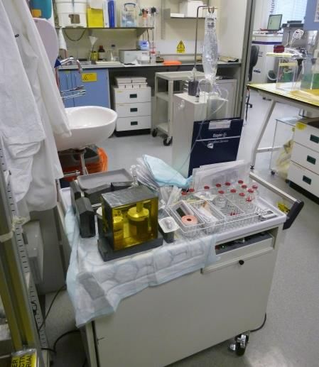
82Rb is a short-lived positron emitting radioisotope (75sec) and a biochemical analogue of potassium. Its short
half-life permits multiple injections into the same subject over a relatively short time period to track disease
progression, or response to therapy. Being generator produced, it does not require access to a cyclotron, and
which greatly enhances its availability. The primary use to date has been as a myocardial perfusion agent. As a
congener for K+, its primary mode of transport into the cell is via the sodium potassium pump (Na+/K+ ATPase).
We have recently investigated the potential of 82Rb as an imaging agent for basic biomedical investigation of
Na/K ATPase activity in the isolated perfused rat heart, tracking the injection, retention, and washout of
radiotracers in response to a variety of drugs, including ouabain (a cardiotonic Na/K ATPase inhibitor) and
isoprenaline (a adrenergic agonist). Isoprenaline increased 82Rb uptake by 30% whilst ouabain reduced
uptake by 10-15%. These experiments point towards the possibility to develop an imaging technique using 82Rb
to report directly on Na/K ATP pump function, providing a new tool for cancer imaging.
(a) (b)
19(c) (d)
1.0
0.9
0.8
Normalised intensity
0.7
0.6
0.5
0.4
0.3
isoprenaline
0.2
control
0.1 ouabain
0.0
100 200
time (s)
Figure. (a) the CardioGen-82 clinical generator. (b) Preclinical perfusion system at KCL for studying the
pharmacokinetics of radiolabelled tracers in the isolated perfused heart. (c) Normalised, decay corrected 82Rb traces
from the heart detector under control perfusion conditions (black), following -adronergic stimulation with
isoprenaline (red) and following NaK inhibition with ouabain (blue). (d) Clinical 82Rb scan of a colon cancer patient,
axial section from pelvic area (courtesy Groves UCLH).
One representative publication from each co-supervisor:
[1] 64Cu-CTS: A Promising Radiopharmaceutical for the Identification of Low-Grade Cardiac Hypoxia by PET.
Medina RA et al. J Nucl Med 56(6):921-6 2015.
[2] Multiple quantum filtered 23Na NMR in the Langendorff perfused mouse heart: Ratio of Triple/ Double
quantum filtered signals correlate with [Na]I, Eykyn TR et al. J Mol Cell Card 86:95-101 2015.
20You can also read