Toxoplasma gondii infection in domestic and wild felids as public health concerns: a systematic review and meta analysis
←
→
Page content transcription
If your browser does not render page correctly, please read the page content below
www.nature.com/scientificreports
OPEN Toxoplasma gondii infection
in domestic and wild felids as public
health concerns: a systematic
review and meta‑analysis
Kareem Hatam‑Nahavandi1, Rafael Calero‑Bernal2, Mohammad Taghi Rahimi3,
Abdol Sattar Pagheh4, Mehdi Zarean5, Asiyeh Dezhkam1 & Ehsan Ahmadpour6,7,8*
Felidae as definitive hosts for Toxoplasma gondii play a major role in transmission to all warm-blooded
animals trough oocysts dissemination. Therefore the current comprehensive study was performed
to determine the global status of T. gondii infection in domestic and wild felids aiming to provide
comprehensive data of interest for further intervention approaching the One Health perspective.
Different databases were searched by utilizing particular key words for publications related to T.
gondii infecting domestic and wild feline host species, worldwide, from 1970 to 2020. The review
of 337 reports showed that the seroprevalence of T. gondii in domestic cats and wild felids was
estimated in 37.5% (95% CI 34.7–40.3) (I2 = 98.3%, P < 0.001) and 64% (95% CI 60–67.9) (I2 = 88%,
P < 0.0001), respectively. The global pooled prevalence of oocysts in the fecal examined specimens
from domestic cats was estimated in 2.6% (95% CI 1.9–3.3) (I2 = 96.1%, P < 0.0001), and that in fecal
samples from wild felids was estimated in 2.4% (95% CI 1.1–4.2) (I2 = 86.4%, P < 0.0001). In addition,
from 13,252 examined soil samples in 14 reviewed studies, the pooled occurrence of T. gondii oocysts
was determined in 16.2% (95% CI 7.66–27.03%). The observed high rates of anti-T. gondii antibodies
seroprevalence levels and oocyst excretion frequency in the felids, along with soil (environmental)
contamination with oocysts may constitute a potential threat to animal and public health, and data
will result of interest in further prophylaxis programs.
Toxoplasma gondii is an opportunistic and successful coccidian parasite capable of infect virtually all homoeo-
thermic vertebrates, including human beings1,2. Domestic cats and other Felidae constitute its specific definitive
hosts3, and all non-feline animals are regarded as intermediate hosts; however, T. gondii can also undergo asexual
reproduction in tissues of Felidae acting as intermediate hosts. First, tachyzoites have active multiplication in
tissues, associated to rapid invasion causing harmful effects. Zoites present a special tropism to central nervous
system and striated muscle, in which they remain latent confined in a cyst as bradyzoites, leading to a long-
term chronic infection until another definitive host ingests the tissue. Then, released bradyzoites penetrate the
ocysts4. Felids
epithelial cells of small intestine, giving rise to schizonts that will form gamonts and, finally, o
excrete oocysts in their faeces, during a limited time lapse, contaminating soil and water5–8. In addition to the
domestic cats, and under the view of the available literature, the role of wild Felidae in the epidemiology of T.
gondii should not be n eglected5,9. Therefore, felids constitute the key element in the epidemiology of T. gondii
since an individual can shed millions of oocyts that can spread the infection to many other susceptible hosts10.
Several important outbreaks of human toxoplasmosis were epidemiologically linked to oocyst contamination of
drinking water11–13. By the way, oocysts were not detected in the samples collected from the water reservoir linked
1
School of Medicine, Iranshahr University of Medical Sciences, Iranshahr, Iran. 2SALUVET, Animal Health
Department, Faculty of Veterinary Sciences, Complutense University of Madrid, Madrid, Spain. 3Center for
Health Related Social and Behavioral Sciences Research, Shahroud University of Medical Sciences, Shahroud,
Iran. 4Infectious Diseases Research Canter, Birjand University of Medical Sciences, Birjand, Iran. 5Department
of Parasitology and Mycology, Faculty of Medicine, Mashhad University of Medical Sciences, Mashhad,
Iran. 6Infectious and Tropical Diseases Research Center, Tabriz University of Medical Sciences, Tabriz,
Iran. 7Immunology Research Center, Tabriz University of Medical Sciences, Tabriz, Iran. 8Department of
Parasitology and Mycology, Tabriz University of Medical Sciences, Tabriz, Iran. *email: ehsanahmadpour@
gmail.com
Scientific Reports | (2021) 11:9509 | https://doi.org/10.1038/s41598-021-89031-8 1
Vol.:(0123456789)www.nature.com/scientificreports/
to a serious Canadian o utbreak14, but viable oocysts were observed in contents of the intestine of a wild trapped
cougar (Felis concolor vancouverensis) and in a fecal pile in close proximity to the r eservoir15. It is important to
highlight that the sporulated oocysts are very resistant and can remain viable and infective for more than 1 year
in favourable conditions11,16–18. In this regard, a recent paper reviewed the environmental pathways by which T.
gondii can infect animals and people mostly driven by water, soil or contaminated fresh produce or s eafood19.
Toxoplasma gondii antibodies have been largely found in cats worldwide, and the seroprevalence degree
increases with the age of the cat, suggesting postnatal transmission of T. gondii20. It is assumed that postnatal
sero-conversion in cats is linked to oocysts excretion episodes. The life style of cats influences the occurrence
of T. gondii infections since feral cats that hunt for their food will present higher rates than domestic cats with
limited access to p arasites21. Seroprevalence level varied among continents, countries and even cities, linked to
many possible environmental factors influencing these variations. As an example, in an urban population of
301 domestic cats in Lyon, France20, the anti-T. gondii seroprevalence was only 18.6%, approximately half the
prevalence in other surveys in E urope22,23. The control of rodents in the area and feeding of cats by people were
considered as protective factors limiting infections. On the other hand, a low income and poor sanitation were
not the determining factors for low seropositivity to T. gondii in cats in Durango, Mexico24. Since a high density
of felines (specially domestic cats) increases the risk of infection and T. gondii prevalence in intermediate hosts,
a gradient of prevalence rate of infection has been demonstrated depending on the anthropization degree of the
environment25,26.
Nearly up to 30% of the world’s human population has had contact with the parasite evidenced by the pres-
ence of anti-T. gondii antibodies; while T. gondii infections are usually asymptomatic, they can lead to harmful
effects, especially in congenital cases and immunocompromissed p ersons27,28. Humans become primarily infected
mostly via oral ingestion of viable tissue cysts present in raw or undercooked meat and oocysts contaminat-
ing water or foodstuffs6,8,29. Nowadays, comprehensive local studies are still necessary to determine the source
attribution of human infections; this constitutes an interesting challenge that should be approached under the
One Health perspective.
To date, different surveys have been focused on domestic and wild felids in order to determine aspects as
seroprevalence rates of anti-T. gondii antibodies, frequency of oocysts excretion and soil presence w orldwide30–36,
but with a certain degree of variance among studies. A systematic review recently assessed the seroprevalence of
T. gondii in felids from 1967 to 2017 with a search strategy restricted to articles in E nglish37. So that, the present
investigation was aimed to determine the global frequency of T. gondii infections in domestic cats and wild felids,
the occurrence of T. gondii-like oocysts shedding, and the frequency of oocysts in soil; such information will
be useful to implement further measures aiming to reduce animal and human infections under a One Health
perspective.
Methods
Search strategy. The review process exactly followed the protocol suggested by the Preferred Report-
ing Items for Systematic Reviews and Meta-analyses (PRISMA) guidelines (Supplementary data: PRISMA/
STROBE)38. We retrieved published studies from the databases of MEDLINE (via PubMed) (https://www.ncbi.
nlm.nih.gov/pubmed/), Scopus (https://www.scopus.com/), Web of Science (https://www.webofknowledge.
com/) and CAB Abstracts (https://www.cabi.org/AHPC) with no restriction on language from Jan 1, 1970, to
Dec 31, 2019. Search terms included a combination of Medical Subject Heading terms (MeSH) and free-text
words in titles, abstracts and full texts.
The systematic search for PubMed accomplished using several Medical Subject Heading terms (Table S1).
In addition, Scopus, Web of Science and CAB Abstracts were searched using the same strategy (Supplementary
DATA). The Google Scholar search engine was used for checking the search strategy. The reference lists of all
included articles and relevant reviews were hand searched for potentially eligible literature. In addition, authors
and experts in the field were consulted to aid in the identification of relevant conference abstracts related to
Toxoplasma and toxoplasmosis. Sometimes, we have had to contact the authors for raw data c ollection39, espe-
cially in old literature.
Selection of studies. Initial screening by manuscript titles and abstracts was performed independently by
two researchers (KHN and EA), that also assessed the full texts of all potentially relevant studies and applied
inclusion criteria. Discrepancies when detected, were resolved after constructive discussion (AD, MZ and MTR).
The studies providing data on the seroprevalence of T. gondii in domestic or wild felids, frequency of oocyst
excretion in felids, and those reporting soil contaminations with oocysts were included. On the other hand,
studies meeting the epidemiology of T. gondii in non-feline hosts, studies where cat faecal samples were collected
from the ground, and data from each animal was not independently retrievable, experimental studies, articles that
only presented the final result and did not provide the raw data, or those without definite sample size, abstracts
presented in congresses without full text, and case–control studies and clinical trials that could not report a cor-
rect estimate of prevalence were excluded. Any duplicated research was also excluded.
Quality assessment. The standard Strengthening the Reporting of Observational Studies in Epidemiology
(STROBE) checklist was u sed40. In present study, articles were evaluated as low quality: less than 16.5, moderate
quality: 16.6–25.5, and high quality: 25.6–34; the articles included in the meta-analysis presented acceptable
quality.
Data extraction. After comprehensive examination of selected articles, the following data were extracted:
the first author’s last name, publication year, country, feline scientific names, keeping status of felids, sample size,
Scientific Reports | (2021) 11:9509 | https://doi.org/10.1038/s41598-021-89031-8 2
Vol:.(1234567890)www.nature.com/scientificreports/
source of soil samples, beginning and end date of study implementation, the number of the positive and negative
cases, cut-off, age groups, as well as information about diagnostic tools. Data were extracted separately if two dif-
ferent populations had been studied. All extracted data from each study were entered into an Excel spreadsheet.
Meta‑analysis. The collected data were entered into the StatsDirect statistical software package (version
2.7.2) (Stata Corporation, College Station, TX, USA) (http://www.statsdirect.com/). Statistical heterogeneity of
the different years among studies was assessed using the Cochrane’s Q test and inconsistency I2 test. To deter-
mine whether there is a significant heterogeneity, a random effect model was used to estimate the pooled preva-
lence’s of cat infection41. In addition, potential publication bias was explored using Funnel plot and Egger’s test.
Results
The initial database search retrieved 14,870 publications. First screening enabled us to exclude 14,441 studies
not meeting the inclusion criteria. Altogether, 429 studies were retained for further investigation. In a secondary
assessment, another 34 documents were excluded because of one of the following reasons: review articles, follow
up studies or case series reports, and papers with insufficient data, or data from each animal were not indepen-
dently retrievable. Eventually, 395 studies which met our eligibility criteria and evaluated soil contamination
with T. gondii oocysts (n = 14), and T. gondii infection in domestic cats (serology, n = 268; oocyst in feces, n = 112)
and wild felids (serology, n = 69; oocyst in feces, n = 15) during five decades were retained for analysis (Fig. 1).
Potential publication bias in the conducted studies regarding the prevalence of T. gondii infection in domestic
cats and wild felids, Toxoplasma-like oocysts shedding, and frequency of oocysts in soil are shown using Funnel
plot and Egger’s test (Fig. 2).
Prevalence of anti‑T. gondii antibodies in blood/serum samples. The global pooled seroprevalence
of T. gondii in domestic cats was estimated in 37.5% (95% CI 34.7–40.3) (I2 = 98.3%, P < 0.001) (Table 1). The
highest rate was observed in Australia (66.6%, 95% CI 62.8–70.3) (I2 = 97.2%, P < 0.0001), followed by Africa
(55.7%, 95% CI 35.6–74.8) (I2 = 98.9%, P < 0.0001), Europe (45.3%, 95% CI 41.1–49.6) (I2 = 96.7%, P < 0.0001),
Central and South America (40.3%, 95% CI 34–49.6) (I2 = 97.7%, P < 0.0001) and North America (31.6%, 95%
CI 27–36.4) (I2 = 96.8%, P < 0.0001). The lowest prevalence was observed in Asia (28.3%, 95% CI 24.1–32.6)
(I2 = 98.1%, P < 0.0001) (Table 1). In the other hand, the worldwide pooled seroprevalence of T. gondii in wild
felids (including lion, jaguarundi, jaguar, ocelot, cougar, leopard, tiger, geoffroy’s cat, oncilla, margay, caracal,
snow leopard, Eurasian lynx, bobcat, cheetah, Prionailurus cats, Iberian lynx, pampas cat, serval, pallas’s cat,
jungle cat, European wildcat, sand cat, Asian golden cat, Canadian lynx, clouded leopard, masked palm civet,
and common genet) was estimated 64% (95% CI 60–67.9) (I2 = 88%, P < 0.0001) (Table 2). The highest and the
lowest seroprevalence rates were related to Panthera leo (87.6%, 95% CI 79–94.3) and Leopardus colocolo (19.6%,
95% CI 6.1–38.3), respectively (Table 2). Heterogeneity was, however, very low I2 = 79.9%. Retrieved information
of the eligible studies based on the of anti-Toxoplasma antibodies prevalence in domestic cats and wild felids
are summarized in Tables S2 and S3. In total, 80,087 blood samples from domestic cats (n = 73,980) and wild
felids (n = 6,107) from 337 eligible studies were examined for the presence of anti-T. gondii antibodies and/or T.
gondii DNA, of which 26,903 subjects were diagnosed as positive (domestic cats n = 23,593; wild felids n = 3,310)
(Tables S2, S3). Different diagnostic methods to evaluate anti-T. gondii antibodies in the cats have been identi-
fied in the studies: MAT (132 studies), IFAT (99 studies), and ELISA (92 studies) (Tables S2, S3). Amongst the
reviewed studies, just one investigation applied PCR method for detection of T. gondii DNA in stray cats using
blood samples42 (Table S2).
Occurrence of T. gondii oocysts in fecal samples. A total number of 137 eligible studies which exam-
ined 66,601 fecal samples from domestic cats (n = 63,458) and wild felids (n = 3,143), 1,330 were positive (domes-
tic cats n = 1,254; wild felids n = 76) for T. gondii oocysts, T. gondii-like oocysts, and/or T. gondii DNA (Table S4,
S5). The global pooled prevalence of oocysts in the fecal examined specimens from domestic cats was estimated
in 2.6% (95% CI 1.9–3.3) (I2 = 96.1%, P < 0.0001) (Table 3). The highest and the lowest prevalence rates were
detected in Africa (9.8%, 95% CI 2.4–21.5) (I2 = 94.1%, P < 0.0001), and North America (0.9%, 95% CI 0.5–1.3),
respectively. Heterogeneity was, however, very low I2 = 50% (Tables 1, 3). The most used methodology for detec-
tion of oocysts was microscopy (99 studies) which was followed by molecular (16 studies) and mouse bioas-
say (10 studies) methods (Table S4). Some studies combined two techniques for detection of oocysts in feline
feces. The highest prevalence was related to the molecular detection method (6.5%, 95% CI 3.7–10) (I2 = 92.1%,
P < 0.0001), followed by bioassay (2.8%, 95% CI 0.6–6.4) (I2 = 95.1%, P < 0.0001), and microscopy (2.1%, 95% CI
1.4–2.8) (I2 = 96.3%, P < 0.0001) (Table 3).
The worldwide pooled prevalence of T. gondii oocysts, T. gondii-like oocysts and T. gondii DNA in fecal speci-
mens from wild felids was estimated in 2.4% (95% CI 1.1–4.2) (I2 = 86.4%, P < 0.0001) (Table 3). The highest and
the lowest prevalence of oocysts in fecal specimens was related to Leopardus pardalis (15.89%, 95% CI 0.2–58.6)
and Panthera onca (3.5%, 95% CI 1.3–13.7), respectively (Table S5). The prevalence of Toxoplasma-like oocysts
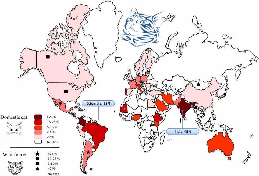
detected in domestic and wild feline stool samples in different countries are shown in Fig. 3. As well India (49%)
and Colombia (33%) had the highest prevalence.
Occurrence of T. gondii oocysts in soil samples. Up to 14 studies reported the examination of 13,252
soil specimens resulting in 3,421 (25.8%) samples positive for T. gondii oocysts (or T. gondii-DNA) using mouse
bioassay and different PCR procedures. Table S6 shows the conducted studies to detect T. gondii oocysts in soil
samples; the pooled prevalence of T. gondii oocysts in those samples was estimated 16.2% (95% CI 7.66–27.03%)
(Q = 1628.10, I2 = 99.2%, P < 0.0001) (Egger’s bias = 5.44, P = 0.3733) (Fig. 4).
Scientific Reports | (2021) 11:9509 | https://doi.org/10.1038/s41598-021-89031-8 3
Vol.:(0123456789)www.nature.com/scientificreports/
Records identified through database searching: Additional records identified
Pubmed (n = 2,224), WoS (n = 2,378), Scopus through other sources (n = 5)
Identification
(conference paper (n = 166); Published data (n = 9,663)),
CAB Abstracts (n = 434)
Records that were seen in the preliminary searched on databases
Records that were selected after a
primary screening (n = 1,221)
Screening
Records after duplicates removed Excluded records based on titles and
(n = 813) abstracts (n = 391)
Included records after references
Articles screened by full-text
checking (n = 7)
(n = 422)
Eligibility
Full-text articles assessed for Full text articles excluded based on
eligibility (n = 429) following reasons (n = 34)
a. Review articles follow up
studies or case series reports
b. Insufficient data
Studies included in qualitative c. Data from each animal was not
synthesis (n = 395) independently retrievable
Included
Studies included in quantitative synthesis (n = 395)
Oocysts in stool samples of domestic cats (n = 112)
Oocysts in stool samples of wild felids (n = 15)
Serology of domestic cats (n = 268)
Serology of wild felids (n = 69)
Oocysts in soil samples (n = 14)
Figure 1. Flowchart of the study design process.
Discussion
Felids as final host play an irreplaceable role for T. gondii life cycle that exclusively yield and excrete oocysts in
their faeces, contaminating soil, water and food6–8,10. According to our findings, 37.5% of domestic cats showed
exposure to T. gondii and 2.6% were actively shedding T. gondii or T. gondii-like oocysts. Similarly, the worldwide
seroprevalence of Toxoplasma in domestic cats had been previously estimated at levels of 30–40%1,43.
Based on the results, the highest value of seroprevalence in domestic cats was observed in Australia, followed
by Africa, Europe, Central/South America, North America and Asia; although the number of studies for Aus-
tralia (n = 6) and Africa (n = 15) were relatively low, whereas only one study in USA included 12,628 animals.
The lowest prevalence was observed in Asia (28.3%), nevertheless, most studies have been conducted in Asian
countries (91 studies). The number of surveys was higher in USA (North America; 34 studies), followed by
Brazil (South America; 30 studies). Our investigation identified a number of countries without any data on T.
gondii infection in cats, emphasizing the need for further studies in this field. According to our findings, 64% of
nondomestic cats showed evidence of exposure to T. gondii and 2.4% were actively shedding T. gondii oocysts.
Accordingly, the highest sero-prevalence of T. gondii in different wild felids were in the following order: lion
(Panthera leo), European wildcat (Felis silvestris), jaguar (Panthera onca), Pallas’s cat (Otocolobus manul), sand
cat (Felis margarita), cheetah (Acinonyx jubatus), caracal (Caracal caracal), leopard (Panthera pardus), Iberian
lynx (Lynx pardinus), ocelot (Leopardus pardalis), tiger (Panthera tigris), serval (Leptailurus serval), Geoffroy’s
cat (Leopardus geoffroyi), bobcat (Lynx rufus), oncilla (Leopardus tigrinus), cougar (Puma concolor), margay
(Leopardus wiedii), snow leopard (Panthera uncial), jaguarundi (Herpailurus yagouaroundi), Asian golden
cat (Catopuma temminckii), Eurasian lynx (Lynx lynx), jungle cat (Felis chaus), Prionailurus cat (Prionailurus
viverrinus), Canadian lynx (Lynx canadiensis), clouded leopard (Neofelis nebulosa), and Pampas cat (Leopardus
Scientific Reports | (2021) 11:9509 | https://doi.org/10.1038/s41598-021-89031-8 4
Vol:.(1234567890)www.nature.com/scientificreports/
Figure 2. Funnel plot of standard error by logit event rate to assess publication or other types of bias across
prevalence studies. (A) Studies based on the seroprevalence of anti-Toxoplasma gondii antibodies in domestic
cats, (B) studies based on detection of T. gondii-like oocyst and T. gondii oocyst DNA in domestic cat feces, (C)
studies based on the seroprevalence of anti-Toxoplasma gondii antibodies in wild felids, (D) studies based on
detection of T. gondii-like oocyst and T. gondii oocyst DNA in wild felids feces.
Heterogeneity Egger’s test
Continent No. of studies Detection method Prevalence % (95% CI) I2 Q P value T P value
6 Stool exam 9.8 (2.4–21.5) 94.1 84.2 < 0.0001 5.1 0.0434
Africa
16 Serology 55.7 (35.6–74.8) 98.9 1408.4 < 0.0001 − 3.8 0.6571
32 Stool exam 4 (1.9–6.9) 96.9 1001.4 < 0.0001 3.2 0.0195
Asia
90 Serology 28.3 (24.1–32.6) 98.1 4730.5 < 0.0001 7.7 < 0.0001
4 Stool exam 1.7 (0.2–4.5) 79.1 14.3 0.0025 1.3 0.3488
Australia
6 Serology 66.6 (62.8–70.3) 97.2 176.1 < 0.0001 − 9.66 0.1256
55 Stool exam 1.21 (0.8–1.6) 89.6 517.1 < 0.0001 1.18 < 0.0001
Europe
61 Serology 45.3 (41.1–49.6) 96.7 1840.1 < 0.0001 2.94 0.1269
16 Stool exam 0.9 (0.5–1.3) 50.0 30.3 0.0108 0.94 0.0583
North America
37 Serology 31.6 (27–36.4) 96.8 1126.2 < 0.0001 0.49 0.7212
12 Stool exam 6.2 (1.8–1.3) 97.1 374.2 < 0.0001 3.4 0.0226
Central/South America
61 Serology 40.3 (34.0–46.8) 97.7 2642.3 < 0.0001 4.5 0.0467
Table 1. Pooled prevalence of Toxoplasma infection in domestic cats and subgroup analyses.
colocolo). Although in general it should be considered that the number of studied nondomestic cats were lower
compared to domestic cats, and keeping status of wilds cats, in captive (n = 2,492) or free ranging (n = 2,949), is
probably associated with how they are fed and how they become infected. Albeit such findings also highlights
the importance of serological and isolation studies on T. gondii infecting their prey (ungulates, birds, etc.) using
validated methodologies for bias reducing.
Scientific Reports | (2021) 11:9509 | https://doi.org/10.1038/s41598-021-89031-8 5
Vol.:(0123456789)www.nature.com/scientificreports/
Heterogeneity Egger’s test
Host species No. of studies Detection method Prevalence (95% CI) I2 Q P value T P value
Asian golden cat (Catopuma
3 Serology 47.1 (8.9–87.4) 72.7 7.3 0.0256 – –
temminckii)
18 Serology 60.5 (47.1–73.1) 91.0 189.8 < 0.0001 1.5 0.3255
Bobcat (Lynx rufus)
5 Stool exam 4.1 (0.2–12.4) 82.5 22.8 0.0001 1.0 0.1297
Canadian Lynx (Lynx
3 Serology 36.4 (10.8–67.2) 91.7 24.1 < 0.0001 – –
canadiensis)
Caracal (Caracal caracal) 7 Serology 69.9 (49.6–86.8) 0.0 5.1 0.5270 − 0.3 0.9283
Cheetah (Acinonyx jubatus) 8 Serology 70.4 (48.1–88.5) 81.0 36.7 < 0.0001 − 0.6 0.7904
Clouded leopard (Neofelis
4 Serology 36.3 (9.0–69.7) 65.8 8.7 0.0325 4.2 0.2131
nebulosa)
24 Serology 56.1 (43.7–68.2) 93.8 371.7 < 0.0001 2.6 0.1155
Cougar (Puma concolor)
6 Stool exam 4.7 (0.5–12.7) 61.5 12.9 0.0236 0.8 0.0955
Eurasian lynx (Lynx lynx) 6 Serology 42.1 (14.9–72.2) 97.4 192.1 < 0.0001 1.7 0.7428
European wildcat (Felis
7 Serology 76.8 (62.6–88.5) 46.0 11.1 0.0848 0.5 0.6714
silvestris)
Geoffroy’s cat (Leopardus
5 Serology 60.7 (39.9–79.6) 60.8 10.1 0.0373 1.7 0.6131
geoffroyi)
Iberian Lynx (Lynx pardi-
4 Serology 66.2 (50.1–80.5) 81.9 16.5 0.0009 2.9 0.6198
nus)
9 Serology 74.4 (63.5–84) 61.6 20.8 0.0076 1.7 0.1306
Jaguar (Panthera onca)
3 Stool exam 3.5 (1.3–13.7) 66.9 6.0 0.0487 – –
Jaguarundi (Herpailurus
10 Serology 47.7 (41.6–53.9) 0.0 8.5 0.4774 1.9 0.0053
yagouaroundi)
Jungle cat (Felis chaus) 2 Serology 44.5 (7.5–97.1) – 7.9 0.0047 – –
17 Serology 68.0 (46.5–86.1) 72.5 58.1 < 0.0001 5.1 0.0010
Leopard (Panthera pardus)
3 Stool exam 3.8 (0.1–17.6) 84.9 13.2 0.0013 – –
20 Serology 87.6 (79–94.3) 79.9 94.6 < 0.0001 − 1.6 0.0175
Lion (Panthera leo)
3 Stool exam 4.9 (0.3–23.8) 85.6 13.8 1.0010 – –
Margay (Leopardus wiedii) 5 Serology 56.0 (46.4–65.4) 29.0 5.6 0.2282 2.3 0.2958
11 Serology 66.2 (58.1–73.8) 45.6 18.3 0.0489 1.6 0.0921
Ocelot (Leopardus pardalis)
3 Stool exam 15.9 (0.2–58.6) 85.5 13.7 0.0010 – –
Oncilla (Leopardus tigrinus) 9 Serology 59.0 (49.7–68) 47.9 15.3 0.0527 1.4 0.2027
Pallas’s cat (Otocolobus
10 Serology 70.6 (43.9–91.3) 89.3 84.1 < 0.0001 − 2.4 0.4067
manul)
Pampas cat (Leopardus
3 Serology 19.6 (6.1–38.3) 0.0 0.7 0.6878 – –
colocolo)
Prionailurus cats (Prionailu- 10 Serology 39.6 (24.3–56.1) 60.0 22.4 0.0075 2.5 0.0606
rus viverrinus) 5 Stool exam 4.0 (1.8–7.1) 0.0 1.4 0.8433 − 0.2 0.4709
Sand cat (Felis margarita) 4 Serology 70.5 (49.9–87.5) 66.8 9.0 0.0287 3.0 0.2817
Serval (Leptailurus serval) 4 Serology 64.3 (35 to88.6) 8.8 3.2 0.3493 -4.1 0.7970
Snow leopard (Panthera
2 Serology 52.6 (10.7–92.2) – 1.9 0.1607 – –
uncial)
16 Serology 66.2 (51.4–79.5) 61.9 39.3 0.0006 1.3 0.3862
Tiger (Panthera tigris)
4 Stool exam 7.4 (0–27.4) 80.2 15.1 0.0017 1.8 0.2827
Table 2. Pooled prevalence of Toxoplasma infection in wild felids and subgroup analyses.
The considerable gap between the prevalence of oocysts in feces and positive serum antibodies can be
explained by the fact that infected felids can shed T. gondii oocysts for a short period (10–15 days), shortly
after primoinfection, and then they become seropositive indefinitely. As, one important point, the activation of
humoral immunity and antibodies production prevent from re-shedding of oocysts; new excretion episodes can
occur when severe immunosuppression appears29,44. The short period of oocyst shedding and the low prevalence
rate of felids which actively excrete oocysts, have led some authors to discuss that direct contact with felids
should not be considered as a risk factor for human infection7,29,45. A systematic review in Iran showed that
humans with history of close contact with cats presented a higher T. gondii seroprevalence rate compared to
those without contact46. In the study conducted by Jones and c olleagues29 in the USA, exposure to kittens was
statistically linked to T. gondii infection. In the study conducted in different European centers45, infections in
pregnant women were attributed to the consumption of undercooked or cured meat products and soil contact
in the 30–63% and 6–17% of cases respectively, but contact with cats was not identified as a risk factor. Similarly,
another study showed that contact with cats is not related to infections, while the ingestion of raw or undercooked
meat highly increased the risk of infection47.
Scientific Reports | (2021) 11:9509 | https://doi.org/10.1038/s41598-021-89031-8 6
Vol:.(1234567890)www.nature.com/scientificreports/
Heterogeneity
Group Number of studies Pooled prevalence (95% CI) P value I2 Cochran Q
Domestic Cat
Serology 271 37.5 (34.7–40.3) < 0.0010 98.3 15,984.3
Stool exam 125 2.6 (1.9–3.3) < 0.0001 96.1 3164.3
Microscopy 99 2.1 (1.4–2.8) < 0.0001 96.3 2664.6
Bioassay 10 2.8 (0.6– 6.4) < 0.0001 95.1 182.7
Molecular 16 6.5 (3.7–10) < 0.0001 92.1 189.3
Wild Feline
Serology 223 64.0 (60–67.9) < 0.0001 88 1854.9
Stool exam 12 2.4 (1.1–4.2) < 0.0001 86.4 227.5
Table 3. The global pooled prevalence of Toxoplasma infection in feline hosts/felids.
Proportion meta-analysis plot [random effects]
Frenkel et al. 1995 Panama 0.011 (0.005, 0.020)
Lass et al. 2009 Poland 0.178 (0.109, 0.267)
dos Santos et al. 2010 Brazil 0.323 (0.167, 0.514)
Du et al. 2012 China 0.379 (0.281, 0.484)
Du et al. 2012 China 0.230 (0.180, 0.287)
Tavalla et al. 2013 Iran 0.087 (0.047, 0.144)
Ajmal et al. 2013 Pakistan 0.048 (0.025, 0.082)
Wang et al. 2014 China 0.127 (0.089, 0.173)
Gotteland et aL. 2014 France 0.292 (0.236, 0.354)
Solymane et al. 2014 Iran 0.050 (0.010, 0.139)
Gao et al. 2016 China 0.303 (0.294, 0.312)
Liu et al. 2017 China 0.010 (0.004, 0.020)
Simon et al. 2017 France 0.498 (0.456, 0.541)
Saki et al. 2017 Iran 0.090 (0.054, 0.139)
combined 0.162 (0.077, 0.270)
0.0 0.2 0.4 0.6 0.8
proportion (95% confidence interval)
Figure 3. Forest plot diagram of the present systematic review and meta-analysis based on studies focused on
detection of soil contamination by Toxoplasma-like oocysts.
Based on the results, 16.2% of soil samples contained T. gondii oocysts (or T. gondii-DNA), those when
sporulated can survive for several months under tough conditions and are resistant to common disinfectants48.
Contaminated soil has been demonstrated as an important source for infection for humans and animals13,19. It
has been shown that gardening and occupations in contact with soil increases the risk of T. gondii infection49, as
previously seen50. In a follow-up study of the toxoplasmosis outbreak during 1977 in G eorgia51, after 25 years,
among 37 individual (exposure to an indoor horse arena), 14 equestrian were tested, that three (21%) were found
to have toxoplasmic retinochoroiditis lesions. Based on the observations is possible that cat feces containing the
Scientific Reports | (2021) 11:9509 | https://doi.org/10.1038/s41598-021-89031-8 7
Vol.:(0123456789)www.nature.com/scientificreports/
Figure 4. Pooled prevalence of Toxoplasma-like oocysts detected in domestic and wild feline stool samples in
different countries (Map created by PowerPoint Microsoft office, source of image: https://commons.wikimedia.
org/wiki/File:BlankMap-World.svg).
organism were most likely stirred up when horses ran on the dirt floor, and were inhaled or ingested by riders
and observers. Based upon number of studies conducted in different European centers, contact with soil or
vegetables or fruit presumably contaminated with soil were highly associated to T. gondii infection in pregnant
women45,52–54. Investigation on sentinels (i.e., molluscs) for environmental c ontamination55 and also the infec-
tion source attribution by using specific t ests56 will be of great interest for integration with data compilation in
definitive and intermediate susceptible hosts.
In the present investigation, the prevalence of soil contamination was highly variable in the selected stud-
ies, which might be influenced by the soil characteristics and the number of infected animals in the area57. The
included studies also reported highly heterogeneous results regarding the prevalence of cat infections, which
could be due to the different risk factors, to note: sex, age, climates, study periods, cat breeds, living conditions
and diagnostic methods as well as other unrecognized confounding factors. Based on our results, T. gondii sero-
prevalence in cats (Felis domesticus) in different countries oscillated from less than 10% in Thailand, Taiwan and
Angola to more than 70% in Qatar, and Ethiopia. This can partly be explained by the different environmental
conditions among the countries58. It has been shown that cat infections present higher occurrence in warm,
moist and low altitude regions, maybe linked to oocysts sporulation and survival of T. gondii oocysts in such
latitudes34. Similarly, T. gondii seroprevalence in pigs was associated with lower geographical latitude and higher
mean annual temperature59, fact that may suggest high environmental contamination with T. gondii sporulated
oocysts. It seems to be clear that oocysts shedding by cats constitute the essential element for sustainment of
the parasite in the environment, this was demonstrated when extremely low seroprevalence of T. gondii (0.9%)
was detected in feral pigs from a remote island lacking cats in the USA60. Furthermore, the time period of study
might influence the results, as the infection rate is higher in autumn, w inter22, and rainy y ears20. One may con-
sider breed as a variable factor for cat T. gondii infection. It has been shown that Toxoplasma seroprevalence is
highly variable in different cat breeds from 18.8% in Burmese cats to 60% in Persian c ats35. Even though, a high
occurrence rate of T. gondii infection in cat may be attributed to some important factors including: uncontrolled
food and access to contaminated sources, wandering outdoor, humid and temperate climate; and cat abundance.
Furthermore, stray cats have been shown to have a higher seroprevalence compared to pet cats, which can be
explained by more access to contaminated source and outdoor living31,34,61. Additionally, pet cats with an outdoor
access are also at an increased risk of infection compared to those kept i ndoor32,35. It has been reported that rural
cats show a higher seroprevalence rate of T. gondii compared to urban ones62.
Scientific Reports | (2021) 11:9509 | https://doi.org/10.1038/s41598-021-89031-8 8
Vol:.(1234567890)www.nature.com/scientificreports/
Furthermore, the different diagnostic methods used to detect T. gondii antibodies and oocysts could influence
the results. While the different techniques used for anti-T. gondii antibodies detection showed comparably good
diagnostic performance, most of the studies aiming to detect T. gondii oocysts employed less reliable microscopic
methods, which might result in false positives, as oocysts and sporocysts of some other coccidia (e.g. Hammondia
hammondi, Besnoitia darlingi) may resemble those of T. gondii48. It shows the necessity of testing environmental
and fecal samples by using specific-PCR aided with amplicon sequencing for identity c onfirmation63.
Felids as key elements in the epidemiology of toxoplasmosis should be considered as a potential threat to
animal and public health, due potential oocysts contamination of the environment; such information is still miss-
ing in several worldwide locations, so further epidemiological investigations on final hosts would be of special
interest for evaluating the status of T. gondii infection and risk assessment implementations. Further investiga-
tions based on QMRA a pproaches64,65 combining raw data in Felidae with those from the environmental side
and those from susceptible hosts will complement the One Health puzzle in defined areas.
In present meta-analysis, it is shown that about one-third of domestic and non-domestic cats have been
exposed to T. gondii, and globally about 1 in 50 cats are actively shedding T. gondii or T. gondii-like oocysts. In
addition, 16.2% of the soil samples examined were contaminated with T. gondii-like oocysts informing on a broad
environmental distribution. Felids are the only final host of T. gondii and play a major role in its life cycle, there-
fore measures aiming to reduce environmental contamination with T. gondii oocysts will be of major interest,
and a One Health perspective covering human, animal and environmental health should be taken into account.
Data availability
The data that supports the findings of this study are available in the supplementary material of this article.
Received: 12 August 2020; Accepted: 13 April 2021
References
1. Webster, J. P. Review of “toxoplasmosis of animals and humans” by JP Dubey. Parasit. Vectors 3, 112 (2010).
2. Dubey, J. P. The history of Toxoplasma gondii—the first 100 years. J. Eukaryot. Microbiol. 55, 467–475 (2008).
3. Martorelli Di Genova, B., Wilson, S. K., Dubey, J. & Knoll, L. J. Intestinal delta-6-desaturase activity determines host range for
Toxoplasma sexual reproduction. PLoS Biol. 17, 20 (2019).
4. Calero-Bernal, R. & Gennari, S. Clinical toxoplasmosis in dogs and cats: An update. Front. Vet. Sci. 6, 54 (2019).
5. Lukesova, D. & Literák, I. Shedding of Toxoplasma gondii oocysts by Felidae in zoos in the Czech Republic. Vet. Parasitol. 74, 1–7
(1998).
6. Dubey, J. Toxoplasmosis—a waterborne zoonosis. Vet. Parasitol. 126, 57–72 (2004).
7. Dubey, J. & Jones, J. Toxoplasma gondii infection in humans and animals in the United States. Int. J. Parasitol. 38, 1257–1278 (2008).
8. Weiss, L. M. & Dubey, J. P. Toxoplasmosis: A history of clinical observations. Int. J. Parasitol. 39, 895–901 (2009).
9. Silva, J. C. R., Ogassawara, S., Marvulo, M. F. V., Ferreira-Neto, J. S. & Dubey, J. Toxoplasma gondii antibodies in exotic wild felids
from Brazilian zoos. J. Zoo Wildl. Med. 32, 349–351 (2001).
10. Dubey, J. et al. All about toxoplasmosis in cats: The last decade. Vet. Parasitol. 20, 109145 (2020).
11. Bowie, W. R. et al. Outbreak of toxoplasmosis associated with municipal drinking water. Lancet 350, 173–177 (1997).
12. De Moura, L. et al. Waterborne toxoplasmosis, Brazil, from field to gene. Emerg. Infect. Dis. 12, 326 (2006).
13. Pinto-Ferreira, F. et al. Patterns of transmission and sources of infection in outbreaks of human toxoplasmosis. Emerg. Infect. Dis.
25, 2177 (2019).
14. Isaac-Renton, J. et al. Detection of Toxoplasma gondii oocysts in drinking water. Appl. Environ. Microbiol. 64, 2278–2280 (1998).
15. Aramini, J., Stephen, C. & Dubey, J. Toxoplasma gondii in Vancouver Island cougars (Felis concolor vancouverensis): Serology and
oocyst shedding. J. Parasitol. 20, 438–440 (1998).
16. Mancianti, F. et al. A retrospective molecular study of select intestinal protozoa in healthy pet cats from Italy. J. Feline Med. Surg.
17, 163–167 (2015).
17. Hatam-Nahavandi, K. et al. Microscopic and molecular detection of Cryptosporidium andersoni and Cryptosporidium xiaoi in
wastewater samples of Tehran Province, Iran. Iran. J. Parasitol. 11, 499 (2016).
18. Nahavandi, K. H. et al. Molecular typing of Eimeria ahsata and E. crandallis isolated from slaughterhouse wastewater. Jundishapur
J. Microbiol. 9, 20 (2016).
19. Shapiro, K. et al. Environmental transmission of Toxoplasma gondii: Oocysts in water, soil and food. Food Waterborne Parasitol.
15, e00049 (2019).
20. Afonso, E., Thulliez, P. & Gilot-Fromont, E. Transmission of Toxoplasma gondii in an urban population of domestic cats (Felis
catus). Int. J. Parasitol. 36, 1373–1382 (2006).
21. DeFeo, M. L., Dubey, J., Mather, T. N. & Rhodes, R. C. III. Epidemiologic investigation of seroprevalence of antibodies to Toxo-
plasma gondii in cats and rodents. Am. J. Vet. Res. 63, 1714–1717 (2002).
22. Simon, J. A. et al. A multi-event capture-recapture analysis of Toxoplasma gondii seroconversion dynamics in farm cats. Parasit.
Vectors 11, 339 (2018).
23. Veronesi, F. et al. Detection of Toxoplasma gondii in faeces of privately owned cats using two PCR assays targeting the B1 gene and
the 529-bp repetitive element. Parasitol. Res. 116, 1063–1069 (2017).
24. Alvarado-Esquivel, C. et al. Seroprevalence of Toxoplasma gondii antibodies in cats from Durango City, Mexico. J. Parasitol. 93,
1214–1216 (2007).
25. Ballash, G. A. et al. Seroprevalence of Toxoplasma gondii in white-tailed deer (Odocoileus virginianus) and free-roaming cats (Felis
catus) across a suburban to urban gradient in northeastern Ohio. EcoHealth 12, 359–367 (2015).
26. Ahlers, A. et al. Survey of Toxoplasma gondii exposure in muskrats in a relatively pristine ecosystem. J. Parasitol. 106, 346–349
(2020).
27. Safarpour, H. et al. Global status of Toxoplasma gondii infection and associated risk factors in people living with HIV. Aids (Lond.,
Engl.) 34, 469–474 (2020).
28. Jafari-Modrek, M., Hasanzadeh, R., Azizi, H. & Hatam-Nahavandi, K. A Seroprevalence Study of Toxoplasmosis in Female Students
in Zahedan, South East of Iran. Iran. J. Public Health 48, 988 (2019).
29. Jones, J. L. et al. Risk factors for Toxoplasma gondii infection in the United States. Clin. Infect. Dis. 49, 878–884 (2009).
30. Aubert, D. & Villena, I. Detection of Toxoplasma gondii oocysts in water: Proposition of a strategy and evaluation in Champagne-
Ardenne Region, France. Mem. Inst. Oswaldo Cruz 104, 290–295 (2009).
Scientific Reports | (2021) 11:9509 | https://doi.org/10.1038/s41598-021-89031-8 9
Vol.:(0123456789)www.nature.com/scientificreports/
31. Kulasena, V., Rajapakse, R., Dubey, J., Dayawansa, P. & Premawansa, S. Seroprevalence of Toxoplasma gondii in cats from Colombo,
Sri Lanka. J. Parasitol. 97, 152–152 (2011).
32. Must, K., Lassen, B. & Jokelainen, P. Seroprevalence of and risk factors for Toxoplasma gondii infection in cats in Estonia. Vector
Borne Zoonot. Dis. 15, 597–601 (2015).
33. Rahimi, M. T. et al. Cats and Toxoplasma gondii: A systematic review and meta-analysis in Iran. Onderstepoort J. Vet. Res. 82, 01–10
(2015).
34. Ding, H., Gao, Y.-M., Deng, Y., Lamberton, P. H. & Lu, D.-B. A systematic review and meta-analysis of the seroprevalence of
Toxoplasma gondii in cats in mainland China. Parasit. Vectors 10, 27 (2017).
35. Must, K., Hytönen, M. K., Orro, T., Lohi, H. & Jokelainen, P. Toxoplasma gondii seroprevalence varies by cat breed. PLoS ONE 12,
20 (2017).
36. Gomez-Rios, A. et al. Toxoplasma gondii in captive wild felids of Mexico: Its frequency and capability to eliminate oocysts. Vector-
Borne Zoonot. Dis. 19, 619–624 (2019).
37. Montazeri, M. et al. The global serological prevalence of Toxoplasma gondii in felids during the last five decades (1967–2017): A
systematic review and meta-analysis. Parasit. Vectors 13, 1–10 (2020).
38. Moher, D., Liberati, A., Tetzlaff, J. & Altman, D. G. Preferred reporting items for systematic reviews and meta-analyses: The PRISMA
statement. Ann. Intern. Med. 151, 264–269 (2009).
39. Bevins, S. N. et al. Three pathogens in sympatric populations of pumas, bobcats, and domestic cats: Implications for infectious
disease transmission. PLoS One 7, 20 (2012).
40. Von Elm, E. et al. The Strengthening the Reporting of Observational Studies in Epidemiology (STROBE) statement: Guidelines
for reporting observational studies. Ann. Intern. Med. 147, 573–577 (2007).
41. DerSimonian, R. & Laird, N. Meta-analysis in clinical trials. Control. Clin. Trials 7, 177–188 (1986).
42. Lee, J., Lee, S., Lee, E. & Song, K. Nested PCR-based detection of Toxoplasma gondii in German shepherd dogs and stray cats in
South Korea. Res. Vet. Sci. 85, 125–127 (2008).
43. Dubey, J. P. & Beattie, C. P. Toxoplasmosis of Animals and Man 2nd edn. (CRC Press, 2010).
44. Dubey, J. Duration of immunity to shedding of Toxoplasma gondii oocysts by cats. J. Parasitol. 20, 410–415 (1995).
45. Cook, A. et al. Sources of toxoplasma infection in pregnant women: European multicentre case–control study. Commentary:
Congenital toxoplasmosis—further thought for food. BMJ 321, 142–147 (2000).
46. Daryani, A. et al. Seroprevalence of Toxoplasma gondii in the Iranian general population: A systematic review and meta-analysis.
Acta Trop. 137, 185–194 (2014).
47. Flegr, J., Hrda, S. & Tachezy, J. The role of psychological factors in questionnaire-based studies on routes of human toxoplasmosis
transmission. Cent. Eur. J. Public Health 6, 45–50 (1998).
48. Elmore, S. A. et al. Toxoplasma gondii: Epidemiology, feline clinical aspects, and prevention. Trends Parasitol. 26, 190–196 (2010).
49. Flegr, J. Predictors of Toxoplasma gondii infection in Czech and Slovak populations: The possible role of cat-related injuries and
risky sexual behavior in the parasite transmission. Epidemiol. Infect. 145, 1351–1362 (2017).
50. Coutinho, S. G., Lobo, R. & Dutra, G. Isolation of Toxoplasma from the soil during an outbreak of toxoplasmosis in a rural area
in Brazil. J. Parasitol. 20, 866–868 (1982).
51. Jones, J. L. et al. Follow-up of the 1977 Georgia outbreak of toxoplasmosis. Am. J. Trop. Med. Hyg. 94, 1299–1300 (2016).
52. Buffolano, W. et al. Risk factors for recent Toxoplasma infection in pregnant women in Naples. Epidemiol. Infect. 116, 347–351
(1996).
53. Kapperud, G. et al. Risk factors for Toxoplasma gondii infection in pregnancy: Results of a prospective case–control study in
Norway. Am. J. Epidemiol. 144, 405–412 (1996).
54. Baril, L., Ancelle, T., Thulliez, P., Goulet, V. & Tirard, V. Facteurs de risque d’acquisition de la toxoplasmose chez les femmes
enceintes en 1995 (France). Bull. Épidémiol. Hebdomadaire 20, 73–75 (1996).
55. Arkush, K. D. et al. Molecular and bioassay-based detection of Toxoplasma gondii oocyst uptake by mussels (Mytilus galloprovin-
cialis). Int. J. Parasitol. 33, 1087–1097 (2003).
56. Liu, X.-Y., Wang, Z.-D., El-Ashram, S. & Liu, Q. Toxoplasma gondii oocyst-driven infection in pigs, chickens and humans in
northeastern China. BMC Vet. Res. 15, 1–7 (2019).
57. Gao, X., Wang, H., Wang, H., Qin, H. & Xiao, J. Land use and soil contamination with Toxoplasma gondii oocysts in urban areas.
Sci. Total Environ. 568, 1086–1091 (2016).
58. VanWormer, E., Fritz, H., Shapiro, K., Mazet, J. A. & Conrad, P. A. Molecules to modeling: Toxoplasma gondii oocysts at the
human–animal–environment interface. Comp. Immunol. Microbiol. Infect. Dis. 36, 217–231 (2013).
59. Foroutan, M. et al. The global seroprevalence of Toxoplasma gondii in pigs: A systematic review and meta-analysis. Vet. Parasitol.
269, 42–52 (2019).
60. Dubey, J., Rollor, E., Smith, K., Kwok, O. & Thulliez, P. Low seroprevalence of Toxoplasma gondii in feral pigs from a remote island
lacking cats. J. Parasitol. 83, 839–841 (1997).
61. Taggart, P. L., Caraguel, C. G. & McAllister, M. M. Fractional seroprevalence rates in common prey species can cause more than
half of feral cats to be exposed to Toxoplasma gondii annually. Vet. Parasitol. 288, 109306 (2020).
62. Wang, Z. T., Verma, S. K., Dubey, J. P. & Sibley, L. D. The aromatic amino acid hydroxylase genes AAH1 and AAH2 in Toxoplasma
gondii contribute to transmission in the cat. PLoS Pathog. 13, e1006272 (2017).
63. Robertson, L. J. et al. Are molecular tools clarifying or confusing our understanding of the public health threat from zoonotic
enteric protozoa in wildlife?. Int. J. Parasitol. Parasit. Wildl. 9, 323–341 (2019).
64. Opsteegh, M., Prickaerts, S., Frankena, K. & Evers, E. G. A quantitative microbial risk assessment for meatborne Toxoplasma gondii
infection in The Netherlands. Int. J. Food Microbiol. 150, 103–114 (2011).
65. Deng, H. et al. Digging into Toxoplasma gondii infections via soil: A quantitative microbial risk assessment approach. Sci. Total
Environ. 20, 143232 (2020).
Acknowledgements
We are especially appreciative of Dr. Jitender P. Dubey and Dr. Solange M. Gennari for their kind help and
constructive comments.
Author contributions
Conceptualization, K.H.N., R.C.B., and E.A.; methodology, A.S.P., K.H.N., M.Z., A.D., and M.T.R.; formal analy-
sis, E.A., and M.T.R.,; investigation, R.C.B., A.S.P., and K.H.N.; data curation, E.A., and A.D.; writing—original
draft preparation, A.S.P., M.T.R., K.H.N.;, and A.D.; writing—review and editing, E.A., R.C.B., and M.Z. All
authors have read and agreed to the published version of the manuscript.
Scientific Reports | (2021) 11:9509 | https://doi.org/10.1038/s41598-021-89031-8 10
Vol:.(1234567890)www.nature.com/scientificreports/
Funding
The article was funded by Research Center for Infectious Diseases and Tropical Medicine, Tabriz University of
Medical (Grant No. 63205). Rafael Calero-Bernal is part of the TOXOSOURCES consortium supported by the
funding from the European Union’s Horizon 2020 Research and Innovation Programme under the grant agree-
ment No 773830: One Health European Joint Programme.
Competing interests
The authors declare no competing interests.
Additional information
Supplementary Information The online version contains supplementary material available at https://doi.org/
10.1038/s41598-021-89031-8.
Correspondence and requests for materials should be addressed to E.A.
Reprints and permissions information is available at www.nature.com/reprints.
Publisher’s note Springer Nature remains neutral with regard to jurisdictional claims in published maps and
institutional affiliations.
Open Access This article is licensed under a Creative Commons Attribution 4.0 International
License, which permits use, sharing, adaptation, distribution and reproduction in any medium or
format, as long as you give appropriate credit to the original author(s) and the source, provide a link to the
Creative Commons licence, and indicate if changes were made. The images or other third party material in this
article are included in the article’s Creative Commons licence, unless indicated otherwise in a credit line to the
material. If material is not included in the article’s Creative Commons licence and your intended use is not
permitted by statutory regulation or exceeds the permitted use, you will need to obtain permission directly from
the copyright holder. To view a copy of this licence, visit http://creativecommons.org/licenses/by/4.0/.
© The Author(s) 2021
Scientific Reports | (2021) 11:9509 | https://doi.org/10.1038/s41598-021-89031-8 11
Vol.:(0123456789)You can also read