Isolation, Characterization, and Application of a Bacteriophage Infecting the Fish Pathogen Aeromonas hydrophila - MDPI
←
→
Page content transcription
If your browser does not render page correctly, please read the page content below
pathogens
Article
Isolation, Characterization, and Application of a
Bacteriophage Infecting the Fish Pathogen
Aeromonas hydrophila
Muhammad Akmal 1 , Aryan Rahimi-Midani 2 , Muhammad Hafeez-ur-Rehman 1 , Ali Hussain 3
and Tae-Jin Choi 2, *
 1   Department of Fisheries and Aquaculture, University of Veterinary and Animal Sciences, Lahore 54000,
     Pakistan; muhammad.akmal@uvas.edu.pk (M.A.); mhafeezurehman@uvas.edu.pk (M.H.-u.-R.)
 2   Department of Microbiology, Pukyong National University, Busan 48513, Korea;
     aryan_rahimi2011@yahoo.com
 3   Department of Wildlife and Ecology, University of Veterinary and Animal Sciences, Lahore 54000, Pakistan;
     ali.hussain@uvas.edu.pk
 *   Correspondence: choitj@pknu.ac.kr

 Received: 21 February 2020; Accepted: 11 March 2020; Published: 13 March 2020

 Abstract: Bacteriophages are increasingly being used as biological control agents against pathogenic
 bacteria. In the present study, we isolate and characterize bacteriophage Akh-2 from Geoje Island,
 South Korea, to evaluate its utility in controlling motile Aeromonas septicemia. Akh-2 lysed four of
 the seven Aeromonas hydrophila strains tested. Transmission electron microscopy analysis showed
 that Akh-2 belongs to the Siphoviridae family, with head and tail sizes of 50 ± 5 and 170 ± 5 nm,
 respectively. One-step growth curve analysis revealed that the phage has a latent period of 50 ± 5 min
 and a burst size of 139 ± 5 plaque-forming units per infected cell. The phage appeared stable in a pH
 range of 6–8 and a temperature range of −80 to 46 ◦ C. Based on next-generation sequencing analysis,
 its genome is 114,901 bp in size, with a 44.22% G + C content and 254 open reading frames. During
 an artificial induction of the disease, loach (Misgurnus anguillicaudatus) treated with Akh-2 showed an
 increased survival rate and time compared with the non-treated control. Our results suggest that
 Akh-2 is a potential biological agent for the treatment of Aeromonas infections in fish.

 Keywords: Aeromonas hydrophila; aquaculture; bacteriophage; phage therapy; phage genomics

1. Introduction
     Aquaculture is the fastest-growing food-production sector worldwide. In 2014, aquaculture
produced nearly 74 million tons of fish, approximately 45% of the global production of fish-based
food [1]. Due to the increasing demand for food protein and the stagnation or reduction of wild
catch, fish production by aquaculture is expanding, with a growth rate exceeding 8% per year [1].
However, the increase in aquaculture has resulted in several problems, including the destruction
of natural ecosystems, water pollution, biological contamination, and the emergence of diverse fish
diseases [2]. Bacterial fish diseases are usually treated with antibiotics, and it is the reason for leakage
to the environment and selection of resistance.
     Aeromonas spp. are the most common bacteria in freshwater habitats and are frequently associated
with severe infections in cultured fish species [3]. Aeromonas hydrophila, a Gram-negative rod-shaped
bacterium, is the main cause of the disease motile Aeromonas septicemia, also known as tail and
fin rot. This bacterium causes serious infections in various freshwater fish species, including loach
(Misgurnus anguillicaudatus), channel catfish (Ictalurus punctatus), and common carp (Cyprinus carpio),
and infects some marine fish species to a lesser extent [4]. Clinical signs of these infections include

Pathogens 2020, 9, 215; doi:10.3390/pathogens9030215                           www.mdpi.com/journal/pathogens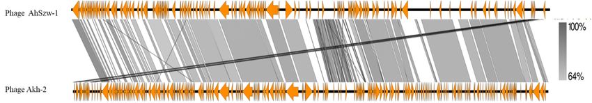
Pathogens 2020, 9, 215                                                                               2 of 13

ulcers, abdominal distension, accumulation of fluid, anemia, and hemorrhaging, resulting in mass
mortality in fishes around the world [4,5].
     In farming practices, various methods such as vaccination, water chlorination, and antibiotic
chemotherapy have been shown to reduce the occurrence of Aeromonas infection. However, excess use
of antibiotics has resulted in the emergence of multidrug-resistant strains of A. hydrophila [6]. In this
context, biological control seems to be a preferable alternative, and one proposed method is phage
therapy. Phages are abundant in nature and can be used to lyse bacteria with the advantages of having
high specificity toward their target and little to no negative impacts on the environment. Several
studies have demonstrated the therapeutic effects of phages as substitutes for antibiotics in human and
animal health problems [7,8].
     Loach (M. anguillicaudatus) is widely cultured in South Korea due to its high demand as a
food fish and its extensive use in traditional medicine and Buddhist ceremonies. A. hydrophila is
one of the main causes of massive mortalities in loach, and the presence of multidrug-resistant
Aeromonas further complicates its aquaculture [9]. Several studies have reported on the phage-based
biocontrol of Aeromonas infection. However, issue with modern regulations and the slow process for
getting phage therapy approval is the hurdle in field application, and the excessive use of antibiotics
continues [6,10,11]. Although A. hydrophila is a major pathogen of freshwater fish, higher densities of
A. hydrophila have been observed in saline habitats compared with freshwater habitats [12]. Considering
the advantages of biological disease control in aquaculture and the prevalence of the host bacterium
in seawater, the present study aims to isolate and characterize a potentially suitable bacteriophage
against A. hydrophila from water samples collected from marine environments. The isolated phage was
characterized and examined for its possible protective effects against experimentally induced motile
Aeromonas septicemia.

2. Results

2.1. Isolation and Characterization of the Bacteriophage
     From a total of 300 soil and water samples tested, two bacteriophages were isolated from one
water sample collected from Wahyeon Beach using A. hydrophila (KCTC 2358) as a host. Plaques of
different sizes were selected, and pure strains were established by three cycles of plaque isolation.
Plaque size is an inherent characteristic of these two phages and showed little variation in each plaque
isolation step. Therefore, we selected a phage designated as Akh-2, which produced bigger plaques for
further characterization and application for phage therapy.

2.2. Specific Host Range and Morphology of Akh-2
     The host range of phage Akh-2 was determined by spot assay. Among the 30 tested strains, Akh-2
lysed only four strains of A. hydrophila but could not lyse the other three strains of A. hydrophila or the
remaining 23 tested bacteria (Table 1).
     Transmission electron microscopy of Akh-2 revealed a phage with an icosahedral capsid of
50 ± 5 nm in diameter and a tail of 170 ± 5 nm in length (Figure 1), suggesting that Akh-2 belongs to
the Siphoviridae family, which was confirmed by genome sequence analysis, as described below.
Pathogens 2020, 9, 215 3 of 13
Table 1. Host range of Akh-2.
Pathogens 2020, 9, x FOR PEER REVIEW 3 of 13
Bacteria Strain ID Lysis by Akh-2 Source *
TableKTCC
1. Host2358
range of Akh-2. YES 1
Bacteria Strain32586
KCCM ID Lysis
NOby Akh-2 Source*
2
KTCC
AH-A6 2358 YESYES 3 1
KCCM 32586 NO 2
Aeromonas hydrophila AH-A8 NO 3
AH-A6 YES 3
Aeromonas hydrophila AH-20
AH-A8 YESNO 3 3
AH-20
AH-21 NOYES 3 3
AH-21
AH-Juwah YESNO 3 3
AH-Juwah YES 3
Pectobacterium spp. 35, 48, 63, 92, E42, E44 NO 4
Pectobacterium spp. 35, 48, 63, 92, E42, E44 NO 4
Streptococcus
Streptococcus aureus
aureus S75,
S75, S86,
S86, S103,
S103, S106
S106 NONO 4 4
Bacillus spp.
Bacillus spp. B5, B6,
B1, B2, B3, B4, B5, B6, B8,
B8, B11,
B11, B12,
B12, B13,
B13, B87,
B87, B97
B97 NONO 4 4
Escherichia coli CJY H7 NO 4
Escherichia coli CJY H7 NO 4
* 1: Korean Collection for Type Cultures, 2: Korean Culture Center of Microorganisms, 3: Seoul
* 1: Korean Collection for Type Cultures, 2: Korean Culture Center of Microorganisms, 3: Seoul National University,
National
4: Microbial University, 4: Microbial
Safety Division, NationalSafety Division,
Institute National
of Agricultural Institute
Science, of Development
Rural Agricultural Administration,
Science, Rural
Wanju-gun,
Development Jeollabuk-do, Republic Wanju-gun,
Administration, of Korea. Jeollabuk-do, Republic of Korea.
Figure 1. Electron
Figure 1. Electron micrograph
micrograph of of bacteriophage
bacteriophage Akh-2.
Akh-2. The two arrows
arrows indicate
indicate the
the beginning
beginning and
and
end
end of
of the
the 170-nm-long
170-nm-long tail. bar =
tail. Scale bar = 50 nm.
2.3.
2.3. One-Step
One-Step Growth
Growth Curve
Curve and
and Phage
Phage Stability
Stability
Based
Based on the one-step growth curve(Figure
on the one-step growth curve (Figure2),2),the
thelatent period
latent and
period burst
and size
burst were
size calculated
were as
calculated
~50 ± 5±min
as ~50 and
5 min and ± 5±PFU/infected
139139 5 PFU/infectedcell, respectively.
cell, respectively.
Phage maintained from −80 ℃ to 37 ℃ showed 99% infectivity after three days of incubation.
When maintained at 45 and 50 ℃ for the same period, the phage titer was decreased by 15% ± 5%
and 47% ± 5%, respectively. When kept at 55 or 60 ℃, no active phage was observed after three
days. The phage survival rate was 100% between 4 and 37 ℃ (Figure 3).
The phage was maintained from pH 4 to 11 for three days to test its pH stability. Phage
maintained at pH 7 showed 100% survival, which did not decrease significantly at pH 8 or 9.Pathogens 2020, 9, x FOR PEER REVIEW 4 of 13
Significant decreases in the phage titer were observed at pH 5 and 10 after three days; no phage
Pathogens 2020, 9, 215 4 of 13
infectivity was observed at pH 4 or 12 (Figure 4).
Pathogens 2020, 9, x FOR PEER REVIEW 4 of 13
Significant decreases in the phage titer were observed at pH 5 and 10 after three days; no phage
infectivity was observed at pH 4 or 12 (Figure 4).
Figure 2. One-step growth curve of phage Akh-2. The results are the average of three replications with
standard deviation as vertical lines.
Figure 2. One-step growth curve of phage Akh-2. The results are the average of three replications
Phagestandard
with maintained fromas
deviation −80 ◦ C tolines.
vertical 37 ◦ C showed 99% infectivity after three days of incubation.
When maintained at 45 and 50 ◦ C for the same period, the phage titer was decreased by 15% ± 5%
and 47% ± 5%,
Figure 2. respectively. When
One-step growth curvekept
of phage or 60 ◦The
at 55 Akh-2. C, no active
results arephage was observed
the average after three days.
of three replications
The phage ◦
withsurvival rate was as
standard deviation 100% between
vertical lines. 4 and 37 C (Figure 3).
Figure 3. Stability of phage Akh-2 at different temperatures. Phage Akh-2 (107 PFU/mL) was
maintained at the indicated temperatures for three days, and then the titer was determined by plaque
assay.
Figure 3.The
Figure results
Stabilityare
3.Stability ofofthe average
phage
phage of at
Akh-2
Akh-2 three replications
at different
different with standard
temperatures.
temperatures. deviation
Phage
PhageAkh-2 as
Akh-2(10 7 vertical
(10 7PFU/mL)
PFU/mL)lines.
was
was
maintained
maintained at at
thethe indicatedtemperatures
indicated temperaturesfor
for three
three days,
days, and
and then
thenthe
thetiter
titerwas
wasdetermined
determined byby
plaque
plaque
assay. The results are the average of three replications with standard deviation as vertical
assay. The results are the average of three replications with standard deviation as vertical lines.lines.
The phage was maintained from pH 4 to 11 for three days to test its pH stability. Phage maintained
at pH 7 showed 100% survival, which did not decrease significantly at pH 8 or 9. Significant decreases
in the phage titer were observed at pH 5 and 10 after three days; no phage infectivity was observed at
pH 4 or 12 (Figure 4).Pathogens 2020, 9, x FOR PEER REVIEW 5 of 13
Pathogens 2020, 9, 215 5 of 13
Pathogens 2020, 9, x FOR PEER REVIEW 5 of 13
Figure 4. Stability of phage Akh-2 at different pH levels. Phage Akh-2 (107 PFU/mL) was maintained at
Figure 4. Stability of phage Akh-2 at different pH levels. Phage Akh-2 (107 PFU/mL) was maintained
the indicated pH levels for three days, and thenlevels.
the titer wasAkh-2
determined by plaque assay. The results
atFigure 4. Stability of phage
levelsAkh-2 at different
days, pH
and thenPhage (10 PFU/mL) was maintained
7
the indicated pH for three the titer was determined by plaque assay. The
at the
are the average
indicatedofpH levels
three for three days,
replications withand then the
standard titer wasas
deviation determined by plaque assay. The
vertical lines.
results are the average of three replications with standard deviation as vertical lines.
results are the average of three replications with standard deviation as vertical lines.
2.4. Genomic Analysis
2.4.
2.4. Genomic Analysis
Genomic Analysis
The sequencing of Akh-2 generated 16,820,356 total reads and 1.070 Gbp with a G + C content
of 45.22%.
The The size of
The sequencing
sequencing of the
ofAkh-2
Akh-2 genome
generated
generated determined
16,820,356
16,820,356 by total
total de novo
reads and assembly
reads and Gbp
1.070 1.070 using
Gbp
with aG Platanus
with a G +was
+ C content Cof 114,901
content of
bp. A total
45.22%.
45.22%. Theofsize
The 254of
size ORFs
of were generated
thegenome
the genome determined
determined using
by debyORF finder,
de novo
novo ofusing
which
assembly
assembly 114 Platanus
using
Platanus werewas encoded
was bp.
114,901 byAthe bp.
114,901 plusA
totalof
strand
total of
and254141
254 ORFs
ORFsbywere
were generated
the minusgenerated using
strand. usingORF
Using
ORFfinder,
BLAST of which
finder, and 114 were
RAST,
of which 187encoded
114 ORFsencoded
were by the
were plus
predictedstrand
by the to encode
plus strand
and 141
hypothetical by the minus
proteins, 31 strand.
to encode Using
tRNAs,BLASTand and
36 toRAST,
encode 187
and 141 by the minus strand. Using BLAST and RAST, 187 ORFs were predicted to encode ORFs
proteins were
with predicted
various to encode
functions (Table S1).
hypothetical
Most proteins,
of the predictions
hypothetical 31 to encode
proteins, 31corresponded tRNAs,
to encode tRNAs, and 36 to
to different encode proteins
and 36 tostructural with
proteins
encode proteins various functions
(i.e.,various
with (Table
tail fiber, S1).
major(Table
functions capsid,
S1).
Most of the predictions corresponded to different structural proteins (i.e., tail fiber, major capsid, and
and phage protein), DNA ligases (repairing new DNA), and proteins involved
Most of the predictions corresponded to different structural proteins (i.e., tail fiber, major capsid, and in viral replication.
phage protein), DNA ligases (repairing new DNA), and proteins involved in viral replication. No
No ORFprotein),
phage encoding DNAa gene involved
ligases the integration
(repairing new DNA), ofand
viralproteins
genomeinvolved
into the in bacterial genome that
viral replication. No
ORF encoding a gene involved the integration of viral genome into the bacterial genome that results
results
ORF in
encodinglysogenic
a infection
gene involved was found.
the The
integration genome
of was
viral submitted
genome
in lysogenic infection was found. The genome was submitted to NCBI under accession number into to NCBI
the under
bacterial accession
genome thatnumber
results
MK318083.1.
lysogenic Among
inMK318083.1. infection
Among the
the was
1717A.A. hydrophila
found.
hydrophilaTheand and
genome Aeromonas
Aeromonaswasphagesphages
of theofSiphoviridae
submitted tothe Siphoviridae
NCBI under
family family
accession
listed bylisted
numberby
BaiBaietetal.al.[13],
MK318083.1. phage
phage Akh-2
[13],Among Akh-2 showed
the 17showed
A. hydrophila
thethe highest homology
and homology
highest Aeromonas to AhSzw-1,
tophages
AhSzw-1, of with with
the Siphoviridae
an an overall
overall familynucleotide
nucleotide listed by
sequence
Bai et al.identity
sequence [13], of
of 74%
phage
identity 74%Akh-2(Figure
(Figure 5). the highest homology to AhSzw-1, with an overall nucleotide
showed
5).
sequence identity of 74% (Figure 5).
Figure 5. Genomic comparison of phage Akh-2 and the reference A. hydrophila phage AhSzw-1
Figure 5. Genomic comparison of phage Akh-2 and the reference A. hydrophila phage AhSzw-1
(GenBank
(GenBank No. MG676225.1),constructed
No. MG676225.1), constructed using
using EasyFigure.
EasyFigure. Arrows
Arrows represent
represent ORFs.
ORFs. The The
level of level of
identity
identityis indicated by
bythe
thegray
grayshading.
shading.
Figure 5.is indicated
Genomic comparison of phage Akh-2 and the reference A. hydrophila phage AhSzw-1
(GenBank No. MG676225.1),
2.5. Protective Effects of Akh-2 constructed using EasyFigure. Arrows represent ORFs. The level of
identity is indicated by the gray shading.
In the trial challenge test with A. hydrophila, loach immersed in bacterial solutions of 1 × 10⁶ (T1),
1 × 10⁷ (T2), and 1 × 10⁸ (T3) CFU/mL for 30 min showed 100% mortality within 72, 48, and 48 h,
respectively. The bacterial dose of 1 × 10⁷ CFU/mL was selected for further trials.Pathogens 2020, 9, x FOR PEER REVIEW 6 of 13
2.4. Protective Effects of Akh-2
2.4. Protective Effects of Akh-2
In the trial challenge test with A. hydrophila, loach immersed in bacterial solutions of 1 × 10⁶ (T1),
1× 10⁷ the
In (T2),trial
and challenge
1 × 10⁸ (T3) test with
CFU/mL A. hydrophila,
for 30 min loach immersed
showed 100%inmortality
bacterial within
solutions 72,of48,1 ×and106 (T1),
48 h,
1× 10⁷ (T2),2020,and
respectively.
Pathogens 9,The
2151 bacterial
× 108 (T3)dose CFU/mL
of 1× 10 for7 CFU/mL
30 min showed
was selected100%for mortality within 72, 48, and 48
further trials. 6 ofh,13
respectively. The bacterial
The protective effect dose of 1× Akh-2
of phage 107 CFU/mL was selected
was assessed over afor96-h further
period trials.
after infection. Group I,
The protective effect of phage Akh-2 was assessed
the control group treated with PBS, showed no mortality or disease signs during over a 96-h period after infection. Group I,
the experiment
the The
control protective
group effect
treated of
withphage
PBS, Akh-2
showed wasno assessed
mortality over
or a 96-h
disease
(Figure 6). Group II, the bacterial infection group challenged with A. hydrophila (1× 10 CFU/mL), period
signs after
during infection.
the experiment
7 Group
I, the
(Figure control
showed6).sudden group
Group increasestreated
II, the bacterialwith PBS, showed
infection
in mortality no
group
of 40% mortality
± challenged or
1.52% and 96.33% disease signs
with A. ±hydrophila during
3.74% after the
(1× 24 experiment
10 and
7 CFU/mL),
48 h of
(Figure 6). Group II, the bacterial infection group challenged with A. hydrophila (1 × 10 7 CFU/mL),
showed sudden increases in mortality of 40% ± 1.52% and 96.33%
infection, respectively (Figures 6 and 7A). A cumulative mortality rate of 100% with abdominal ± 3.74% after 24 and 48 h of
showed
infection,
hemorrhagingsudden increases
respectively
and the(Figures in mortality
presence6ofand of 40%
red 7A). ± 1.52%
spotsAoncumulativeand
the body was 96.33%
mortality± 3.74%
observed rate after 24
of 100%
within and 48
72 towith h of infection,
96 h abdominal
(Figure 7B).
respectively
hemorrhaging (Figures
and the 6 and
presence7A). ofA cumulative
red spots on mortality
the body rate
was of 100%
observed
Group III, treated by immersion in water containing 1.0 × 10 PFU/mL phage Akh-2 after challenge
8 with
within abdominal
72 to 96 hemorrhaging
h (Figure 7B).
and
Group the presence
III, treated of
by red spots
immersion on the
in body
water was observed
containing 1.0 within
× 10 72 to
8 PFU/mL
with A. hydrophila, showed cumulative mortality rates of 16% ± 3.60%, 53% ± 1.50%, 57% ± 3.60%, and96 h (Figure
phage 7B).
Akh-2 Group
after III, treated
challenge
by immersion × h, 8 PFU/mL phage Akh-2 after challenge with A. hydrophila,
with
56.67% ± 3.78%inafter
A. hydrophila, water
showed 24,containing
cumulative
48, 72, and 1.096 10respectively
mortality rates of (Figure
16% ± 3.60%,
6). Most53%of± the1.50%, 57% ± 3.60%,
surviving fish inandthis
showed
56.67% ± cumulative
3.78% after mortality
24, 48, 72,rates
and of
96 16%
h, ± 3.60%,
respectively 53% ±
(Figure1.50%,
group showed no disease symptoms, but very small red spots could be observed on a few of the6). 57%
Most ±of 3.60%,
the and
surviving56.67% fish ±
in3.78%
this
after 24,
group
surviving 48,fish
showed 72,noand
by 96 h, respectively
disease
careful examination(Figure
symptoms, but very6).small
(Figure MostGroup
7C). redof the
spotssurviving
IV, could
treatedbefish in phage
this group
observed
with onAkh-2 showed
a few of the
without no
disease
surviving symptoms, but
fish by careful
bacterial infection, showedvery small
examination red
100% survival spots could
(Figure
with no be
7C). observed
Group IV,
symptoms, on a few of
treated with
confirming the surviving
phageofAkh-2
the safety fish
the phage by careful
without
(data
examination
bacterial (Figure
infection, 7C).
showed Group
100% IV, treated
survival with
with nophage Akh-2
symptoms, without
confirming
not shown). At the end of the experiment, A. hydrophila was re-isolated from the kidneys of dead bacterial
the infection,
safety of the showed
phage 100%
(data or
survival
not shown).
diseased with no
At but
fish, symptoms,
the end of thethe
not from confirming
experiment, the safety
survivingA.phage-treated of the
hydrophila was phage (data
re-isolated
fish, confirmingnot shown).
fromthat thethe At
kidneys the end
of dead
mortalities of the
or
were
experiment,
diseased
caused by A.A.
fish, buthydrophila
not fromwas
hydrophila. there-isolated
surviving from the kidneys
phage-treated of dead
fish, or diseased
confirming fish,mortalities
that the but not from werethe
surviving
caused by A. hydrophila. fish, confirming that the mortalities were caused by A. hydrophila.
phage-treated
Figure 6. Protective effect of Akh-2 against A. hydrophila
hydrophila in
in inoculated
inoculated loach.
loach. Af-clipped loach were
Figure 6. Protective
immersed in PBS (), effect
(□), of Akh-2 (1
A. hydrophila
hydrophila (1 ×× 1077 A.
against hydrophila
CFU/mL) ( ),in
(▨), orinoculated
A. hydrophila
hydrophila loach.
(1 1077 CFU/mL),
(1 ×× Af-clipped loach were
followed
immersed 8
by Akh-2 in
(1 PBS108(□), A. hydrophila (1 ×the10survival
CFU/mL) (▨), or measured
A. hydrophila (1 × 10 CFU/mL), followed
7
×
× 10 PFU/mL)
PFU/mL) (), and
(■), and the survival rate was within 967 h. The results are the
by Akh-2 of
average (1 three PFU/mL) (■),
× 108 replications and
with the survival
standard
standard rate was
deviation
deviation as measured
as vertical
vertical lineswithin
lines (Table96
(Table h. The results are the
S2).
S2).
average of three replications with standard deviation as vertical lines (Table S2).Pathogens 2020, 9, 215 7 of 13
Pathogens 2020, 9, x FOR PEER REVIEW 7 of 13
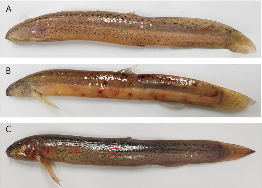
Protectionofofloach
Figure7.7.Protection
Figure loach(M. anguillicaudatus)from
(M.anguillicaudatus) fromA.A.hydrophila
hydrophilainfection
infectionbybybacteriophage
bacteriophage Akh-
Akh-2.
(A) Non-infected negative control. (B) Loach inoculated with A. hydrophila showing hemorrhagic
2. A: Non-infected negative control. B: Loach inoculated with A. hydrophila showing hemorrhagic red red
spots. (C) Loach treated with phage Akh-2 after inoculation with A. hydrophila showing
spots. C: Loach treated with phage Akh-2 after inoculation with A. hydrophila showing fewer and fewer and
smallerhemorrhagic
smaller hemorrhagicred redspots
spots(indicated
(indicatedbybyred
redboxes).
boxes).The
Theaverage
averagefish
fishsize was5 5± ±2 2cm.
sizewas cm.
3. Discussion
3. Discussion
The appearance of antibiotic-resistant pathogenic bacteria is a severe problem in aquaculture,
The appearance of antibiotic-resistant pathogenic bacteria is a severe problem in aquaculture,
especially when there are few treatment options, such as in A. hydrophila infection of cyprinid loach [6,9].
especially when there are few treatment options, such as in A. hydrophila infection of cyprinid loach
In some cases, 50% mortality was observed even with the use of antibiotics, indicating the presence
[6,9]. In some cases, 50% mortality was observed even with the use of antibiotics, indicating the
of antibiotic-resistant strains of Aeromonas species [9]. Bacteriophages have been used for the control
presence of antibiotic-resistant strains of Aeromonas species [9]. Bacteriophages have been used for
of bacterial infection since their discovery. Their therapeutic effects have been confirmed in animals,
the control of bacterial infection since their discovery. Their therapeutic effects have been confirmed
humans, crops, and aquaculture [7,8,10]. Several studies have reported the isolation of Aeromonas
in animals, humans, crops, and aquaculture [7,8,10]. Several studies have reported the isolation of
phages and examined their protective effects against disease [3,14,15].
Aeromonas phages and examined their protective effects against disease [3,14,15].
In the present study, we isolated bacteriophage Akh-2 infecting A. hydrophila (KTCT 2358) from
In the present study, we isolated bacteriophage Akh-2 infecting A. hydrophila (KTCT 2358) from
seawater collected from Geoje Island, South Korea, and analyzed its potential as a phage therapy agent.
seawater collected from Geoje Island, South Korea, and analyzed its potential as a phage therapy
Different phages that infect pathogenic bacteria of fish have been isolated from sea, riverine, and pond
agent. Different phages that infect pathogenic bacteria of fish have been isolated from sea, riverine,
waters [16–19]. Although A. hydrophila is a major pathogen of freshwater fish, a high abundance of
and pond waters [16–19]. Although A. hydrophila is a major pathogen of freshwater fish, a high
this bacterium in seawater has been reported [12]; thus, we attempted to isolate A. hydrophila-infecting
abundance of this bacterium in seawater has been reported [12]; thus, we attempted to isolate A.
phages from seawater samples. Yuan et al. [20] also reported the isolation of two A. hydrophila-infecting
hydrophila-infecting phages from seawater samples. Yuan et al. [20] also reported the isolation of two
Siphoviridae members from seawater. Transmission electron microscopy suggested that Akh-2 belongs
A. hydrophila-infecting Siphoviridae members from seawater. Transmission electron microscopy
to the Siphoviridae family due to its icosahedral head and long non-contractile tail. Among the 60 viruses
suggested that Akh-2 belongs to the Siphoviridae family due to its icosahedral head and long non-
that infect the Aeromonas species, including nine novel phages reported recently [13], only six belong
contractile tail. Among the 60 viruses that infect the Aeromonas species, including nine novel phages
to the Siphoviridae family, and only 3 (AhSzq-1, AhSzw-1, and 4L372X) infect A. hydrophila [13,20].
reported recently [13], only six belong to the Siphoviridae family, and only 3 (AhSzq-1, AhSzw-1, and
Although possible applications of these viruses in fish disease control have been suggested, they have
4L372X) infect A. hydrophila [13,20]. Although possible applications of these viruses in fish disease
not been tested. However, different studies have reported that bacteriophages from other families can
control have been suggested, they have not been tested. However, different studies have reported
be used for the control of fish disease caused by A. hydrophila [3], and Siphoviridae family phages can be
that bacteriophages from other families can be used for the control of fish disease caused by A.
used for the treatment of Pseudomonas plecoglossicida infections in fish [21]. Therefore, we conducted
hydrophila [3], and Siphoviridae family phages can be used for the treatment of Pseudomonas
plecoglossicida infections in fish [21]. Therefore, we conducted molecular characterization of aPathogens 2020, 9, 215 8 of 13
molecular characterization of a Siphoviridae family phage that infects A. hydrophila and examined the
therapeutic application of this phage.
Most bacteriophages are very specific for the receptors required for attachment and penetration of
the host cell [18]. In the Akh-2 host range analysis, it infected only four of the seven tested Aeromonas
strains, and no lytic effect was observed against the other non-Aeromonas species. Similar specific
host range results were described by Kim et al. [22]. The high specificity of Akh-2 can be considered
beneficial in terms of its effect on the healthy flora of aquatic organisms.
The one-step growth curve describes each of the different stages involved in the multiplication of
a bacteriophage. For every environmental condition, there is a strong relationship between the latent
period and burst size; that is, a high level of phage fitness is associated with an optimal latent time [23].
Phage Akh-2 has a latent period of 50 ± 5 min, which is almost the same as those of phages AhSzq-1
(50 min) and AhSzw-1 (60 min). Meanwhile, the burst size of Akh-2 was ~139 ± 5 PFU/infected cell,
which is higher than that of phages AhSzq-1 and AhSzw-1 (45 PFU/infected cell) [20]. The burst size of
a phage mainly depends on its plaque size and latent period, while the latent period can be affected by
the type of phage, the host, and the environmental conditions [24]. The short latent period combined
with the higher burst size makes Akh-2 a suitable candidate for phage therapy and could be used in
combination with other phages as a cocktail for better control effects.
Many factors can affect the normal processes of phage infection, mainly attachment, penetration,
and multiplication, but temperature and pH also play vital roles [25]. During the farming of warm
water fish species, the optimal temperature range is 25 to 37 ◦ C. The survival rate of phage Akh-2 was
100% between –80 ◦ C and 37 ◦ C, making it more suitable for long-term preservation and farm trails
(Figure 2). With the highest survival rate observed at pH 7, Akh-2 shows reasonable stability around
neutral pH, which is the optimum range for warm water fish farming.
The genome size (114,901 bp) and G + C content (45.22%) of Akh-2 are similar to those of
A. hydrophila phages AhSzq-1 (112,558 bp and 43.86%, respectively) and AhSzw-1 (115,739 bp and
43.82%, respectively). AhSzw-1 encodes 32 predicted tRNA genes, whereas the genome of Akh-2
contains 31 tRNA genes. The presence of large numbers of tRNA genes in A. hydrophila phages is
presumed to complement the codon usage bias of the host, which has a G + C content of 61% and more
frequent usage of G or C at the third position [20].
In the genome of AKh-2, no ORF encoding a protein involved in the integration of viral genome
such as an integrase was found. Temperate or lysogenic phages are generally not recommended for
phage therapy [7]. The lysogenic phages can grant immunity against the same or related phages to the
host. Also, the host bacteria can acquire new genetic traits such as phage-encoded toxins and resistance
to antibiotic resistance determinants by phage conversion [7,17,19].
Several studies have reported the isolation of Aeromonas phages and the investigation of their
protective effects against Aeromonas salmonicida [26–28] and A. hydrophila [3,14]. Although the results of
these studies suggested their protective effects against Aeromonas infection, the degrees of protection
are different. The methods of bacteria inoculation and phage administration, such as intraperitoneal
injection [3], intramuscular injection [26], addition to water [28], and feeding phage impregnated
feed [3], can affect the degree of protection. For example, in a phage therapy experiment against
A. hydrophila in cyprinid loach (M. anguillicaudatus), the same fish species used in this experiment,
the fish were infected by intraperitoneal injection with A. hydrophila at 2.6 × 10⁷ CFU/fish, causing
100% cumulative mortality in 7 days, and were treated by injection of phage or feeding with pellets
that had been impregnated with phage suspensions [3]. In the case of intraperitoneal injection with
phage pAh1-C (3.0 × 10⁷ PFU/fish) or pAh6-C (1.7 × 10⁷ PFU/fish), the cumulative mortalities were
43.33% ± 2.89% and 16.67% ± 3.82%, respectively. When the fish were given phage-impregnated feed,
the cumulative mortality rates were 46.67% ± 3.82% (pAh1-C) and 26.67% ± 2.89% (pAh6-C), which
indicated that injection provided better protection than phage feeding. In our experiment, we used the
Af-clipping methods developed by Zhang et al. [29], which is more similar to the natural infection route.
Immersion of Af-clipped fish for 30 min in water containing 1 × 10⁷ CFU/mL A. hydrophila resultedPathogens 2020, 9, 215 9 of 13
in 100% cumulative mortality at 48 h. Other factors such as fish species, bacterial strain, the order of
bacteria inoculation, and phage administration and environmental conditions can also affect the degree
of protection. One factor that must be considered is the MOI. MOIs of 6.5, 100, and 10,000 were used
by Kim et al. [26], Silva et al. [27], and Imbeault et al. [28], respectively, and we used an MOI of 10.
Due to multiplication and the resulting increase in phage titer in the presence of the host bacterium,
the precise initial dose of phage has not been considered critical. In fact, Verner-Jeffreys [30] reported
that phage therapy with an MOI of 100,000 was not effective for controlling A. salmonicida.
One consideration in phage therapy is the appearance of phage-resistant host bacteria. Although
the exact mechanism of resistance, such as resistance by the CRISPR-CAS system, has not been
determined, the occurrence of phage-resistant mutants has been reported in phage therapy against
Aeromonas [3,27,31]. It has been suggested that phage resistance can be overcome by the phage due to
the co-evolution of the phage and host [27]. Moreover, the slow growth of phage-resistant strains has
been observed, which can limit the bacterial population to a level controllable by the host immune
system [27,32]. One possible method to overcome the presence or occurrence of phage-resistant strains
is the application of a phage cocktail rather than a single phage isolate, as shown by Chen et al. [33].
Although we isolated only two phages that can infect A. hydrophila from seawater, isolation of more
A. hydrophila phages is promising, as shown by Hassan et al. [34], and further isolation, characterization,
and application of A. hydrophila phages are necessary.
4. Materials and Methods
4.1. Isolation, Culture, and Characterization of the Bacteriophage
A. hydrophila strain KCTC 2358, which has been reported as a fish pathogen, was obtained from
the Korean Collection for Type Cultures and used as a host bacterium for phage isolation. A total
of 300 samples of seawater near a beach and soil from a beach were collected from nine locations
on Geoje Island, South Korea. For phage isolation, 10 g of soil was mixed with 10 mL of sterilized
distilled water and agitated 20 m at room temperature. The mixture or collected water samples were
centrifuged at 3000× g for 20 min at 4 ◦ C, and the resulting supernatant was filtered using a 0.2-µm
filter (GVS Filter Technology, Indianapolis, Indiana). For phage enrichment, 100 µL filtrate, 4 mL
nutrient broth, and 300 µL A. hydrophila overnight culture were mixed and incubated at 37 ◦ C for 24 h.
The cultures were centrifuged (3000× g for 20 min), and the supernatants were filtered as above. Then,
an overlay mixture of 0.5 µL of the filtrate, 300 µL A. hydrophila overnight culture, and molten nutrient
agar (0.7% agar) was poured over solidified nutrient agar (1.5%). After incubation at 37 ◦ C for 24 h,
clear phage plaques were picked using sterile end-cut pipette tips and stored at 4 ◦ C in 1 M SM buffer
(100 mM NaCl, 8 mM MgSO4 ·7H2 O, and 50 mM Tris-Cl (pH 7.5)). Phage purification was performed
as reported by Gencay et al. [35]. Phage titers were determined by the double-layer agar method.
For long-term preservation, phages were preserved in 1 M SM buffer and stored at −80 ◦ C.
4.2. Specific Host Range and Morphology of the Bacteriophage
A total of 30 strains, including seven strains of A. hydrophila (Table 1), were used for spot tests to
confirm the host range of the phage. Purified phage particles (109 plaque-forming units [PFU]/mL)
preserved in SM buffer were adsorbed onto a carbon-coated copper grid and negatively stained with
2% uranyl acetate. After drying for 20 min at room temperature, the grids were observed using
the JEM1010 electron microscope (Jeol, Tokyo, Japan) at the National Instrumentation Center for
Environmental Management (NICEM) of Seoul National University, South Korea.
4.3. One-Step Growth Curve and Stability Analyses
One-step growth curve analysis was conducted according to Verma et al. [36] with some
modifications. Briefly, 11 mL phage suspension (109 PFU/mL) was added to a 20-mL overnight
culture of the host bacterium for a multiplicity of infection (MOI) of 0.001, and the mixture wasPathogens 2020, 9, 215 10 of 13
incubated at 37 ◦ C for 5 min. After centrifugation at 35,000× g for 30 min, the supernatant was carefully
removed, and the pellet was suspended in 20 mL nutrient broth. The mixture was incubated at 37 ◦ C
for 90 min, and samples were collected at 5-min intervals. The aliquots were diluted, and phage titers
were determined by the double-layer agar method.
Phage stability was determined, according to Verma et al. [36], with slight modifications. Briefly,
1-mL aliquots of phage (107 PFU/mL) were stored at various temperatures (−80, −20, 4, 25, 37, 45, 50,
and 55 ◦ C) in Eppendorf tubes for three days, and samples were collected every 24 h to determine the
titer by the double-layer agar method. For the pH stability test, Tris-Cl was adjusted from pH 4 to
12. At each pH level, the phage was inoculated to a final concentration of 107 PFU/mL and incubated
at 37 ◦ C for three days. Samples were taken every 24 h to determine the titer by the double-layer
agar method.
4.4. Genomic Analysis
4.4.1. Phage Genome Extraction
For nucleic acid isolation, 1 mL purified phage (109 PFU/mL) was mixed with 0.5 µL DNase
(1 U/µL) and 1 µL RNase A (10 mg/mL) and incubated for 1 h at 37 ◦ C to remove any bacterial DNA
contamination. Next, 20 µL EDTA (0.5 M), 1.25 µL proteinase K (20 mg/mL), and 100 µL 10% SDS were
added to the phage solution, followed by a further incubation at 60◦ C for 1 h. After incubation, a
500-µL mixture of phenol, chloroform, and isoamyl alcohol (25:24:1) was added to the solution and
mixed, followed by centrifugation at 12,000× g for 10 min. The supernatant was transferred to a new
Eppendorf tube with a 1/10 volume of 3 M sodium acetate and a 2× volume of 100% isopropanol for
nucleic acid precipitation, followed by incubation at −20 ◦ C for 20 min and centrifugation at 12,000× g
and 4 ◦ C for 15 min. The nucleic acid pellet was washed with 70% ethanol, dried at room temperature,
dissolved in 30 µL TE buffer, and stored at 4 ◦ C until further use. The obtained genome was dissolved
in distilled water, treated with DNase, RNase, and exonuclease II, and subjected to electrophoresis on
an agarose gel to determine the property of the genome.
4.4.2. Whole-Genome Sequencing, Assembly, and Annotation
Whole-genome sequencing was conducted using next-generation sequencing technology on the
Illumina Hiseq sequencer at the Theragen Etex Bio Institute (Suwon, South Korea). The genomic DNA
sample from the isolated phage was processed further for library preparation under the sample ID V2
TN1809D0396. The sequence reads were assembled de novo using Platanus version 1.2.2. Open reading
frames (ORFs) were predicted using GeneMark.hmm, and their functions were annotated using the
National Center for Biotechnology Information (NCBI) BLAST server and Rapid Annotations using
Subsystems Technology (RAST) [37]. The genomes of Akh-2 and Ahszw-1 were compared by using
Easyfig [38].
4.5. Protective Effects of Akh-2 in Infected Loach Fish
4.5.1. Challenge Test and Estimation of the Lethal Dose
To simulate a more natural infection route similar to that in the aquatic environment,
the methodology described by Zhang et al. [29] was adopted. A total of 120 healthy adipose
fin-clipped (Af-clipped) loach with an average body weight of 10 g was distributed into four groups
(each group consisting of three replicates of 10 fish) in four 30-L tanks filled with 15 L tap water that
had been aerated overnight, which were labeled as control, T1, T2, and T3.
A. hydrophila (KCTC 2358) was cultured in NA medium, harvested by centrifugation at 3000× g
for 20 min, resuspended in saline, and counted by plating. To determine the optimal challenge dose,
T1, T2, and T3 groups were inoculated with final concentrations of 1 × 10⁶ , 1 × 10⁷ , and 1 × 10⁸ colony
forming units (CFU)/mL, respectively. No bacteria were inoculated in the control group.Pathogens 2020, 9, 215 11 of 13
After a 30-min immersion with the respective treatment, fish were transferred to the experimental
tanks with tap water and observed for 96 h for determination of clinical signs and mortality.
4.5.2. Phage Treatment of Infected Fish
A total of 135 healthy loach with an average body weight of 10 g were divided into three groups
(each consisting of three replicates of 15 fish) and placed in 30-L tanks filled with 15 L water. Af-clipping
and the immersion method described above were used for the inoculation of bacteria and phage.
Group I was the control group in which loach were immersed in phosphate-buffered saline
(PBS) for 30 min. Group II was the bacterial challenge group in which loach were immersed in water
containing A. hydrophila at a concentration of 1 × 10⁷ CFU/mL for 30 min, as described above. Group
III was the phage-treated group in which loach were immersed in water containing A. hydrophila
(1 × 10⁷ CFU/mL) for 30 min, and then immediately immersed in water containing phage Akh-2 at
a final concentration of 1 × 10⁸ PFU/mL for 30 min, maintaining an MOI of 10. In Group IV, loach
were immersed for 30 min in water containing phage Akh-2 (1.0 × 10⁸ PFU/mL) without bacterial
infection. After each immersion, loach were raised in separate 15-L water-filled experimental tanks
and observed for 96 h without water exchange. Continuous aeration was provided, but food was not
provided to maintain the water quality. The experiments were conducted three times on different
occasions. The water was maintained at 28 ± 2 ◦ C and pH 7 throughout the experimental trails.
At the end of experiments, kidneys of dead or diseased fish were taken, ground in PBS and spread
on LB plates after dilution. After the culture of grown bacterial colonies in LB medium, DNA was
extracted and used as a template for PCR with universal primers for bacterial 16S rRNA gene, 27F
(5’-AGAGTTTGATCCTGGCTCAG-3’) and 1492R (5’-CGGTTACCTTGTTACGACTT-3’). The PCR
products were sequenced for the identification of bacteria.
4.6. Statistical Analysis
Statistically significant differences in all experiments were determined by Student’s t-test. A p-valuePathogens 2020, 9, 215 12 of 13
3. Jun, J.W.; Kim, J.H.; Shin, S.P.; Han, J.E.; Chai, J.Y.; Park, S.C. Protective effects of the Aeromonas phages
pAh1-C and pAh6-C against mass mortality of the cyprinid loach (Misgurnus anguillicaudatus) caused by
Aeromonas hydrophila. Aquaculture 2013, 416, 289–295. [CrossRef]
4. Austin, B.; Austin, D.A. Aeromonadaceae Representative (Aeromonas salmonicida). In Bacterial Fish
Pathogens; Springer: Dordrecht, The Netherlands, 2012; pp. 147–228.
5. Lilley, J.; Hart, D.; Richards, R.H.; Roberts, R.J.; Cerenius, L.; Söderhäll, K. Pan-Asian spread of single fungal
clone results in large scale fish kills. Veter Rec. 1997, 140, 653–654. [CrossRef]
6. Kaskhedikar, M.; Chhabra, D. Multiple drug resistance in Aeromonas hydrophila isolates of fish. Food
Microbiol. 2010, 28, 157–168.
7. Altamirano, F.L.G.; Barr, J.J. Phage Therapy in the Postantibiotic Era. Clin. Microbiol. Rev. 2019, 32, e00066-18.
[CrossRef]
8. McCallin, S.; Sacher, J.C.; Zheng, J.; Chan, B.K. Current State of Compassionate Phage Therapy. Viruses 2019,
11, 343. [CrossRef] [PubMed]
9. Jun, J.W.; Kim, J.H.; Gomez, D.; Choresca, C.H., Jr.; Han, J.E.; Shin, S.P.; Park, S.C. Occurrence of
tetracycline-resistant Aeromonas hydrophila infection in Korean cyprinid loach (Misgurnus anguillicaudatus).
African J. Microbiol. Res. 2010, 4, 849–855.
10. Richards, G.P. Bacteriophage remediation of bacterial pathogens in aquaculture: A review of the technology.
Bacteriophage 2014, 4, e975540. [CrossRef]
11. Fauconnier, A. Phage Therapy Regulation: From Night to Dawn. Viruses 2019, 11, 352. [CrossRef] [PubMed]
12. Hazen, T.; Fliermans, C.B.; Hirsch, R.P.; Esch, G.W. Prevalence and distribution of Aeromonas hydrophila in
the United States. Appl. Environ. Microbiol. 1978, 36, 731–738. [CrossRef] [PubMed]
13. Bai, M.; Cheng, Y.-H.; Sun, X.-Q.; Wang, Z.-Y.; Wang, Y.-X.; Cui, X.-L.; Xiao, W. Nine Novel Phages from a
Plateau Lake in Southwest China: Insights into Aeromonas Phage Diversity. Viruses 2019, 11, 615. [CrossRef]
[PubMed]
14. Easwaran, M.; Dananjaya, S.H.S.; Park, S.C.; Lee, J.; Shin, H.; De Zoysa, M. Characterization of bacteriophage
pAh-1 and its protective effects on experimental infection of Aeromonas hydrophila in Zebrafish (Danio
rerio). J. Fish Dis. 2016, 40, 841–846. [CrossRef] [PubMed]
15. Da, E.-A.; Th, G.E.-D.A.M.M. New Approach to Use Phage Therapy against Aeromonas hydrophila Induced
Motile Aeromonas Septicemia in Nile Tilapia. J. Mar. Sci. Res. Dev. 2016, 6, 6. [CrossRef]
16. Phumkhachorn, P.; Rattanachaikunsopon, P. A lytic podophage specific to fish pathogenic Edwardsiella
tarda. Pak. J. Biotechnol. 2018, 15, 117–121.
17. Vincent, A.T.; Paquet, V.E.; Bernatchez, A.; Tremblay, D.M.; Moineau, S.; Charette, S. Characterization and
diversity of phages infecting Aeromonas salmonicida subsp. salmonicida. Sci. Rep. 2017, 7, 7054. [CrossRef]
18. Haq, I.U.; Chaudhry, W.N.; Andleeb, S.; Qadri, I. Isolation and Partial Characterization of a Virulent
Bacteriophage IHQ1 Specific for Aeromonas punctata from Stream Water. Microb. Ecol. 2011, 63, 954–963.
[CrossRef]
19. Luo, L.; Liao, G.; Liu, C.; Jiang, X.; Lin, M.; Zhao, C.; Tao, J.; Huang, Z. Characterization of bacteriophage HN48
and its protective effects in Nile tilapia Oreochromis niloticus against Streptococcus agalactiae infections.
J. Fish Dis. 2018, 41, 1477–1484. [CrossRef]
20. Yuan, S.; Chen, L.; Liu, Q.; Zhou, Y.; Yang, J.; Deng, D.; Li, H.; Ma, Y. Characterization and genomic analyses
of Aeromonas hydrophila phages AhSzq-1 and AhSzw-1, isolates representing new species within the
T5virus genus. Arch. Virol. 2018, 163, 1985–1988. [CrossRef]
21. Park, S.C.; Shimamura, I.; Fukunaga, M.; Mori, K.-I.; Nakai, T. Isolation of Bacteriophages Specific to a Fish
Pathogen, Pseudomonas plecoglossicida, as a Candidate for Disease Control. Appl. Environ. Microbiol. 2000,
66, 1416–1422. [CrossRef]
22. Kim, J.H.; Son, J.S.; Choi, Y.J.; Choresca, C.H.; Shin, S.P.; Han, J.E.; Jun, J.W.; Kang, D.H.; Oh, C.; Heo, S.J.;
et al. Isolation and Characterization of a Lytic Myoviridae Bacteriophage PAS-1 with Broad Infectivity in
Aeromonas salmonicida. Curr. Microbiol. 2012, 64, 418–426. [CrossRef] [PubMed]
23. Shivu, M.M.; Rajeeva, B.C.; Girisha, S.K.; Karunasagar, I.; Krohne, G.; Karunasagar, I. Molecular
characterization of Vibrio harveyi bacteriophages isolated from aquaculture environments along the coast of
India. Environ. Microbiol. 2007, 9, 322–331. [CrossRef]
24. Weinbauer, M. Ecology of prokaryotic viruses. FEMS Microbiol. Rev. 2004, 28, 127–181. [CrossRef] [PubMed]Pathogens 2020, 9, 215 13 of 13
25. Jończyk, E.; Kłak, M.; Mi˛edzybrodzki, R.; Górski, A. The influence of external factors on
bacteriophages—review. Folia Microbiol. 2011, 56, 191–200. [CrossRef] [PubMed]
26. Kim, J.H.; Choresca, C.H.; Shin, S.P.; Han, J.E.; Jun, J.W.; Park, S.C. Biological Control ofAeromonas
salmonicidasubsp.salmonicidaInfection in Rainbow Trout (Oncorhynchus mykiss) UsingAeromonasPhage
PAS-1. Transbound. Emerg. Dis. 2013, 62, 81–86. [CrossRef]
27. Silva, Y.J.; Moreirinha, C.; Pereira, C.S.G.; Costa, L.; Rocha, R.; Cunha, A.; Gomes, N.C.M.; Calado, R.;
Almeida, A. Biological control of Aeromonas salmonicida infection in juvenile Senegalese sole (Solea
senegalensis) with Phage AS-A. Aquaculture 2016, 450, 225–233. [CrossRef]
28. Imbeault, S.; Parent, S.; Lagacé, M.; Uhland, C.F.; Blais, J.-F. Using Bacteriophages to Prevent Furunculosis
Caused by Aeromonas salmonicida in Farmed Brook Trout. J. Aquat. Anim. Health 2006, 18, 203–214.
[CrossRef]
29. Zhang, D.; Xu, D.; Shoemaker, C. Experimental induction of motile Aeromonas septicemia in channel catfish
( Ictalurus punctatus ) by waterborne challenge with virulent Aeromonas hydrophila. Aquac. Rep. 2016, 3,
18–23. [CrossRef]
30. Verner–Jeffreys, D.W.; Algoet, M.; Pond, M.J.; Virdee, H.K.; Bagwell, N.J.; Roberts, E.G. Furunculosis in
Atlantic salmon (Salmo salar L.) is not readily controllable by bacteriophage therapy. Aquaculture 2007, 270,
475–484.
31. Horvath, P.; Barrangou, R. CRISPR/Cas, the Immune System of Bacteria and Archaea. Science 2010, 327,
167–170. [CrossRef]
32. Jun, S.Y.; Jung, G.M.; Yoon, S.J.; Oh, M.-D.; Choi, Y.-J.; Lee, W.J.; Kong, J.-C.; Seol, J.G.; Kang, S.H. Antibacterial
properties of a pre-formulated recombinant phage endolysin, SAL-1. Int. J. Antimicrob. Agents 2013, 41,
156–161. [CrossRef] [PubMed]
33. Chen, L.; Yuan, S.; Liu, Q.; Mai, G.; Yang, J.; Deng, D.; Zhang, B.; Liu, C.; Ma, Y. In Vitro Design and Evaluation
of Phage Cocktails Against Aeromonas salmonicida. Front. Microbiol. 2018, 9, 9. [CrossRef] [PubMed]
34. Hassan, S.W.M.; Ali, S.M.; Almisherfi, M.M. Isolation and Molecular Characterization of Some Marine
Aeromonas phages: Protective Effects for Nile Tilapia Infected with Aeromonas hydrophila. J. Pure Appl.
Microbiol. 2018, 12, 1175–1185. [CrossRef]
35. Gencay, Y.E.; Birk, T.; Sørensen, M.C.H.; Brøndsted, L. Methods for Isolation, Purification, and Propagation
of Bacteriophages of Campylobacter jejuni. In Methods in Molecular Biology; Butcher, J., Stintzi, A., Eds.;
Humana Press: New York, NY, USA, 2017; Volume 1512, pp. 19–28.
36. Verma, V.; Harjai, K.; Chhibber, S. Characterization of a T7-Like Lytic Bacteriophage of Klebsiella pneumoniae
B5055: A Potential Therapeutic Agent. Curr. Microbiol. 2009, 59, 274–281. [CrossRef]
37. RAST. Available online: https://rast.theseed.org/FIG/rast.cgi. (accessed on 18 July 2018).
38. Sullivan, M.J.; Petty, N.; Beatson, S.A. Easyfig: A genome comparison visualizer. Bioinformatics 2011, 27,
1009–1010. [CrossRef]
© 2020 by the authors. Licensee MDPI, Basel, Switzerland. This article is an open access
article distributed under the terms and conditions of the Creative Commons Attribution
(CC BY) license (http://creativecommons.org/licenses/by/4.0/).You can also read