Transient Receptor Potential Ankyrin 1 Activation within the Cardiac Myocyte Limits Ischemia-reperfusion Injury in Rodents
←
→
Page content transcription
If your browser does not render page correctly, please read the page content below
Transient Receptor Potential Ankyrin 1 Activation within
the Cardiac Myocyte Limits Ischemia–reperfusion Injury
in Rodents
Yao Lu, M.D., Honit Piplani, Ph.D., Stacy L. McAllister, Ph.D., Carl M. Hurt, M.D., Ph.D.,
Eric R. Gross, M.D., Ph.D.
ABSTRACT
Background: Recent evidence suggests that cross talk exists between cellular pathways important for pain signaling and
ischemia–reperfusion injury. Here, the authors address whether the transient receptor potential ankyrin 1 (TRPA1) channel,
important in pain signaling, is present in cardiac myocytes and regulates cardiac ischemia–reperfusion injury.
Downloaded from http://pubs.asahq.org/anesthesiology/article-pdf/125/6/1171/272784/20161200_0-00023.pdf by guest on 21 October 2020
Methods: For biochemical analysis of TRPA1, techniques including quantitative polymerase chain reaction, Western blot,
and immunofluorescence were used. To determine how TRPA1 mediates cellular injury, the authors used an in vivo model of
rat cardiac ischemia–reperfusion injury and adult rat–isolated cardiac myocytes subjected to hypoxia–reoxygenation.
Results: The authors’ biochemical analysis indicates that TRPA1 is within the cardiac myocytes. Further, using a rat in vivo
model of cardiac injury, the TRPA1 activators ASP 7663 and optovin reduce myocardial injury (45 ± 5%* and 44 ± 8%,*
respectively, vs. control, 66 ± 6% infarct size/area at risk; n = 6 per group; mean ± SD; *P < 0.001). TRPA1 inhibition also
blocked the infarct size–sparing effects of morphine. In isolated cardiac myocytes, the TRPA1 activators ASP 7663 and opt-
ovin reduce cardiac myocyte cell death when given during reoxygenation (20 ± 3%* and 22 ± 4%* vs. 36 ± 3%; percentage of
dead cells per field, n = 6 per group; mean ± SD; *P < 0.05). For a rat in vivo model of cardiac injury, the infarct size–sparing
effect of TRPA1 activators also occurs during reperfusion.
Conclusions: The authors’ data suggest that TRPA1 is present within the cardiac myocytes and is important in regulating
myocardial reperfusion injury. The presence of TRPA1 within the cardiac myocytes may potentially explain why certain pain
relievers that can block TRPA1 activation, such as cyclooxygenase-2 inhibitors or some nonsteroidal antiinflammatory drugs,
could be associated with cardiovascular risk. (Anesthesiology 2016; 125:1171-80)
M ore than 200 million opioid prescriptions are writ-
ten annually in the United States to treat pain.1 These
drugs have serious unwanted side effects including abuse,
What We Already Know about This Topic
• Transient receptor potential ankyrin 1 (TRPA1) receptors func-
dependence, and addiction. As a result, an opioid epidemic tion as pain receptors within the nervous system.
• TRPA1 receptor antagonists are novel targets for nonopioid
within the United States is shifting medical practice toward pain management. However, a physiologic role for TRPA1
prescribing nonopioid analgesics or opioid adjuvants to receptors during myocardial ischemia–reperfusion injury has
reduce opioid use intraoperatively, postoperatively, and in not been investigated.
the clinic.2,3 Further, developing novel analgesics to replace What This Article Tells Us That Is New
opioids within medical practice is a continued focus for
• The authors report that the TRPA1 receptors exist within the
scientists. cardiac myocyte, and TRPA1 activation protects the heart
For these reasons, understanding how the pathways from myocardial ischemia–reperfusion injury.
of pain signaling may contribute to protection of organs • The results reported have the potential to impact novel targets
from ischemia–reperfusion injury is important since cel- for cardiac protection and have significant implications regard-
ing the safety of TRPA1 antagonists being developed as pain
lular cross talk exists between these two pathways.3 Non- medications.
narcotic analgesics may also have a deleterious effect by
blocking the endogenous mechanisms that reduce the COX-2 inhibitors, the cardiac safety of some nonsteroidal
ischemia–reperfusion injury of organs. For example, antiinflammatory drugs (NSAIDs) has also been recently
inhibition of cyclooxygenase-2 (COX-2) experimentally questioned.2,3
blocks the natural ability of the heart to protect against Since the heart unlike other organs also possesses neu-
ischemia–reperfusion injury.4,5 Clinically, in addition to roendocrine qualities, pain receptors from the transient
Supplemental Digital Content is available for this article. Direct URL citations appear in the printed text and are available in both the
HTML and PDF versions of this article. Links to the digital files are provided in the HTML text of this article on the Journal’s Web site www.
anesthesiology.org.
Submitted for publication April 13, 2016. Accepted for publication August 29, 2016. From the Department of Anesthesiology, Perioperative
and Pain Medicine, Stanford University, Stanford, California.
Copyright © 2016, the American Society of Anesthesiologists, Inc. Wolters Kluwer Health, Inc. All Rights Reserved. Anesthesiology 2016; 125:1171-80
Anesthesiology, V 125 • No 6 1171 December 2016
Copyright © 2016, the American Society of Anesthesiologists, Inc. Wolters Kluwer Health, Inc. Unauthorized reproduction of this article is prohibited.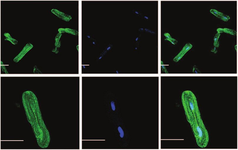
TRPA1 and Cardioprotection

         receptor potential family could potentially exist within the             For qPCR, adult rat cardiac myocytes, left ventricle heart
         cardiac myocytes. In particular, transient receptor poten-           tissue, and H9C2 cell samples were stored in RNA later
         tial ankyrin 1 (TRPA1) inhibitors are being explored and             (Ambion, USA) at −80°C and homogenized in 1 ml TRI
         developed as an alternative to opioids for pain control.6            reagent (Molecular Research Center, USA). Total RNA was
         Further, the TRPA1 receptor is also modulated by different           isolated from tissue and cell homogenates using RNeasy Mini
         pain relievers including NSAIDs, COX-2 inhibitors, and               Kit 50 (Qiagen, Germany). Complementary DNA (cDNA)
         acetaminophen.7–9                                                    synthesis was performed with the high capacity RNA-to-
             The TRPA1 receptor, besides regulating pain, also serves         cDNA kit (Applied Biosystems, USA). qPCR was performed
         multiple functions within the cell.10,11 In particular, TRPA1        using TaqMan Gene Expression Assays (Applied Biosys-
         functions as a sensor that is activated by reactive aldehydes        tems). The 20 μl reactions contained 10 μl TaqMan Fast
         and is modulated when intracellular changes in oxygen lev-           Universal PCR Master Mix (2×; Thermo Fischer Scientific,

                                                                                                                                                  Downloaded from http://pubs.asahq.org/anesthesiology/article-pdf/125/6/1171/272784/20161200_0-00023.pdf by guest on 21 October 2020
         els occur.12,13 Both factors are important in regard to organ        USA), 1 μl of the specific TaqMan assay, 1 μl cDNA, and 8
         ischemia–reperfusion injury. In particular, the production           μl water. Cycling parameters were 30-s initial setup at 95°C,
         of reactive aldehydes as a result of the breakdown of lipid          followed by 40 cycles at 95°C for 3 s and 30 s at 61°C (ABI
         membranes is considered a critical mediator of cellular              7500 Fast; Applied Biosystems). Primers used were as fol-
         injury.13                                                            lows: TRPA1 forward: GCTTCTGCAAGACATCAGCG,
             However, little is known about whether the TRPA1 recep-          TRPA1 reverse: CCTCTCCATCTGGCAGCAAA, glycer-
         tor exists in the cardiac myocyte and if TRPA1 contributes           aldehyde 3-phosphate dehydrogenase (GAPDH) forward:
         to regulating injury during cardiac ischemia–reperfusion. If         CTCAGTTGCTGAGGAGTCCC, and GAPDH reverse:
         TRPA1 is present in the cardiac myocyte, this could explain          ATTCGAGAGAAGGGAGGGCT.
         why some nonopioid analgesics block natural pathway(s) of                For Western blot, adult cardiac myocytes, left ventricle
         protection from ischemia–reperfusion injury. This would              tissue, and H9C2 cells were used. The left ventricles of male
         also be important to understand when developing drugs                Sprague–Dawley rats were excised, finely minced with scis-
         targeting TRPA1 to provide analgesia. Here, we address the           sors, and homogenized with mannitol–sucrose lysis buffer
         question whether TRPA1 is present in the cardiac myocyte             (210 mM mannitol, 70 mM sucrose, 5 mM 3-N-morpholino
         and further if this receptor is important in mediating isch-         propanesulfonic acid, 1 mM EDTA with pH 7.4, protease/
         emia–reperfusion injury.                                             phosphatase inhibitors, and 1% Triton-X [Sigma, USA]).
                                                                              Homogenates were centrifuged at 800g for 5 min to remove
                                                                              cellular debris, and the supernatant was kept as the total frac-
         Materials and Methods
                                                                              tion. Adult cardiac myocytes and H9C2 cells were also lysed in
         Procedures and protocols were approved by the Animal                 mannitol–sucrose buffer. For the three types of samples used
         Care and Use Committee at Stanford University, Stanford,             (adult cardiac myocytes, left ventricle heart tissue, and H9C2
         California. All animal studies conformed to the National             cells), total protein content was determined by using Bradford
         Institutes of Health Guide for the Care and Use of Laboratory        assay with 35 μg of each homogenate run on 10% sodium
         Animals (eighth edition, 2011). Eight- to 10-week-old male           dodecyl sulfate polyacrylamide gel electrophoresis gels. Mem-
         Sprague–Dawley rats (Charles River, USA) were used for the           brane proteins were transferred to a polyvinylidene difluoride
         studies outlined.                                                    membrane and probed overnight at 4°C for specific antibod-
                                                                              ies to TRPA1 (1:500 dilution in 5% milk-tris-buffered saline
         Pharmacologic Agents                                                 with tween-20; Novus, USA) and GAPDH (1:1,000 dilution
         The TRPA1 receptor activators, ASP 7663 (ASP; 3 mg/kg in             in 5% milk-tris-buffered saline with tween-20; Sigma). The
         vivo and 3 μM in vitro) and optovin (1 mg/kg in vivo and             next day, membranes were washed and incubated in second-
         1 μM in vitro), in addition to the TRPA1 inhibitors, TCS             ary anti-rabbit antibody for 2 h (1:1,000 for TRPA1 and
         5861528 (TCS; 1 mg/kg in vivo and 1 mM in vitro) and                 1:3,000 for GAPDH in 5% milk; ThermoScientific, USA).
         AP 18 (AP; 1 mg/kg in vivo and 1 mM in vitro), were dis-             Membranes were developed in enhanced chemiluminescent
         solved in dimethyl sulfoxide (DMSO). Doses were chosen               reagent, and images were acquired by using an Azure Biosys-
         for TRPA1 activators and inhibitors based on data for these          tems c300 (Azure Biosystems, USA).
         compounds provided in the manufacturer insert (Tocris,                   For immunofluorescence, isolated cardiomyocytes were
         United Kingdom). Morphine (0.3 mg/kg IV bolus) was dis-              plated on precoated laminin (Invitrogen, USA) glass cover-
         solved in saline, and the dose was determined from our pre-          slips (2 μg/cm2) for 24 h and fixed with 2% buffered parafor-
         vious studies.14,15                                                  maldehyde for 10 min at room temperature. The fixed cells
                                                                              were washed three times at 5-min intervals with phosphate-
         Biochemical Studies                                                  buffered saline (PBS). The cells were blocked and permeabi-
         Biochemical studies consisted of quantitative poly-                  lized with blocking buffer (40 mM HEPES, 3% dry milk,
         merase chain reaction (qPCR), Western blot, and                      PBS, and 0.1% Triton-X 100) for 30 min at room tempera-
         immunofluorescence.                                                  ture. Cells were incubated with TRPA1 primary antibody

         Anesthesiology 2016; 125:1171-80                              1172                                                          Lu et al.

Copyright © 2016, the American Society of Anesthesiologists, Inc. Wolters Kluwer Health, Inc. Unauthorized reproduction of this article is prohibited.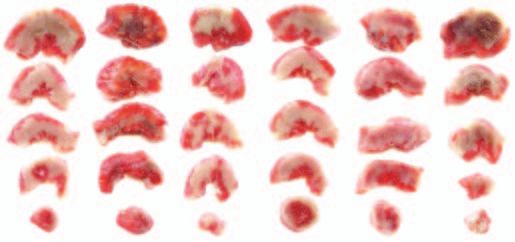
PERIOPERATIVE MEDICINE

         (1:250 dilution) in blocking buffer for 1 h at room tem-               and calculated throughout the experimental protocol using
         perature. Excess antibody was removed by washing samples               PowerLab monitoring system (MLS060/8 PowerLab 4/35;
         with PBS three times at 5-min intervals. The cells were then           AD Instruments, USA). We defined rate pressure prod-
         incubated with donkey anti-rabbit Alexa Fluor 488 (1:500               uct (RPP) as the product of heart rate and systolic blood
         dilution; Invitrogen) in blocking buffer for 2 h at room tem-          pressure.
         perature. To remove excess secondary antibody, the cells were
         washed six times at 5-min intervals. The nucleus was stained           In Vitro Cardiac Myocytes Hypoxia and Reoxygenation
         with dye 4′,6-diamidino-2-phenylindole (1 μg/ml) diluted               Injury Model
         in PBS for 20 min. Cells were then washed for 10 min with                  Adult rat primary cardiac myocytes were isolated from
         PBS and mounted in ProLong gold reagent antifade reagent               male Sprague–Dawley rat hearts by enzymatic dissociation
         (Invitrogen) for microscopic imaging.                                  as previously described.16 The isolated cardiac myocytes were

                                                                                                                                                  Downloaded from http://pubs.asahq.org/anesthesiology/article-pdf/125/6/1171/272784/20161200_0-00023.pdf by guest on 21 October 2020
                                                                                plated in 4% fetal bovine serum (Gemini Bioproducts, USA)
         In Vivo Myocardial Infarction Rodent Model                             and medium 199 (Gibco-Thermo Fischer Scientific, USA)
             The model has been described in a number of pub-                   on laminin coated plates (2 μg/cm2) for 2 h. The plating
         lications.1,14,15 After obtaining body weight, rats were               medium was changed to serum-free medium (1% bovine
         anesthetized with Inactin (thiobutabarbital, 100 mg/kg                 serum albumin medium 199) to remove nonmyocytes. Car-
         intraperitoneal; Sigma). A tracheotomy was performed in                diomyocytes were incubated at 37°C in 5% CO2 for 24 h
         addition to cannulation of the carotid artery and internal             before experiments in 24-well plates with a seeding density
         jugular vein to measure blood pressure and to administer               of 5 × 10⁴ per milliliter.
         drugs, respectively. Rats were placed on a ventilator (30 to 40            After 24 h, serum-free media 199 was changed the morn-
         breaths/min; tidal volume, 8 ml/kg) and adjusted to maintain           ing of the study 1 h before hypoxia and remained on the cells
         a normal pH (7.35 to 7.45) and end-tidal carbon dioxide                until completion of the study. Experiments were divided
         (35 to 45 mmHg) by using a blood gas machine (Radiom-                  into two subsets: a normoxic control group and a hypoxia–
         eter ABL-80; Radiometer America, USA). Body tempera-                   reoxygenation group. In the hypoxia–reoxygenation groups,
         ture was monitored with a rectal thermometer (Thermalert               cardiac myocytes were subjected to DMSO, a TRPA1 acti-
         TH-5; Physiotemp Instruments, USA) and maintained at                   vator (optovin, 1 μM or ASP, 3 μM), or a TRPA1 inhibitor
         36° to 38°C by using heating pads and heat lamps. The heart            (TCS, 1 μM; AP, 1 μM) immediately before reoxygenation.
         was exposed by an incision in the fourth intercostal space,            For the normoxic control group, cardiac myocytes were
         the pericardium was excised, and a suture was placed around            also treated with DMSO, a TRPA1 activator, or a TRPA1
         the left anterior descending coronary artery (6-0 prolene;             inhibitor. Hypoxia was induced by placing the plates into
         Ethicon, USA). After surgical manipulation and adjustment              an anaerobic gas pouch (GasPak EZ Gas Generating Pouch
         of the ventilator settings based on blood gas analysis, rodents        Systems; BD Biosciences, USA) for 2 h. The pouch creates
         were allowed to stabilize for 30 min before initiation of the          an anaerobic environment where the oxygen level within the
         experimental protocol.                                                 pouch is less than 0.1%.17 The cells were then removed from
             The experimental protocol had 14 treatment groups,                 the anaerobic gas pouches and reoxygenated within the cell
         which are described in detail throughout the manuscript.               culture incubator for an additional 4 h.
         Based on our previous studies where a power analysis with                  To determine cell death in this model, trypan blue exclu-
         α = 0.05 and 80% power to detect at least a 15% difference,            sion and lactate dehydrogenase (LDH) release were used
         a minimum of six experiments are required.14 Rodents were              in separate biologic replicates. For the trypan blue experi-
         randomized to experimental treatment groups, and all rats              ments after 4 h of reoxygenation, trypan blue was added to
         were subjected to 30 min of left anterior descending coro-             adult cardiac myocytes at a final concentration of 0.04%. To
         nary artery occlusion followed by 2 h of reperfusion. After            distinguish viable cells from dead cells, two digital images
         reperfusion, the left anterior descending coronary artery              for each experiment were acquired using a camera (Nikon
         was again occluded, and the heart was negatively stained               Coolpix 8800; Nikon, USA) attached to an adapter (MM99
         for the area at risk by injection of patent blue dye (Sigma)           adapter S/N: 1925; Martin Microscope Company, USA)
         given through the internal jugular vein. The heart was then            connected to the microscope (Motic AE21; China). All
         excised, both atria and the right ventricle were removed, and          images were taken within 3 min of trypan blue application
         the left ventricle was cut into five equal slices to create cross      to minimize the variability associated with changes in the
         sections from apex to base. The slices were separated into             ratio of stained/unstained cells over time. Cell death was
         normal zone and area at risk, both followed by incubation in           determined by a person blinded to the experimental groups
         1% triphenyl tetrazolium chloride to measure the viability of          that were provided the digital images. The number of trypan
         myocardial tissue. Viable tissue stained red, while nonviable          blue–positive cells were counted and further expressed as a
         tissue remained unstained or white. Infarct size as a percent-         percentage of the total cells in the image. Approximately 300
         age of area at risk was determined gravimetrically. Heart rate,        cells were counted per well from two images. A total of six
         blood pressure, and rate pressure product were monitored               experiments per group were performed from two biologic

         Anesthesiology 2016; 125:1171-80                                1173                                                        Lu et al.

Copyright © 2016, the American Society of Anesthesiologists, Inc. Wolters Kluwer Health, Inc. Unauthorized reproduction of this article is prohibited.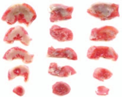
TRPA1 and Cardioprotection

         replicates. The LDH release was measured as previously                          reperfusion, significant differences were noted in the heart
         described and quantified as the ratio of LDH release after                      rate, mean arterial pressure, and rate pressure product at 2 h
         4 h of reoxygenation to total LDH.18                                            of reperfusion when compared to baseline values (table 1).
                                                                                             To initially address whether TRPA1 is present in the
         Statistical Analysis                                                            heart, we performed qPCR and Western blot on the cardiac
            All data were shown as mean ± SD. For analysis of in                         myocytes, the left ventricle–derived stable cell line, H9C2
         vivo and cardiac myocyte models of ischemia–reperfusion                         cells, and heart homogenates from the left ventricle. Both
         or ischemia–reoxygenation, a one-way ANOVA followed                             PCR and protein expression detected TRPA1 in the cardiac
         by Bonferroni correction for multiplicity was used in order                     myocytes (fig. 1, A and B). Further, by immunofluores-
         to compare each group to the control group. A two-way                           cence, isolated adult cardiac myocytes also displayed TRPA1
         ANOVA was used to determine significance for hemody-                            expression (fig. 1C). These findings were further supported

                                                                                                                                                                    Downloaded from http://pubs.asahq.org/anesthesiology/article-pdf/125/6/1171/272784/20161200_0-00023.pdf by guest on 21 October 2020
         namic parameters. For differences between two groups, a                         by detecting TRPA1 in the H9C2 cell line and homogenate
         two tailed Student’s t test was performed. Statistical analysis                 of left ventricle (Supplemental Digital Content 1, qPCR
         was performed using GraphPad Prism 6 (GraphPad Software                         showing the presence of TRPA1 in the H9C2 cell line and
         Inc., USA). P < 0.05 was considered statistically significant.                  homogenate of left ventricle, and Supplemental Digital
                                                                                         Content 2, which shows Western blot validation, http://
         Results                                                                         links.lww.com/ALN/B321).
         A total of 107 rats were used for the study. Twenty-one rats                        TRPA1 activators, including ASP 7663 and optovin, are
         were used to isolate adult cardiac myocytes to determine                        selective for TRPA1 (fig. 2A). We gave these agents before
         the TRPA1 expression, conduct ischemia–reoxygenation                            ischemia in an in vivo rodent model of heart attack injury
         experiments, and obtain heart left ventricle. One prep was                      (fig. 2B). ASP 7663 and optovin reduced myocardial damage
         excluded due to complications with cannulating the aorta                        when compared to the vehicle DMSO (ASP 7663, 45 ± 5%*
         for cardiac myocyte isolation. Further, 86 rats were used                       and optovin, 44 ± 8% vs. DMSO, 66 ± 6%; percentage
         for the in vivo experiments for completion of 84 success-                       of infarct size/area of risk; n = 6 per group; mean ± SD;
         ful experiments. Two rats were excluded for this portion of                     *P < 0.001 vs. DMSO; fig. 2, C and D). Together, these data
         the study with one rat due to intractable ventricular fibrilla-                 suggest that TRPA1 is present in the cardiac myocyte and
         tion during reperfusion (ASP in the reperfusion group) and                      TRPA1 activators can modulate injury from a heart attack.
         another rat secondary to a small area at risk per left ventricle                    TRPA1 is also selectively modulated by the reactive alde-
         (AP plus morphine group). No differences in the percent-                        hyde, cinnamaldehyde (Supplemental Digital Content 3A,
         age of the area at risk per left ventricle were noted for any                   which shows the chemical structure of cinnamaldehyde,
         of the groups (table 1). Further, no differences in hemody-                     http://links.lww.com/ALN/B321). Unlike the other TRPA1
         namics including heart rate, blood pressure, and rate pres-                     activators tested that are not reactive aldehydes (ASP 7663
         sure product occurred between any of the treatment groups                       and optovin), cinnamaldehyde given at 0.01 or 0.1 mg/kg
         (table 1). Within the group where DMSO was given during                         increased blood pressure dose-dependently immediately on

         Table 1. Number of Animals Used, Area at Risk Per Left Ventricle (%), and Hemodynamic Values Measured

                                                              Baseline                          15-min Ischemia                       2-h Reperfusion

         Groups              n    AAR/LV%           HR           MAP         RPP           HR          MAP          RPP          HR           MAP         RPP

         DMSO                6       41 ± 9       398 ± 20      98 ± 12     47 ± 5       407 ± 37     89 ± 21     42 ± 7      397 ± 27      80 ± 11      43 ± 8
         Optovin             6       40 ± 6       419 ± 32     104 ± 21     54 ± 9       437 ± 22    102 ± 22     52 ± 10     425 ± 13      79 ± 14      43 ± 7
         ASP                 6       41 ± 9       422 ± 41     114 ± 11     60 ± 5       428 ± 33    105 ± 28     52 ± 13     415 ± 25      89 ± 12      49 ± 6
         MOR                 6       37 ± 5       410 ± 35     109 ± 14     53 ± 9       407 ± 37    104 ± 14     49 ± 9      374 ± 19      73 ± 10      37 ± 6
         TCS + MOR           6       42 ± 3       420 ± 38     104 ± 12     53 ± 9       407 ± 35     92 ± 26     44 ± 11     398 ± 44      73 ± 8       42 ± 8
         AP + MOR            6       40 ± 9       423 ± 14     107 ± 14     54 ± 5       378 ± 28    107 ± 31     48 ± 15     388 ± 29      77 ± 11      40 ± 8
         TCS                 6       36 ± 4       427 ± 30     111 ± 16     57 ± 8       424 ± 25    109 ± 27     54 ± 10     404 ± 23      83 ± 15      47 ± 7
         AP                  6       40 ± 8       425 ± 32     103 ± 22     53 ± 13      421 ± 34    100 ± 43     50 ± 22     397 ± 25      75 ± 14      41 ± 10
         DMSO at rep         6       39 ± 3       421 ± 33     105 ± 11     50 ± 8       447 ± 36     96 ± 15     48 ± 9      370 ± 28*     73 ± 18*     34 ± 9*
         Optovin at rep      6       40 ± 8       413 ± 43     108 ± 16     54 ± 8       434 ± 36    100 ± 24     54 ± 13     388 ± 31      84 ± 20      42 ± 9
         ASP at rep          6       45 ± 7       421 ± 12     104 ± 8      52 ± 4       423 ± 22     94 ± 25     49 ± 14     395 ± 33      81 ± 15      42 ± 8

         Described are the groups, number of animals per group (n), AAR/LV, and hemodynamics acquired for the in vivo studies. HR, MAP, and RPP defined as the
         product of HR and systolic blood pressure were assessed at baseline, during ischemia, and at 2 h of reperfusion. RPP was calculated as the product of HR
         and systolic blood pressure. Data are presented as mean ± SD (n = 6). No significant differences were found between groups.
         *P < 0.05 vs. baseline.
         AAR/LV = area at risk per left ventricle percent; AP = AP18; ASP = ASP 7663; DMSO = dimethyl sulfoxide; HR = heart rate; MAP = mean arterial pressure;
         MOR = morphine; n = number of animals per group; rep = reperfusion; RPP = rate pressure product; TCS = TCS 5861528.

         Anesthesiology 2016; 125:1171-80                                         1174                                                                 Lu et al.

Copyright © 2016, the American Society of Anesthesiologists, Inc. Wolters Kluwer Health, Inc. Unauthorized reproduction of this article is prohibited.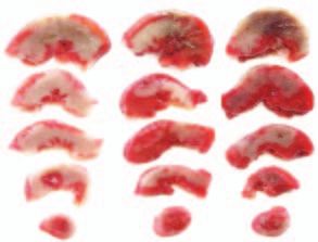
PERIOPERATIVE MEDICINE

                                 A                                   B

                                               30.9±0.3

                                                          20.2±1.3
                                          40

                                                                                        150

                                                                                  kDa
                                                                         TRPA1
                                          30
                                                                                        100

                                     Ct   20
                                                                         GAPDH

                                          10   TRPA1

                                                          GAPDH

                                                                                                                                                  Downloaded from http://pubs.asahq.org/anesthesiology/article-pdf/125/6/1171/272784/20161200_0-00023.pdf by guest on 21 October 2020
                                 C             TRPA1                       DAPI                     Merged

         Fig. 1. Biochemical evidence transient receptor potential ankyrin 1 (TRPA1) is present in cardiac myocytes. (A) Quantitative PCR
         of cardiac myocytes with four biologic replicates. (B) Western blot of cardiac myocytes representative of four biologic replicates.
         (C) Immunofluorescence for TRPA1 in cardiac myocytes and nuclear staining with 4′,6-diamidino-2-phenylindole (DAPI). Top,
         Multiple cardiomyocytes per field (×20). Bottom, A single cardiomyocyte under higher magnification (×63). White bar = 30 μm.
         Ct = cycle threshold; GAPDH = glyceraldehyde 3-phosphate dehydrogenase.

         administration (cinnamaldehyde, 0.01 mg/kg: 124 ± 10*                        Transient receptor potential channel family members are
         mmHg; cinnamaldehyde, 0.1 mg/kg: 134 ± 12* mmHg                          also known to colocalize with opioid receptors in the ner-
         compared to DMSO, 92 ± 2 mmHg; 5 min after administra-                   vous system, and opioids activate TRPA1.19 Thus, we further
         tion; n = 6 per group; *P < 0.01 vs. DMSO; Supplemen-                    questioned whether morphine decreases myocardial injury
         tal Digital Content 3B, which shows mean arterial blood                  by TRPA1 activation. Selective inhibitors of TRPA1 include
         pressures after cinnamaldehyde administration, http://links.             AP 18 and TCS 5861528 (fig. 3A). We tested whether these
         lww.com/ALN/B321). Further, cinnamaldehyde did not                       TRPA1 inhibitors affect the ability of morphine to reduce
         affect myocardial infarct size (cinnamaldehyde, 0.01 mg/kg:              myocardial injury in an in vivo rodent heart attack injury
         61 ± 3%; cinnamaldehyde, 0.1 mg/kg: 61 ± 5% vs. control,                 model (fig. 3B). When either TRPA1 inhibitor was given
         62 ± 3%; n = 6 per group; Supplemental Digital Content 3,                before morphine, the ability of morphine to decrease heart
         C and D, which shows infarct size data and representative                damage was blocked (morphine, 44 ± 5%*; TCS plus mor-
         images of infarct size, http://links.lww.com/ALN/B321).                  phine, 62 ± 5%#; AP plus morphine, 65 ± 6%# vs. DMSO,
         For these studies, no differences were noted between groups              66 ± 6%; *P < 0.001 vs. all groups; #P < 0.001 vs. morphine;
         for the area at risk per left ventricle percentage. In addition,         fig. 3, C and D). No effect on myocardial infarct size was
         no changes were noted between groups for the recorded                    noted when either TRPA1 inhibitor was alone given (fig. 3,
         hemodynamics at baseline, 15 min of ischemia, and at 2 h of              C and D).
         reperfusion (Supplemental Digital Content 4, which shows                     Since the data we obtained were from an in vivo rodent
         a table of hemodynamics for the cinnamaldehyde portion of                model, the effect seen could potentially be due to modu-
         the study, http://links.lww.com/ALN/B321).                               lation of the nervous system rather than a direct effect on

         Anesthesiology 2016; 125:1171-80                                  1175                                                       Lu et al.

Copyright © 2016, the American Society of Anesthesiologists, Inc. Wolters Kluwer Health, Inc. Unauthorized reproduction of this article is prohibited.
TRPA1 and Cardioprotection
A TRPA1 Agonists: B 25 5 30 120 min
ASP 7663 (ASP) Optovin DMSO Baseline Ischemia Reperfusion
DMSO
ASP Baseline Ischemia Reperfusion
ASP
Optovin Baseline Ischemia Reperfusion
Optovin
C 80 D
Infarct / Area at risk (%)
Downloaded from http://pubs.asahq.org/anesthesiology/article-pdf/125/6/1171/272784/20161200_0-00023.pdf by guest on 21 October 2020
40
44±8*
45±5*
66±6
0
Optovin
ASP
DMSO
DMSO ASP Optovin
Fig. 2. Transient receptor potential ankyrin 1 (TRPA1) activators reduce myocardial infarct size. (A) Chemical structure of TRPA1
activators. (B) Experimental protocol for myocardial ischemia–reperfusion studies. Rats were given a TRPA1 activator (ASP
[ASP7663] or optovin) or vehicle (dimethyl sulfoxide [DMSO]) 5 min before 30 min of left anterior descending coronary artery liga-
tion to cause ischemia followed by 2 h of reperfusion. (C) Infarct size per area at risk percentage for each experimental group.
Data points represent individual biologic results for each experiment in addition to values presented as mean ± SD (n = 6);
*P < 0.001 versus DMSO group. (D) Representative images of left ventricle area at risk. Infarcted areas are unstained and remain
white, while viable tissue is stained red by triphenyl tetrazolium chloride.
A TRPA1 Antagonists:
B 15 10 5 30 120 min
DMSO Baseline Ischemia Reperfusion
TCS 5861528 (TCS) AP 18 (AP)
DMSO
MOR Baseline Ischemia Reperfusion
MOR
Inhibitor Baseline Ischemia Reperfusion
+MOR TCS
MOR
or AP
Inhibitor Baseline Ischemia Reperfusion
TCS
or AP
C D
80
Infarct / Area at risk (%)
40
# #
MOR 44±5*
AP 60±4
TCS+MOR 62±5
AP+MOR 65±6
64±4
DMSO 66±6
0
TCS
DMSO MOR TCS+ AP+ TCS AP
MOR MOR
Fig. 3. Opioid-induced reduction of myocardial infarct size is mediated by transient receptor potential ankyrin 1 (TRPA1). (A)
Chemical structure of two TRPA1 inhibitors (TCS [TCS 5861528] and AP [AP18]). (B) Experimental protocol for myocardial
ischemia–reperfusion studies. Dimethyl sulfoxide (DMSO) or morphine (MOR) was given 5 min before ischemia. The two TRPA1
inhibitors (TCS and AP) were given 10 min before morphine treatment. (C) Infarct size per area at risk percentage. Data are pre-
sented as mean ± SD (n = 6); *P < 0.001 versus DMSO; #P < 0.001 versus MOR. For comparison purposes, the DMSO group data
presented in figure 2 are also presented here in this figure. (D) Representative images of left ventricle area at risk for each group.
Anesthesiology 2016; 125:1171-80 1176 Lu et al.
Copyright © 2016, the American Society of Anesthesiologists, Inc. Wolters Kluwer Health, Inc. Unauthorized reproduction of this article is prohibited.PERIOPERATIVE MEDICINE
the cardiac myocyte itself. Therefore, we further determined optovin, 10 ± 2; AP, 10 ± 2; TCS, 11 ± 2; percentage of trypan
how TRPA1 modulates cellular death of cardiac myocytes blue–positive cells; n = 6 per group; Supplemental Digital
when independent of the nervous system. We isolated pri- Content 5, C and D, which provides images and quantifi-
mary cardiac myocytes from adult rat hearts and the fol- cation of trypan blue exclusion assay, http://links.lww.com/
lowing day subjected the cardiac myocytes to either sham ALN/B321). We also tested the TRPA1 inhibitors when
or hypoxia–reoxygenation. Primary adult cardiac myocytes given before reoxygenation (Supplemental Digital Con-
are viable in cell culture for 48 h, and as also shown by oth- tent 6A, which describes the experimental protocol, http://
ers,20 a percentage of cell death will occur for a sham-treated links.lww.com/ALN/B321). Similar to the in vivo model
group. We administered the TRPA1 activators and inhibitors findings, the TRPA1 inhibitors did not affect LDH release
without the presence of hypoxia–reoxygenation (Supple- (sham, 13 ± 2; DMSO, 49 ± 11*; AP, 49 ± 6*; TCS, 49 ± 10*;
mental Digital Content 5A, which describes the experimen- n = 6 per group; *P < 0.05 vs. sham; Supplemental Digi-
Downloaded from http://pubs.asahq.org/anesthesiology/article-pdf/125/6/1171/272784/20161200_0-00023.pdf by guest on 21 October 2020
tal protocol, http://links.lww.com/ALN/B321). Without tal Content 6B, which describes LDH release data, http://
hypoxia–reoxygenation, these agents showed no differences links.lww.com/ALN/B321). The TRPA1 inhibitors also did
in cell death by percentage of LDH release (sham, 14 ± 6; not change the number of trypan blue–positive cells when
DMSO, 14 ± 3; ASP, 13 ± 4; optovin, 13 ± 2; AP, 15 ± 7; TCS, compared to DMSO-treated adult cardiac myocytes (sham,
14 ± 7; percentage of LDH release of total amount in cells; 9 ± 3; DMSO, 38 ± 3*; AP, 33 ± 3*; TCS, 33 ± 4*; n = 6 per
n = 6 per group; Supplemental Digital Content 5B, which group; *P < 0.05 vs. sham; Supplemental Digital Content 6,
describes LDH release data, http://links.lww.com/ALN/ C and D, which provides images and quantification of try-
B321). Further, no differences were noted in trypan blue pan blue exclusion assay, http://links.lww.com/ALN/B321).
exclusion when compared to untreated or DMSO-treated Further, we tested whether the TRPA1 activators affected
cardiac myocytes (sham, 11 ± 2; DMSO, 11 ± 2; ASP, 11 ± 4; cardiac myocyte viability when the TRPA1 activators were
# #
42±3*
19±4
24±6
26±2
A B 50
1 hrs 2 hrs 4 hrs
Sham Baseline Normoxia
LDH release
(% of total)
DMSO Baseline Hypoxia Reoxygenation
DMSO
25
ASP Baseline Hypoxia Reoxygenation
ASP
Optovin Baseline Hypoxia Reoxygenation
Optovin 0
Sham
DMSO
ASP
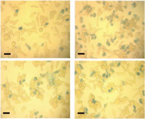
Optovin
C Sham DMSO D
# #
50
36±3*
20±3
22±4
10±2
Trypan blue positive cells
(% of total cells)
25
Asp Optovin
0
Sham
DMSO
ASP
Optovin
Fig. 4. Activation of transient receptor potential ankyrin 1 (TRPA1) at reoxygenation in isolated adult cardiac myocytes reduces
cell death. (A) Experimental protocol for cardiac myocyte hypoxia–reoxygenation studies. The two TRPA1 activators (ASP 7663
[ASP] and optovin) were given immediately after hypoxia. (B) Percentage of lactate dehydrogenase (LDH) release for each exper-
imental group (n = 3 per group). (C) Representative images of trypan blue–positive and trypan blue–negative cardiac myocytes
for each group (black bar represents 50 μm). (D) Percentage of dead cells for each experimental group (n = 6 per group). Data
points represent individual biologic results for each experiment in addition to values presented as mean ± SD; *P < 0.05 versus
sham; #P < 0.05 versus dimethyl sulfoxide (DMSO).
Anesthesiology 2016; 125:1171-80 1177 Lu et al.
Copyright © 2016, the American Society of Anesthesiologists, Inc. Wolters Kluwer Health, Inc. Unauthorized reproduction of this article is prohibited.TRPA1 and Cardioprotection
given before reoxygenation (fig. 4A). We determined that were given before ischemia (fig. 2C), implying that the
when either TRPA1 activator was given, the amount of LDH beneficial effect of TRPA1 activation occurs mainly during
released was less when compared to vehicle-treated adult rat reperfusion to limit reperfusion injury.
cardiac myocytes (ASP, 26 ± 2%# and optovin, 24 ± 6%# vs.
DMSO, 42 ± 3%* and sham, 19 ± 4%; percentage of LDH Discussion
release; n = 3 per group; mean ± SE; *P < 0.05 vs. all other Here, we report that TRPA1 is present within the cardiac
groups; #P < 0.05 vs. sham or DMSO; fig. 4B). Further, the myocytes and regulates cardiac reperfusion injury. This find-
percentage of cardiac myocytes that died was significantly ing is important since it suggests that the TRPA1 receptor,
reduced by almost 40% when assessed by using a trypan blue largely considered only in the cells of the nervous system,23
exclusion assay (ASP, 20 ± 3%# and optovin, 22 ± 4%# vs. also contributes an important physiologic role in the heart
DMSO, 36 ± 3*% and sham, 10 ± 2%, percentage of trypan
that regulates cellular damage from ischemia–reperfusion
Downloaded from http://pubs.asahq.org/anesthesiology/article-pdf/125/6/1171/272784/20161200_0-00023.pdf by guest on 21 October 2020
blue–positive cells; n = 6 per group; mean ± SE; *P < 0.05
injury (fig. 6).
vs. all other groups; #P < 0.05 vs. sham or DMSO; fig. 4, C
TRPA1 can also be modulated by different pain relievers
and D). Together, these data suggest that TRPA1 activators
including NSAIDs, acetaminophen, and COX-2.7–9 The pres-
are effective agents to reduce injury from hypoxia–reoxygen-
ence and function of TRPA1 within the cardiac myocyte must
ation when TRPA1 activators are directly applied to the car-
be considered since drugs specifically targeting TRPA1 for pain
diac myocyte before reoxygenation.
relief may be detrimental to the heart when at risk of ischemia–
About 50% of the myocardial damage can be attributed
reperfusion. If TRPA1 is inhibited, this should be considered
to reperfusion, and accordingly, drugs to limit reperfusion
when selecting pain relievers for patients during the periopera-
injury are importantly needed.21,22 Therefore, we further
tive period and when treating acute or chronic pain conditions.
determined whether administration of a TRPA1 activator
This will be particularly important in the future when more
just before reperfusion also reduces cellular death in an in
specific TRPA1 small molecules are designed as analgesics.
vivo rodent heart attack injury model (experimental pro-
Prostaglandins including A1, A2, and J2 can activate neu-
tocol; fig. 5A). Interestingly, TRPA1 activators when given
ronal TRPA1.9 One can surmise that some NSAIDs, COX-2
before reperfusion reduced myocardial injury (ASP, 42 ± 8,*
inhibitors, and acetaminophen modulate TRPA1 by altering
optovin, 43 ± 7,* and DMSO 60 ± 5, n = 6 per group;
the production of arachidonic acid metabolites that occur dur-
*P < 0.001 vs. DMSO; fig. 5, B and C). The reduction in
ing reperfusion injury. This is supported since either prosta-
heart damage seen was also equal to that when the agents
glandin A1 or J2, when given exogenously, reduces myocardial
infarct size in experimental models.24,25 Although further stud-
A 30 25 5 120 min ies are needed, NSAIDs or acetaminophen, by limiting the
Baseline Ischemia Reperfusion
production of prostaglandins interacting with TRPA1, may
DMSO
DMSO potentially block this mechanism that reduces cellular injury.
ASP Baseline Ischemia Reperfusion Opioid-dependent activation of TRPA1 is reported in
ASP neuronal cells.19 Receptors associated with pain relief, such
Optovin Baseline Ischemia Reperfusion as the opioid receptor family, are also present in the cardiac
Optovin myocytes.26,27 When activated by opioids, opioid receptors
B C
80
Infarct / Area at risk (%)
Direct Agonists:
ASP7663
Optovin
40
43±7*
42±8*
60±5
TRPA1
0
DMSO ASP Optovin
DMSO
Optovin
ASP
Fig. 5. Transient receptor potential ankyrin 1 (TRPA1) acti-
vation reduces infarct size at reperfusion. (A) Experimental Myocardial
protocol for myocardial ischemia–reperfusion studies. TRPA1
Indirect Agonists: Salvage
activator (ASP 7663 [ASP] and optovin) was given 5 min be- Opioids
fore reperfusion. (B) Infarct size per area at risk percentage.
Data points represent individual biologic results for each Fig. 6. Hypothetical pathway summary. Activation of tran-
experiment in addition to values presented as mean ± SD sient receptor potential ankyrin 1 (TRPA1) before or during
(n = 6). *P < 0.05 versus dimethyl sulfoxide (DMSO). (C) Rep- ischemia within the cardiac myocyte reduces ischemia–
resentative images of left ventricle area at risk for each group. reperfusion injury.
Anesthesiology 2016; 125:1171-80 1178 Lu et al.
Copyright © 2016, the American Society of Anesthesiologists, Inc. Wolters Kluwer Health, Inc. Unauthorized reproduction of this article is prohibited.PERIOPERATIVE MEDICINE
contribute an initiation of a signaling cascade that reduces within TRPA1 that are important in regulating cellular injury.
damage from ischemia–reperfusion injury.28,29 Our study Further, we administered optovin intravenously without opto-
suggests that opioids, frequently given by anesthesiologists genetic activation of the drug before delivery as described in a
for analgesia, reduce myocardial injury via a TRPA1-depen- previous study.31 Since ultraviolet light in this previous study
dent mechanism. This finding suggests that adjuvants to was used to activate optovin, we can only assume that the effect
opioids, which may block TRPA1, may limit the beneficial we see for our studies, which is similar to another TRPA1 ago-
effects of opioids in regard to reducing reperfusion injury. nist, ASP 7763, is possibly secondary to the reactive oxygen
We previously showed that aspirin, unlike ibuprofen, can species produced during reperfusion, converting optovin into
block the ability of opioids to reduce myocardial reperfusion an active form. This is an interesting idea that will require fur-
injury.30 Although more studies are needed, potentially other ther study and could suggest a means to develop therapeutic
myocardial salvaging techniques (such as using volatile anes- modulators of cellular injury that are selectively cleaved into
Downloaded from http://pubs.asahq.org/anesthesiology/article-pdf/125/6/1171/272784/20161200_0-00023.pdf by guest on 21 October 2020
thetics31 or implementing remote conditioning31) may also an active form only during ischemia–reperfusion injury.
have a reduced efficacy for limiting myocardial ischemia– Our findings can also lead to developing more cardiac safe
reperfusion injury when TRPA1 is inhibited. analgesics that will not block endogenous pathways involv-
Reactive aldehydes, produced at the highest levels during ing TRPA1, which are important for cardiac protection. Fur-
the initial minutes of reperfusion, are established to modify ther, once the pathway is understood, design of agents may
critical cysteines present on TRPA1, which in turn regulate be possible to provide a beneficial effect of pain relief, with a
function.12 These cysteine modifications, which occur through secondary benefit of reducing reperfusion injury. This could
Michael adduct addition, can change the cellular gating of cal- improve upon using opioids to reduce myocardial ischemia–
cium by TRPA1.32 For our study, the binding site for optovin reperfusion injury, which have deleterious side effects that
is identified to be within a region of critical cysteines present can lead to addiction, overdose, and death.34
on TRPA1 that is also essential for reactive aldehyde-induced In summary, this report describes the presence of TRPA1
TRPA1 activation.12,31 Although further studies will be in the heart and how TRPA1 agonists can reduce damage
needed, the TRPA1 activators we used may reduce injury dur- from cardiac reperfusion injury. This may need to be consid-
ing a heart attack by occupying critical cysteines of TRPA1, ered when developing drugs to target TRPA1 for analgesia.
limiting the amount of irreversible reactions that occur by Further, these findings identify that adjuvants given with opi-
reactive aldehydes during the initial minutes of reperfusion. oids that target TRPA1 may block the ability for opioids to
In contrast to the reversible TRPA1 activators used, cin- reduce myocardial injury. This is important when considering
namaldehyde, the fragrant component of cinnamon oil, is treatment strategies for patients with acute or chronic pain
a reactive aldehyde. Administering this reactive aldehyde who may have a potential risk of suffering a heart attack.
before ischemia–reperfusion injury resulted in an increase in
blood pressure, which was previously shown by a previous Research Support
study from others in rodents. This particular study described Supported by the Department of Anesthesiology, Periop-
that the cardiovascular hemodynamic responses when cin- erative and Pain Management, Stanford University, Stanford,
California (Dr. Gross), National Institute of General Medi-
namaldehyde was given were lost in TRPA1 knockout mice, cine T32 training award GM089626 (Bethesda, Maryland; Dr.
suggesting the specificity for cinnamaldehyde within the McAllister), and grant HL109212 from the National Heart
cardiovascular system to interact with TRPA1.33 We further Lung and Blood Institute (Bethesda, Maryland; Dr. Gross).
extend the findings of this study by describing how cinnam-
aldehyde does not reduce myocardial injury unlike the revers- Competing Interests
ible TRPA1 agonists given. It is also interesting to note that The authors declare no competing interests.
the blood pressure elevation seen for cinnamaldehyde did not
occur for the reversible TRPA1 activators optovin and ASP Correspondence
7663. Although more work is needed, we believe that this dif- Address correspondence to Dr. Gross: Department of Anes-
ference in effect seen by cinnamaldehyde (with an end result thesiology, Perioperative and Pain Medicine, School of Med-
of increased blood pressure and lack of infarct size reduction icine, Stanford University, 300 Pasteur Drive, Grant Building
compared to the other TRPA1 agonists optovin and ASP Room S268b, Stanford, California 94305. ergross@stanford.
edu. Information on purchasing reprints may be found at
7663) is secondary to the aldehyde that is part of the chemi- www.anesthesiology.org or on the masthead page at the be-
cal structure of cinnamaldehyde. The aldehyde may form an ginning of this issue. Anesthesiology’s articles are made free-
irreversible adduct by Michael addition as opposed to the ly accessible to all readers, for personal use only, 6 months
other activators, optovin and ASP 7663, which may instead from the cover date of the issue.
cause a reversible modification of TRPA1.
Our study should be interpreted within the context of References
several potential limitations. With the discovery of TRPA1 1. Gross ER, Hsu AK, Urban TJ, Mochly-Rosen D, Gross GJ:
Nociceptive-induced myocardial remote conditioning is
within the cardiac myocyte, more in-depth molecular analy- mediated by neuronal gamma protein kinase C. Basic Res
sis is needed. This includes determining the critical cysteines Cardiol 2013; 108:381
Anesthesiology 2016; 125:1171-80 1179 Lu et al.
Copyright © 2016, the American Society of Anesthesiologists, Inc. Wolters Kluwer Health, Inc. Unauthorized reproduction of this article is prohibited.TRPA1 and Cardioprotection
2. Mukherjee D, Nissen SE, Topol EJ: Risk of cardiovascular 17. Keysar SB, Trncic N, Larue SM, Fox MH: Hypoxia/reoxygen-
events associated with selective COX-2 inhibitors. JAMA ation-induced mutations in mammalian cells detected by the
2001; 286:954–9 flow cytometry mutation assay and characterized by mutant
3. Bhala N, Emberson J, Merhi A, Abramson S, Arber N, Baron spectrum. Radiat Res 2010; 173:21–6
JA, Bombardier C, Cannon C, Farkouh ME, FitzGerald GA, 18. Lee YS, Song YS, Giffard RG, Chan PH: Biphasic role of
Goss P, Halls H, Hawk E, Hawkey C, Hennekens C, Hochberg nuclear factor-kappa B on cell survival and COX-2 expres-
M, Holland LE, Kearney PM, Laine L, Lanas A, Lance P, sion in SOD1 Tg astrocytes after oxygen glucose deprivation.
Laupacis A, Oates J, Patrono C, Schnitzer TJ, Solomon S, J Cereb Blood Flow Metab 2006; 26:1076–88
Tugwell P, Wilson K, Wittes J, Baigent C; Coxib and tradi- 19. Forster AB, Reeh PW, Messlinger K, Fischer MJ: High con-
tional NSAID Trialists’ (CNT) Collaboration: Vascular and centrations of morphine sensitize and activate mouse dor-
upper gastrointestinal effects of non-steroidal anti-inflamma- sal root ganglia via TRPV1 and TRPA1 receptors. Mol Pain
tory drugs: Meta-analyses of individual participant data from 2009; 5:17
randomised trials. Lancet 2013; 382:769–79
20. Gomez L, Thiebaut PA, Paillard M, Ducreux S, Abrial M, Crola
4. Shinmura K, Tang XL, Wang Y, Xuan YT, Liu SQ, Takano H, Da Silva C, Durand A, Alam MR, Van Coppenolle F, Sheu SS,
Downloaded from http://pubs.asahq.org/anesthesiology/article-pdf/125/6/1171/272784/20161200_0-00023.pdf by guest on 21 October 2020
Bhatnagar A, Bolli R: Cyclooxygenase-2 mediates the car- Ovize M: The SR/ER-mitochondria calcium crosstalk is regu-
dioprotective effects of the late phase of ischemic precon- lated by GSK3β during reperfusion injury. Cell Death Differ
ditioning in conscious rabbits. Proc Natl Acad Sci USA 2000; 2015; 22:1890
97:10197–202
21. Hausenloy DJ, Yellon DM: Targeting myocardial reperfusion
5. Rossoni G, Muscara MN, Cirino G, Wallace JL: Inhibition injury–the search continues. N Engl J Med 2015; 373:1073–5
of cyclo-oxygenase-2 exacerbates ischaemia-induced acute
22. Yellon DM, Hausenloy DJ: Myocardial reperfusion injury.
myocardial dysfunction in the rabbit. Br J Pharmacol 2002;
N Engl J Med 2007; 357:1121–35
135:1540–6
23. Story GM, Peier AM, Reeve AJ, Eid SR, Mosbacher J, Hricik
6. Wei H, Karimaa M, Korjamo T, Koivisto A, Pertovaara A:
TR, Earley TJ, Hergarden AC, Andersson DA, Hwang SW,
Transient receptor potential ankyrin 1 ion channel contrib-
McIntyre P, Jegla T, Bevan S, Patapoutian A: ANKTM1, a TRP-
utes to guarding pain and mechanical hypersensitivity in a rat
model of postoperative pain. Anesthesiology 2012; 117:137–48 like channel expressed in nociceptive neurons, is activated
by cold temperatures. Cell 2003; 112:819–29
7. Andersson DA, Gentry C, Alenmyr L, Killander D, Lewis
SE, Andersson A, Bucher B, Galzi JL, Sterner O, Bevan S, 24. Wayman NS, Hattori Y, McDonald MC, Mota-Filipe H,
Hogestatt ED, Zygmunt PM: Trpa1 mediates spinal antino- Cuzzocrea S, Pisano B, Chatterjee PK, Thiemermann C:
ciception induced by acetaminophen and the cannabinoid Ligands of the peroxisome proliferator-activated receptors
delta(9)-tetrahydrocannabiorcol. Nat Commun 2011; 2:551 (PPAR-gamma and PPAR-alpha) reduce myocardial infarct
size. FASEB J 2002; 16:1027–40
8. Hu H, Tian J, Zhu Y, Wang C, Xiao R, Herz JM, Wood JD, Zhu
MX: Activation of TRPA1 channels by fenamate nonsteroidal 25. Zingarelli B, Hake PW, Mangeshkar P, O’Connor M,
anti-inflammatory drugs. Pflugers Arch 2010; 459:579–92 Burroughs TJ, Piraino G, Denenberg A, Wong HR: Diverse
cardioprotective signaling mechanisms of peroxisome
9. Materazzi S, Nassini R, Andre E, Campi B, Amadesi S, proliferator-activated receptor-gamma ligands, 15-deoxy-
Trevisani M, Bunnett NW, Patacchini R, Geppetti P: Cox- delta12,14-prostaglandin J2 and ciglitazone, in reperfusion
dependent fatty acid metabolites cause pain through activa- injury: Role of nuclear factor-kappaB, heat shock factor 1,
tion of the irritant receptor trpa1. Proc Natl Acad Sci USA and Akt. Shock 2007; 28:554–63
2008; 105:12045–50
26. Sobanski P, Krajnik M, Shaqura M, Bloch-Boguslawska
10. Macpherson LJ, Xiao B, Kwan KY, Petrus MJ, Dubin AE, E, Schäfer M, Mousa SA: The presence of mu-, delta-, and
Hwang S, Cravatt B, Corey DP, Patapoutian A: An ion chan- kappa-opioid receptors in human heart tissue. Heart Vessels
nel essential for sensing chemical damage. J Neurosci 2007; 2014; 29:855–63
27:11412–5
27. Bell SP, Sack MN, Patel A, Opie LH, Yellon DM: Delta opi-
11. Andersson DA, Gentry C, Moss S, Bevan S: Transient receptor oid receptor stimulation mimics ischemic preconditioning in
potential A1 is a sensory receptor for multiple products of
human heart muscle. J Am Coll Cardiol 2000; 36:2296–302
oxidative stress. J Neurosci 2008; 28:2485–94
28. Gross ER, Gross GJ: Ligand triggers of classical precondition-
12. Trevisani M, Siemens J, Materazzi S, Bautista DM, Nassini
ing and postconditioning. Cardiovasc Res 2006; 70:212–21
R, Campi B, Imamachi N, Andre E, Patacchini R, Cottrell
GS, Gatti R, Basbaum AI, Bunnett NW, Julius D, Geppetti 29. Tanaka K, Kersten JR, Riess ML: Opioid-induced cardiopro-
P: 4-Hydroxynonenal, an endogenous aldehyde, causes tection. Curr Pharm Des 2014; 20:5696–705
pain and neurogenic inflammation through activation of 30. Gross ER, Hsu AK, Gross GJ: Acute aspirin treatment abol-
the irritant receptor trpa1. Proc Natl Acad Sci USA 2007; ishes, whereas acute ibuprofen treatment enhances mor-
104:13519–24 phine-induced cardioprotection: Role of 12-lipoxygenase.
13. Takahashi N, Kuwaki T, Kiyonaka S, Numata T, Kozai D, J Pharmacol Exp Ther 2004; 310:185–91
Mizuno Y, Yamamoto S, Naito S, Knevels E, Carmeliet P, Oga 31. Kokel D, Cheung CY, Mills R, Coutinho-Budd J, Huang L,
T, Kaneko S, Suga S, Nokami T, Yoshida J, Mori Y: TRPA1 Setola V, Sprague J, Jin S, Jin YN, Huang XP, Bruni G, Woolf
underlies a sensing mechanism for O2. Nat Chem Biol 2011; CJ, Roth BL, Hamblin MR, Zylka MJ, Milan DJ, Peterson RT:
7:701–11 Photochemical activation of TRPA1 channels in neurons and
14. Gross ER, Hsu AK, Gross GJ: Acute methadone treatment animals. Nat Chem Biol 2013; 9:257–63
reduces myocardial infarct size via the delta-opioid receptor 32. Macpherson LJ, Dubin AE, Evans MJ, Marr F, Schultz PG,
in rats during reperfusion. Anesth Analg 2009; 109:1395–402 Cravatt BF, Patapoutian A: Noxious compounds activate
15. Small BA, Lu Y, Hsu AK, Gross GJ, Gross ER: Morphine TRPA1 ion channels through covalent modification of cyste-
reduces myocardial infarct size via heat shock protein 90 in ines. Nature 2007; 445:541–5
rodents. Biomed Res Int 2015; 2015:129612 33. Pozsgai G, Bodkin JV, Graepel R, Bevan S, Andersson DA,
16. Patel HH, Head BP, Petersen HN, Niesman IR, Huang D, Brain SD: Evidence for the pathophysiological relevance
Gross GJ, Insel PA, Roth DM: Protection of adult rat cardiac of TRPA1 receptors in the cardiovascular system in vivo.
myocytes from ischemic cell death: Role of caveolar micro- Cardiovasc Res 2010; 87:760–8
domains and delta-opioid receptors. Am J Physiol Heart Circ 34. Okie S: A flood of opioids, a rising tide of deaths. N Engl J
Physiol 2006; 291:H344–50 Med 2010; 363:1981–5
Anesthesiology 2016; 125:1171-80 1180 Lu et al.
Copyright © 2016, the American Society of Anesthesiologists, Inc. Wolters Kluwer Health, Inc. Unauthorized reproduction of this article is prohibited.You can also read