Treatment of coral Wounds by combining an Antiseptic Bilayer film and an injectable Antioxidant Biopolymer - Nature
←
→
Page content transcription
If your browser does not render page correctly, please read the page content below
www.nature.com/scientificreports

            OPEN           Treatment of Coral Wounds by
                           Combining an Antiseptic Bilayer
                           Film and an Injectable Antioxidant
                           Biopolymer
                           Marco Contardi1*, Simone Montano2,3, Giulia Liguori3, José A. Heredia-Guerrero1,4,
                           Paolo Galli2,3, Athanassia Athanassiou1 & Ilker S. Bayer1*
                           Coral reefs are vital for the marine ecosystem and their potential disappearance can have unequivocal
                           consequences on our environment. Aside from pollution-related threats (changes in water temperature,
                           plastics, and acidity), corals can be injured by diseases, predators, humans and other invasive species.
                           Diseases play an important role in this decline, but so far very few mitigation strategies have been
                           proposed and developed to control this threat. In this work, we demonstrate that recently developed
                           bi-layer human skin wound treatment patches containing antiseptics and natural antioxidants with
                           controlled-release capacity can be adapted to treat scleractinian coral wounds effectively. A hydrophilic
                           bilayer film based on polyvinylpyrrolidone (PVP) and hyaluronic acid was used to cover the open
                           wounds while delivering the antiseptics for rapid action. Afterwards, the hydrophilic bi-layer covered
                           wound was sealed with an antioxidant and hydrophobic ε-caprolactone-p-coumaric acid copolymer
                           by melt injection at low temperatures. Treated coral injuries were monitored both in aquaria system
                           and in natural environment in Maldives for over 4 months to reduce the number of entry points for
                           organisms that could lead to diseases. The corals well-tolerated both biomaterials as well as the
                           antiseptics incorporated in these materials. The treatments displayed self-adhering properties,
                           tuneable dissolution time, and biocompatibility and stimulated regeneration properties within the coral
                           wound. As such, this work demonstrates that certain human skin wound treatment materials can be
                           successfully adapted to the curing of coral wounds and delivery of specific drugs to slow down, reduce or
                           even stop the spread of diseases in scleractinian corals as well as in all other benthic organisms affected
                           by uncontrolled pathologies.

                           Coral reefs are declining worldwide with coral diseases emerging as one of the most distressing threats1. In the
                           last 50 years, this ecosystem is subjected to constant and extensive degradation, and a reduction of 25% of the
                           global functional coral reefs has been recorded2. Among the coral reefs dwellers, reef-building corals are one
                           of the most fragile components affected by changes in the marine realm3. Environmental stressors, including
                           rising of the seawater temperatures, nutrient input of runoff, and sedimentation exacerbate the declining health
                           of corals. Diseases play an important role in this apocalyptic scenario with the increased sea temperature that
                           expedites the frequent diffusion of infections caused by microorganisms3,4. Currently, the number of known and
                           identified coral diseases varies from 24 to 40, but it might be even more due to limited analytical characterization
                           methods, deprived knowledge of the putative pathogens and the consequent deficiency of epizootiological data5,6.
                           Therefore, the lack of well-defined knowledge makes the control of coral diseases a real challenge, especially when
                           they are caused by bacteria6. One of the most dramatic and recent examples of such diseases is the Stony Coral
                           Tissue Loss Disease (SCTLD), which has been destroying an entire ecosystem on the Florida Reef Tract since
                           2014. So far, its effect is unprecedented, and all the efforts carried out by researches have not been successful in

                           1
                            Smart Materials, Istituto Italiano di Tecnologia, Genova, Italy. 2Department of Earth and Environmental Sciences
                           (DISAT), University of Milan – Bicocca, Milan, Italy. 3MaRHE Center (Marine Research and High Education Center),
                           Magoodhoo Island, Faafu Atoll, Republic of Maldives. 4IHSM La Mayora, Departamento de Mejora Genética y
                           Biotecnología, Consejo Superior de Investigaciones Científicas, Málaga, Spain. *email: marco.contardi@iit.it; ilker.
                           bayer@iit.it

Scientific Reports |   (2020) 10:988 | https://doi.org/10.1038/s41598-020-57980-1                                                            1
www.nature.com/scientificreports/                                                              www.nature.com/scientificreports

                           discovering the relevant agents and taking the appropriate countermeasures7. For instance, mitigation techniques
                           have already been proposed to reduce or slow down the progression of many coral diseases, however, most of
                           them have not yielded and effective solution on a global scale8 yet.
                               In particular, coral wounds can be considered as the starting point of some coral diseases. Since the wound
                           practically exposes the coral skeleton, pathogens settle on the skeleton and transform it into a proliferation sub-
                           strate, increasing the likelihood of infections9. The first method to treat and manage a coral disease was developed
                           in the late 1980s to control an outbreak of black-band disease (BBD) in Looe Key National Marine Sanctuary10
                           (lower Florida Keys, USA). Later on, Hudson et al.10 developed an aspirator device to remove the filament mats
                           produced by the BBD bacterium, and then they covered the wound with a clay-based sealant. However, as they
                           reported, in some cases, this passive covering did not ensure efficient disinfection of the underlying area leading
                           to post-treatment infections of the corals. In the meantime, several other treatments and strategies have been pro-
                           posed to manage and mitigate the damage inflicted by BBD and similar syndromes11,12. One of the most common
                           strategy suggested was the removal of the lesion, considered as a common form of medical intervention to cure
                           some diseases affecting both vertebrates and invertebrates11,12. Other than this, researchers used biological control
                           techniques such as probiotics and phage therapy to manage bacterial pathogens13, the mechanical removal of dis-
                           ease vectors, such as the corallivore Coralliophila abbreviata14, and the excision of healthy coral branch tips from
                           diseased Acropora cervicornis colonies15. In addition, researchers also used epoxy adhesives to mechanically block
                           the progression of a tissue-loss disease on A. cervicornis15. Aeby et al. reported the application of antibacterial
                           chlorine powder embedded within epoxy to treat and mechanically block the progression of BBD in Montipora12,
                           but considering the polymicrobial origin of several coral diseases, the employment of more specific type of drugs
                           can be a better strategy to complete eradicate the infection.
                               A recent work of Sweet et al.16 showed as some antibiotics can be employed for the treatment of the White
                           band disease (WBD), a polymicrobial infection of the corals. Here, the authors simply added the antibiotics at a
                           suitable concentration in the water of the aquarium. Although the results were positive in mitigating the WBD
                           diffusion, the same authors highlighted how this type of treatment could not be applied at a large-scale level.
                           Indeed, to reach an effective concentration of antibiotic in the sea, we would risk compromising the marine eco-
                           system, limiting this approach to emergency cases in the aquarium environment.
                               Therefore, the ever-increasing spread of coral diseases and the lack of adequate healing strategies bring up the
                           urgent necessity to design new treatments for the in situ delivery of drugs such as antiseptics, antibacterial agents
                           or anti-protozoans, and, at the same time, avoiding the spread of potential pollutions in the sea.
                               One of the approaches could be the fabrication of biocompatible, biodegradable, and bioactive polymeric drug
                           delivery systems suitable for the coral application. In particular, the modern strategies and treatments applied
                           for human wounds can be a point of inspiration and constitute some potential guidelines also for the coral dis-
                           ease prevention17–20. In humans, the main role of the skin tissue is to protect the body from external pathogenic
                           agents, acting as a shield21. After a skin injury, the loss of this barrier can cause rapid infections and bacterial
                           colonization. The presence of bacteria in the wound bed is one of the reasons why the healing process is delayed21.
                           Therefore, in order to avoid bacterial infection and promote the healing, an ideal wound dressing, firstly, should
                           ensure a covering of the wound. At the same time, the wound dressing should be able to kill the bacteria that have
                           already infected the wound22. Similarly, for coral injuries or wounds, the potential wound dressings must be made
                           from materials that are not bio-persistent and toxic for marine environment in general. Intelligent wound dress-
                           ings can be made from several polymers and active compounds that can be infused with medicinal compounds to
                           accelerate and manage the healing process. Among them, the biocompatible synthetic polymer polyvinylpyrro-
                           lidone (PVP) has gained interest in the topical application not only because it can modulate the crystallinity of
                           several drugs favourably but also for its capacity to form transparent films and ensure good self-adhesion to the
                           moist skin23–25. In recent years, the natural polymer hyaluronic acid (HA) has also played an important role in
                           the pharmaceutical field due to its anti-inflammatory, regenerative and anti-age properties. By the same token,
                           polycaprolactone (PCL) is a biodegradable synthetic polyester26 that has been broadly applied in biomedical fields
                           for the preparation of controlled drug delivery systems such as implants and scaffolds27,28. It has been recently
                           combined or co-polymerized with effective natural antioxidants such as vanillic, ferulic, p-coumaric and syringic
                           acids29.
                               In this work, we attempted to use polymeric smart wound dressings in tandem, inspired by human
                           skin wound care and management to cure coral wounds. In particular, we used a dual-drug carrying
                           PVP-HA-based bilayer construct, loaded with eosin-based antiseptic and the antibiotic Ciprofloxacin, as well as a
                           ε-caprolactone-p-coumaric acid copolymer (PCL-PCA) developed for skin regeneration25,30.
                               Dual drug-carrying bilayer construct was applied directly onto the wound and acted as material for the in situ
                           delivery of two antibacterial drugs25 (see Fig. 1). Afterwards, the PCL-PCA copolymer with antioxidant capacity
                           was melted to seal and shield the wound. The two materials were applied and tested in combination (see Fig. 1)
                           and separately as control samples, and monitored during two distinct phases: ex-situ and in-situ. The first part
                           of the experiments consisted of 10-days of observation ex-situ, and we focused on understanding the ability of
                           the two materials to adhere to the coral surface, to resist underwater dissolution and to evaluate potential toxic
                           effects on the coral, if any. Afterwards, the survival and the health of the corals were followed for four months
                           continuously in-situ.

                           Results and Discussion
                           Coral wounds may increase susceptibility to some diseases31, hence, the materials developed and used in this
                           study are expected to prevent disease outbreaks in the short term relevant to the early stages of the coral wound.
                           During the monitoring period, the applied materials were well-tolerated by the coral system and exhibited proper
                           and healthy restoration properties. In total, 52 coral wounds were created and they were monitored within 4

Scientific Reports |   (2020) 10:988 | https://doi.org/10.1038/s41598-020-57980-1                                                             2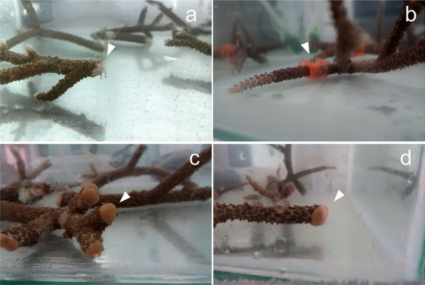
www.nature.com/scientificreports/ www.nature.com/scientificreports
Figure 1. Schematic representation of a coral wound and its potential treatment. Illustration of a coral wound
in the sea. In the first close-up, all the potential risks when a coral loses its protection are presented. In the other
two close-ups, the two potential steps of the treatment suggested in this work are shown. The bilayer should
act as a bioactive dressing for the delivery of drugs in order to disinfect and treat the wound (Step 1). Instead,
the PCL-PCA paste should act as a shield against all the external agents and the fast dissolution of the bilayer
dressing (Step 2).
Figure 2. Coral wounds. Photographs in tank immediately after inflicted injuries of the control (a), bilayer-
treated sample (b), PCL-PCA-treated sample (c) and bilayer + PCL-PCA-treated sample. (d) The arrows
indicate the position of the injuries.
experimental groups: untreated, treated with only bilayer, treated with only PCL-PCA copolymer, and treated
with both bilayer and PCL-PCA copolymer (Figs. 2 and S1).
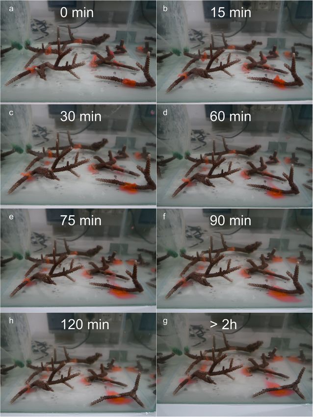
The first treatment tested consisted of the application of the dual drug-carrying PVP-HA-based bilayer film
directly on the open coral wounds. In total, 8 lesions have been treated with this material, where each of them
has been carefully covered by a strip of bilayer film wrapped over the wound (red-colored foils in Fig. 3a). As
expected, due to the hydrophilic nature of its components PVP and HA, the bilayer dissolved and disintegrated
in few hours after the total immersion in the water. The dissolution process started about 10–15 minutes after
the immersion (Fig. 3b) in all coral-induced wounds where the material has been located. Eventually, the bilayer
film disappeared, revealing the bare skeleton underneath after about 3 hours (Fig. 3b–g). The treatment has been
monitored for 10 days to observe any further changes on the coral fragments before termination.
Scientific Reports | (2020) 10:988 | https://doi.org/10.1038/s41598-020-57980-1 3
www.nature.com/scientificreports/                                                              www.nature.com/scientificreports

                           Figure 3. Application of bilayer materials on coral wounds. Photographs of bilayer applied on different coral
                           wound monitored for the first 3 hours.

                              For the other 3 experimental groups, during this ex-situ phase, we took into account different parameters,
                           reported in Table S1, to investigate the state of health of the corals. In particular, we evaluated the injury condi-
                           tion, the colony health, and the biopaste condition. For each condition, three variables were ranked with values
                           from 0 to 4 (see Table S1).
                              In Fig. 4, we reported the main results of the monitoring parameters at different time points. For the injury
                           condition, we reported results about the percentage of covered injuries, the general condition of the injuries, and
                           the progression of the healing. As colony health, we evaluated the formation of mucus, the bleaching, and the
                           necrosis. Instead, regarding the biopaste condition, we monitored the hardening, the adhesion, and the dissolu-
                           tion parameters.
                              The PCL-PCA copolymer treatment consisted of the application of a melted material directly on the 16
                           induced coral wounds. The PCL-PCA copolymer is a solid film at room temperature, thus to obtain a confor-
                           mal coverage of the coral wound, it was heated to 60 °C and immediately applied in the molten state. After the
                           immersion in water, the material solidified, leaving a cup-shaped self-adherent structure on all coral wounds
                           (Fig. 5b). Immediately after the application and during the whole initial phase of monitoring period (10 days), the
                           PCL-PCA copolymer remained adherent to the wounds and no signs of dissolution and detachment were noticed

Scientific Reports |   (2020) 10:988 | https://doi.org/10.1038/s41598-020-57980-1                                                             4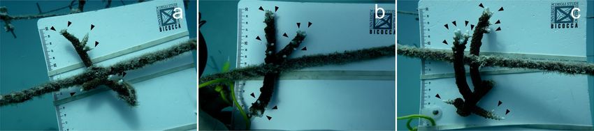
www.nature.com/scientificreports/                                                            www.nature.com/scientificreports

                           Figure 4. Monitoring parameters. Graphs of the observed parameters during the ex-situ phase for the PCL-
                           PCA, PCL-PCA + Bilayer and for the Control expressed as “Injury Condition” (a–c), “Colony Health” (d–f), and
                           “Biopaste Condition” (g–i).

                           Figure 5. Coral wounds status after 10 days. Photographs in tank after 10 days from the injuries of the control
                           (a), PCL-PCA sample (b) and bilayer + PCL-PCA sample. (c) The arrows indicate the position of the injuries.

Scientific Reports |   (2020) 10:988 | https://doi.org/10.1038/s41598-020-57980-1                                                            5
www.nature.com/scientificreports/                                                               www.nature.com/scientificreports

                           Figure 6. Coral wounds status after 2 months. Photographs underwater after 2 months from the injuries of the
                           control (a), PCL-PCA-treated sample (b) and bilayer + PCL-PCA-treated sample. (c) The arrows indicate the
                           position of the injuries.

                           for all the coral wounds under analysis except for a single wound that lost few small pieces from the top portion of
                           the cup without exposing the wound. Furthermore, even if no detectable anomalies have been observed regard-
                           ing the mucus production and presence of necrosis tissue, a slight variation in color in three of 16 fragments was
                           observed between day 8 and day 10. In particular, those fragments showed a slightly pale tissue diffuse spread
                           throughout their surface (Fig. 4d–f).
                                Based on these observations, it appears that the application of the melt copolymer does not injure the coral.
                           Note that the copolymer is not constantly kept at its melting point during its application on the coral. Hence, it
                           already cools down during application. However, we are aware that the melted copolymer can prematurely close
                           the wound or kill some bacteria. At this stage, it is difficult to quantify this. However, during the application, the
                           wound does not experience a constant high temperature. In relation to this, some works32,33 demonstrated that
                           a similar temperature maintained at a longer time can have a weak antibacterial activity. We cannot exclude that
                           the molten biopolymer injection process can affect the microorganisms present in the wound. Hence, further
                           in-depth studies will be planned and conducted to this effect.
                                For the third set of experiments, a dual treatment of bilayer and PCL-PCA was applied on 12 coral-induced
                           wounds (Fig. 5c). This treatment combines the possibility of a short time in situ drug delivery ensured by the
                           bilayer film and the sealing and shielding activity of the PCL-PCA cup. This tandem treatment demonstrated
                           superior self-adhesion to the wound surface with respect to the PCL-PCA treatment. In support of this, none
                           of the applied PCL-PCA structures came off the induced coral wounds and, moreover, no variations in the con-
                           sistency or dissolution of the copolymer have been detected (Fig. 4g,h). In line with this, the condition of the
                           lesion zones after 10 days of monitoring remained at an optimal state, with no changes in the coverage status
                           and including the backside of the lesion zone. A small alteration observed was related to three fragments that
                           appeared slightly paled between day 9 and day 10. No relevant signs of mucus production and the presence of
                           tissue necrosis were noticed; see Figs. 4d,f and 5b,c.
                                On the untreated coral wounds, no significant changes occurred in the inflicted wounds during 10 days of
                           monitoring (Fig. 5a). However, light pale conditions and patches over the wound surfaces formed almost on all
                           the fragments monitored (Fig. 4e).
                                During these 10 days of indoor aquaria system monitoring, all fragments survived, and no clear differences
                           were observed among the untreated control, the PCL-PCA and bilayer/PCL-PCA-treated samples in terms of
                           healing or progression of the injuries, see Fig. 4. This may be attributed to the particular coral species and its
                           resistance, the number of wounds produced, absence of infection, use of aquaria controlled systems and more
                           likely to a combination of these scenarios. Moreover, to the best of our knowledge, several causes can trigger
                           the host’s immune responses involved in wound healing such as the toll-like pathway, the melanin synthesis for
                           tissue regeneration, the complementary system resulting in apoptosis of damaged cells, and cell activation to
                           move amoebocytes to the wound34. Furthermore, tissue regeneration and its associated immune pathways require
                           extensive cellular resources (e.g., carbon products and amoebocytes) to be translocated to the wound35. Therefore,
                           to further explore the effectiveness of the treatments, in the second part of the study, all the coral fragments were
                           attached on a rope and dipped 5–20 meters down the ocean waters (December 2018, Maldives) and monitored
                           till April 2019 (see Figure S2). The in-situ monitoring revealed that all treated fragments survived and that most
                           of the injuries showed complete tissue recovery after about 4 months.
                                By contrast, after 2 months of observations on untreated wounds (the control samples) demonstrated some
                           lesions with algal overgrowth (see white arrow in Fig. 6a), suggesting a failure in the natural healing process com-
                           pared to the treated samples where no algae growth and invasion were noticed (Fig. 6b,c).
                                After 4 months underwater, we used the generation of new tips “branchiness” as measure of a positive healing
                           effect on the coral fragments treated with PCL-PCA and bilayer/PCL-PCA materials and compared the results
                           with untreated coral fragments. All fragments showed formation of new tips (including the control samples),
                           confirming the suitability of Maldivian lagoons for the restoration of branching Acropora corals, see Fig. 7.
                                An average of 16.4 new tips per fragment for the untreated corals was observed. Instead, in the case of cor-
                           als treated with PCL-PCA and PCL-PCA/bilayer an average of 19.4 and 21.4 new tips per coral fragment were
                           recorded, respectively (Fig. 8).
                                Although the branchiness per fragment was not statistically significant between the treatments (H- Kruskal
                           Wallis p = 0.323), this trend was also observed in the total number of new tips formed. Indeed, the untreated coral
                           produced in total 82 new tips, while the corals treated with PCL-PCA and PCL-PCA/bilayer, generated 97 and

Scientific Reports |   (2020) 10:988 | https://doi.org/10.1038/s41598-020-57980-1                                                              6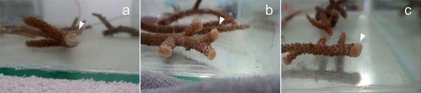
www.nature.com/scientificreports/                                                               www.nature.com/scientificreports

                           Figure 7. Coral wounds status after 4 months. Photographs underwater after 4 months from the injuries of the
                           control (a), PCL-PCA sample (b) and bilayer + PCL-PCA sample (c). The arrows indicate the position of the
                           new tips.

                           Figure 8. Branchiness. The number of new tips for fragment formed after 4 months from the wound injury.

                           107 new tips, respectively. Moreover, the highest number of new tips (n = 34) has been observed on a fragment
                           treated with PCL-PCA/bilayer.
                               As such, although the tandem treatment appears to have a positive impact on the coral fragment restoration,
                           the next steps will be focused on verifying the efficacy of the treatment in an infected coral system. However, the
                           slight difference observed may be indicative of possible future development of similar tools for the large-scale
                           treatment of coral diseases. In the real ocean habitat, the number of new tips may be influenced by many factors
                           such as the number of starting branches, the total size of the fragment, the presence of predators, nonetheless,
                           present observations indicate that the fragments treated with PCL-PCA/bilayer grow and heal faster than the
                           untreated and PCL-PCA-treated samples. It might be possible that the fragments without treatments needed
                           more energy to cover the injuries with the new tissue. In fact, corals use the same finite cellular resources for both
                           tissue regeneration and reproduction35, creating a tradeoff between these metabolic processes. Immediately after
                           the damage, polyps neighboring lesions experience a limited supply of cellular resources, reducing local repro-
                           ductive output within the colony as resources are shunted toward wound healing36.
                               On the other hand, we cannot exclude that the competition between algae invasion or other organisms that
                           colonize the open wound and healing may slow the recovery process in the control coral fragments compared
                           with the other two treatments. In line with this, the use of injectable PCL-PCA copolymer as a seal for injuries not
                           only closes the injury but may also hinder the settlement of other organisms, including the algae. Although more
                           observations and data would be required, this may be attributed to the inherent antibacterial, antifungal and anti-
                           oxidant properties of p-coumaric acid37. More habitat observations will be needed to confirm these preliminary
                           but encouraging observations toward controlling coral reef damage. Similarly, finding the proper coral wound
                           dressing treatments may not only reduce the corallivores activity but also may function to enhance coral growth.
                           Indeed, protection from corallivores with various foraging strategies can increase coral growth38 and coral repro-
                           ductive potential39. Moreover, beyond affecting corals through mechanical damage, corallivores provide entry
                           points for opportunistic bacteria (e.g., coral diseases) and can aid in parasite transmission on the reef40. Further
                           understanding of how the use of antiseptic bilayer and the injectable PCL-PCA copolymer might shape the effects
                           of corallivores on coral health will also be important and will need to be elucidated in the next future.
                               It is widely acknowledged that the systems of large-scale coral reefs are declining 41. Strategies for
                           ecosystem-scale reefs conservation is now an indispensable component for the recovery of this ecosystem. Given
                           the inability of these species2 to adapt to rapid environmental changes42, vigorous efforts have been initiated
                           in various areas of the world to enhance the recovery of reefs or increase their resilience43. One example is the
                           coral reef restoration practice that consists of the active recovery of an area that has been degraded, damaged, or

Scientific Reports |   (2020) 10:988 | https://doi.org/10.1038/s41598-020-57980-1                                                              7
www.nature.com/scientificreports/                                                              www.nature.com/scientificreports

                           destroyed by direct transplantation, gardening and seeding approach44,45. This requires open wound coverage to
                           avoid settlement of pathogens or reducing the spread of infectious disease as well as the algal overgrowth. Thus,
                           the use of new mitigation tools aimed to cure open wounds will be at the forefront of these efforts.
                               In conclusion, our preliminary tests reveal that treating scleractinian coral injuries/wounds by combining a
                           rapidly dissolving antiseptic bilayer film with an injectable antioxidant copolymer can be a promising method
                           to reduce the number of wound entry points for organisms that could lead to diseases. Scleractinian coral
                           well-tolerated both biomaterials tested as well as the antiseptics incorporated in these materials. In addition, the
                           treatments had very good self-adhering properties, tuneable dissolution time, and biocompatibility and promoted
                           regeneration of coral tissues. PCL-PCA copolymer not only acts as a hydrophobic seal for the wound but also
                           keeps the treated zone free from invasive proliferation. This work could be a first step toward applying active
                           biomaterials developed for human wound treatment to the coral restoration field.
                               However, it is stressed that future work related to this tandem treatment should be conducted in more detail
                           on already infected coral wounds in their real habitats in order to assess its efficiency. Nonetheless, these prelim-
                           inary observations are encouraging in the sense that certain human skin wound treatment soft materials can be
                           adapted to the sealing and subsequent curing cure of coral wounds while delivering specific drugs to the potential
                           infection zones. This would pave the way to slow down, reduce or even stop the spread of diseases in scleractinian
                           corals as well as in all other benthic organisms affected by uncontrolled pathologies.

                           Methods
                           Bilayer fabrication. The details of the preparation of bilayer wound dressing films were given in a recent
                           study25. In summary, the first layer was easily obtained by solvent casting method starting from an acetic
                           acid (AcOH)/water solution (1% v/v in AcOH) containing hyaluronic acid 2% (w/v), polyvinylpyrrolidone
                           2% (w/v), ciprofloxacin 0.014% (w/v), and glycerol (to a concentration of 10 wt% with respect to total poly-
                           mer dry film weight). For the fabrication of the second layer, an aqueous solution based on PVP 20% (w/v),
                           NEOMERCUROCROMO 10% (v/v) and glycerol (10 wt% dry basis) was spread on the first layer using two
                           different methodologies: spin-coating and rod-coating. Here, we decided to use the rod-coating method because
                           it allowed us to obtain larger size samples that can be easily adapted to different coral wound sizes. Therefore,
                           samples of the first layer with a rectangular shape (15 × 30 cm) were prepared. Afterward, the second layer was
                           fabricated using a Rod-coater system (model EZ coater EC-200; ChemInstruments) over the initial dry film.

                           PCL-PCA copolymer synthesis. The synthesis of the ɛ-caprolactone-p-coumaric acid copolymers follows
                           the protocol described by Contardi et al.30. Briefly, the monomer ɛ-caprolactone was melted and maintained at
                           150 °C and then p-coumaric and the catalyst 4-dodecylbenzene sulfonic acid (DBSA) were added. This mixture
                           was stirred at 150 °C for 24 h. After that, the blend was cooled at room temperature to obtain a solid material.
                           This step was followed by the dissolution of the solid material in 25 mL of chloroform and precipitation of the
                           copolymers with an excess of cold methanol. Then, the polyesters were washed with water and methanol (50 mL
                           per gram of film) three times each one and dried at room temperature for 24 h under vacuum. Finally, in order to
                           form homogeneous films, the samples were melted at 75 °C for a few minutes and cast into Teflon Petri dishes at
                           room temperature.

                           Coral injuries preparation.           The study was carried out between December 2018 and April 2019 in
                           the marine laboratories facility at the Marine Research and High Education Center (MaRHE), placed in the
                           Magoodhoo Island, Faafu Atoll, Republic of Maldives (see Figure S3). To test if the mitigation tool can be used in
                           coral reef habitats, the staghorn coral Acropora muricata was chosen for this study since it represents one of the
                           most abundant coral species in the Indo-Pacific reef46, including in the Maldives where their populations have
                           severely suffered from intense mortality events due algal overgrowth47, and coral diseases48–51. Before sampling,
                           the presence of A. muricata was recorded qualitatively by applying a roving dive technique with SCUBA, in
                           which a 1 h dive served as the sampling unit, by starting at the maximum depth at each dive site (15–25 m) and
                           moving from there to shallower water52. For documentary purposes, underwater photographs of A. muricata
                           were taken using a Canon GX7 camera in a Fantasea underwater housing. Afterward that four colonies of the
                           scleractinian corals A. muricata, of approximatively of the same size (30 × 30 × 20 cm), were sampled in the coral
                           reefs surrounding Magoodhoo by SCUBA diving. Coral fragments (n = 5–6 per colony) were broken off with a
                           side cutter, (approximately 8–12 cm in length), creating at the same time from 1 to 4 injuries of about one square
                           centimeter (Fig. S1). Corals were further scratched to create additional open wounds on the coral surface if nec-
                           essary. All coral colonies were identified at the species level, following the last revised taxonomic classifications
                           for Acroporidae53.

                           Experimental design. Before setting up the experiments, the coral colonies were acclimated for 48–72 hours
                           in an aquaria system consisting of four 60-l seawater tanks, connected to a 330-l sump containing gravel-bed fil-
                           ter, protein skimmer and a 500 W Titanium Heater (Aqua Medic) connected to a temperature controller. In all
                           the tanks, light was provided by 400 W metal halide lamps (Powerstar HQI-T, Osram), which were turned on at 9
                           a.m. for the day cycle and turned off at 9 p.m. (12:12 light:dark cycle).
                               The experimental design was divided into an ex-situ phase, conducted in standard controlled aquaria system
                           (tank), and an in-situ phase, moving the samples in the ocean ecosystem (Maldives).
                               After the acclimation period, in the ex-situ phase, three different treatments (bilayer only, PCL-PCA only,
                           Bilayer + PCL-PCA) plus control have been monitored. The treatments consist of (1) Bilayer, coral wounds have
                           been covered by bilayer only. It was applied out of the water, gently covering as much possible the wounds until
                           complete adhesion; (2) PCL-PCA, the PCL-PCA has a melting point of ≈50 °C, before application, it was kept
                           at 60 °C in order to ensure a homogeneous liquidity and to delay rapid solidification during injection. Once the

Scientific Reports |   (2020) 10:988 | https://doi.org/10.1038/s41598-020-57980-1                                                             8
www.nature.com/scientificreports/ www.nature.com/scientificreports
biomaterial was melted, the wounds were covered out of the water and then the coral fragments were put back in
the tank (seawater at 27 °C); (3) Bilayer + PCL-PCA, it consisted in the application of both biomaterials on the
induced coral wounds. In this case, the bilayer has been applied first, followed immediately by the PCL-PCA used
to cover and seal the bilayer in the injuries. Finally, a tank with induced wounds on Acropora muricata without
cure has been used as untreated. The condition of each single coral fragments, for each treatment, of both lesion
and materials have been monitored every 15 minutes in the first two hours, then each hour for the next 10 hours,
and then every 12 hours for the following 10 days.
After this period, the in-situ phase followed. Here, in order to see the efficacy of the treatments, the coral
fragments were deployed in a rope on coral nurseries hanging at 5–20 meters’ depth (Fig. S2). In this phase, we
collected data about the presence of new tips, herein called as “branchiness”, in order to assess the additional
health condition of the fragments.
The branchiness was statistically compared between the treatments using the Kruskal-Wallis test because the
data did not meet the normality assumption. Statistical analyses were performed using SPSS computer software
(IBM, NY, USA). All data are presented as arithmetic means ± standard error (SE) unless stated otherwise.
Statement of source of figures. The authors want to clarify the authorship and the source of the Figs. 1–3,
5–7, S1–S3. In particular, the Fig. 1 was realized by Dr. Marco Contardi by means of the software Illustrator.
Instead the Figs. 2, 3, 5–7, S1, S2, S3 were assembled by Dr. Simone Montano using the software CorelDraw. In
particulare the photographes were taken in Dr. Simone Montano’s laboratory and in the Maldives sea by means
of a camera Canon G7X. Therefore, the authorship of the figures belongs to Dr. Marco Contardi and Dr. Simone
Montano.
Received: 5 August 2019; Accepted: 24 December 2019;
Published: xx xx xxxx
References
1. Sutherland, W. J. et al. A horizon scan of global conservation issues for 2015. Trends in ecology & evolution 30, 17–24 (2015).
2. Carpenter, K. E. et al. One-third of reef-building corals face elevated extinction risk from climate change and local impacts. Science
321, 560–563 (2008).
3. Zaneveld, J. R. et al. Overfishing and nutrient pollution interact with temperature to disrupt coral reefs down to microbial scales.
Nature communications 7, 11833 (2016).
4. Vega Thurber, R. L. et al. Chronic nutrient enrichment increases prevalence and severity of coral disease and bleaching. Global
change biology 20, 544–554 (2014).
5. Work, T. & Meteyer, C. To understand coral disease, look at coral cells. EcoHealth 11, 610–618 (2014).
6. Bourne, D. G., Ainsworth, T., Pollock, F. & Willis, B. Towards a better understanding of white syndromes and their causes on Indo-
Pacific coral reefs. Coral Reefs 34, 233–242 (2015).
7. Meyer, J. L. et al. Microbial community shifts associated with the ongoing stony coral tissue loss disease outbreak on the Florida Reef
Tract. bioRxiv, 626408 (2019).
8. Randall, C. J. et al. Testing methods to mitigate Caribbean yellow-band disease on Orbicella faveolata. PeerJ 6, e4800 (2018).
9. Willis, B. L., Page, C. A. & Dinsdale, E. A. In Coral health and disease 69–104 (Springer, 2004).
10. Hudson, J. First aid for massive corals infected with black band disease, Phormidium corallyticum: An underwater aspirator and
post-treatment sealant to curtail reinfection. (2000).
11. Williams, G. J. Contrasting recovery following removal of growth anomalies in the corals Acropora and Montipora. Diseases of
aquatic organisms 106, 181–185 (2013).
12. Aeby, G. S. et al. First record of black band disease in the Hawaiian archipelago: response, outbreak status, virulence, and a method
of treatment. PLoS One 10, e0120853 (2015).
13. Teplitski, M. & Ritchie, K. How feasible is the biological control of coral diseases? Trends in ecology & evolution 24, 378–385 (2009).
14. Gignoux-Wolfsohn, S., Marks, C. J. & Vollmer, S. V. White Band Disease transmission in the threatened coral, Acropora cervicornis.
Scientific reports 2, 804 (2012).
15. Miller, M. W., Lohr, K. E., Cameron, C. M., Williams, D. E. & Peters, E. C. Disease dynamics and potential mitigation among restored
and wild staghorn coral, Acropora cervicornis. PeerJ 2, e541 (2014).
16. Sweet, M. J., Croquer, A. & Bythell, J. C. Experimental antibiotic treatment identifies potential pathogens of white band disease in
the endangered Caribbean coral Acropora cervicornis. Proceedings of the Royal Society B: Biological Sciences 281, 20140094 (2014).
17. Suarato, G., Bertorelli, R. & Athanassiou, A. Borrowing from Nature: biopolymers and biocomposites as smart wound care materials.
Frontiers in bioengineering and biotechnology 6 (2018).
18. Boateng, J. S., Matthews, K. H., Stevens, H. N. E. & Eccleston, G. M. Wound Healing Dressings and Drug Delivery Systems: A
Review. Journal of Pharmaceutical Sciences 97, 2892–2923, https://doi.org/10.1002/jps.21210 (2008).
19. Ovington, L. G. Advances in wound dressings. Clinics in Dermatology 25, 33–38, https://doi.org/10.1016/j.clindermatol.2006.09.003
(2007).
20. Borda, L. J., Macquhae, F. E. & Kirsner, R. S. Wound Dressings: A Comprehensive Review. Current Dermatology Reports 5, 287–297
(2016).
21. Gurtner, G. C., Werner, S., Barrandon, Y. & Longaker, M. T. Wound repair and regeneration. Nature 453, 314–321 (2008).
22. Simões, D. et al. Recent advances on antimicrobial wound dressing: A review. European Journal of Pharmaceutics and
Biopharmaceutics (2018).
23. Contardi, M. et al. Combining Dietary Phenolic Antioxidants with Polyvinylpyrrolidone: Transparent Biopolymer Films based on
p-Coumaric Acid for Controlled Release. Journal of Materials Chemistry B (2019).
24. Contardi, M. et al. Transparent ciprofloxacin-povidone antibiotic films and nanofiber mats as potential skin and wound care
dressings. European Journal of Pharmaceutical Sciences 104, 133–144 (2017).
25. Contardi, M. et al. Polyvinylpyrrolidone/hyaluronic acid-based bilayer constructs for sequential delivery of cutaneous antiseptic and
antibiotic. Chemical Engineering Journal 358, 912–923 (2019).
26. Żółtowska, K., Sobczak, M. & Olędzka, E. Novel zinc-catalytic systems for ring-opening polymerization of ε-caprolactone. Molecules
20, 2816–2827 (2015).
27. Dash, T. K. & Konkimalla, V. B. Poly-є-caprolactone based formulations for drug delivery and tissue engineering: A review. Journal
of Controlled Release 158, 15–33 (2012).
28. Ciardelli, G. et al. Blends of poly-(ε-caprolactone) and polysaccharides in tissue engineering applications. Biomacromolecules 6,
1961–1976 (2005).
Scientific Reports | (2020) 10:988 | https://doi.org/10.1038/s41598-020-57980-1 9www.nature.com/scientificreports/ www.nature.com/scientificreports
29. Nguyen, H. T. H., Short, G. N., Qi, P. & Miller, S. A. Copolymerization of lactones and bioaromatics via concurrent ring-opening
polymerization/polycondensation. Green Chemistry 19, 1877–1888 (2017).
30. Contardi, M. et al. Low molecular weight ε-caprolactone-p-coumaric acid copolymers as potential biomaterials for skin regeneration
applications. PloS one 14, e0214956 (2019).
31. Page, C. & Willis, B. Epidemiology of skeletal eroding band on the Great Barrier Reef and the role of injury in the initiation of this
widespread coral disease. Coral Reefs 27, 257–272 (2008).
32. Vanderzant, C. & Nickelson, R. Survival of Vibrio parahaemolyticus in shrimp tissue under various environmental conditions. Appl.
Environ. Microbiol. 23, 34–37 (1972).
33. Spinks, A. T., Dunstan, R., Harrison, T., Coombes, P. & Kuczera, G. Thermal inactivation of water-borne pathogenic and indicator
bacteria at sub-boiling temperatures. Water Research 40, 1326–1332 (2006).
34. Toledo-Hernández, C. & Ruiz-Diaz, C. The immune responses of the coral. Invertebrate Survival Journal 11, 319–328 (2014).
35. Henry, L. A. & Hart, M. Regeneration from injury and resource allocation in sponges and corals–a review. International Review of
Hydrobiology: A Journal Covering all Aspects of Limnology and Marine Biology 90, 125–158 (2005).
36. Rotjan, R. D. & Lewis, S. M. Impact of coral predators on tropical reefs. Marine Ecology Progress Series 367, 73–91 (2008).
37. Lou, Z. et al. p-Coumaric acid kills bacteria through dual damage mechanisms. Food Control 25, 550–554 (2012).
38. Shantz, A., Stier, A. & Idjadi, J. Coral density and predation affect growth of a reef-building coral. Coral Reefs 30, 363–367 (2011).
39. Enochs, I. C. & Glynn, P. W. In Coral Reefs of the Eastern Tropical Pacific 315–337 (Springer, 2017).
40. Nicolet, K., Chong-Seng, K., Pratchett, M., Willis, B. & Hoogenboom, M. Predation scars may influence host susceptibility to
pathogens: evaluating the role of corallivores as vectors of coral disease. Scientific reports 8, 5258 (2018).
41. Hughes, T. P. et al. Global warming transforms coral reef assemblages. Nature 556, 492 (2018).
42. Hughes, T. P. et al. Coral reefs in the Anthropocene. Nature 546, 82 (2017).
43. Young, C., Schopmeyer, S. & Lirman, D. A review of reef restoration and coral propagation using the threatened genus Acropora in
the Caribbean and Western Atlantic. Bulletin of Marine Science 88, 1075–1098 (2012).
44. Edwards, A. J. et al. Direct seeding of mass-cultured coral larvae is not an effective option for reef rehabilitation. Marine Ecology
Progress Series 525, 105–116 (2015).
45. dela Cruz, D. W. & Harrison, P. L. Enhanced larval supply and recruitment can replenish reef corals on degraded reefs. Scientific
reports 7, 13985 (2017).
46. Veron, J. Corals of the World (Australian Institute of Marine Science, Townsville, Australia). Vol 1, 204–205 (2000).
47. Montano, S., Seveso, D., Strona, G., Arrigoni, R. & Galli, P. Acropora muricata mortality associated with extensive growth of
Caulerpa racemosa in Magoodhoo Island, Republic of Maldives. Coral Reefs 31, 793–793 (2012).
48. Montano, S., Strona, G., Seveso, D. & Galli, P. Prevalence, host range, and spatial distribution of black band disease in the Maldivian
Archipelago. Diseases of Aquatic Organisms 105, 65–74 (2013).
49. Montano, S., Strona, G., Seveso, D. & Galli, P. First report of coral diseases in the Republic of Maldives. Diseases of Aquatic Organisms
101, 159–165 (2012).
50. Montano, S., Strona, G., Seveso, D., Maggioni, D. & Galli, P. Slow progression of black band disease in Goniopora cf. columna
colonies may promote its persistence in a coral community. Marine Biodiversity 45, 857–860 (2015).
51. Montano, S., Chou, W.-H., Chen, C. A., Galli, P. & Reimer, J. D. First record of the coral-killing sponge Terpios hoshinota in the
Maldives and Indian Ocean. Bull Mar Sci 91, 000–000 (2015).
52. Hoeksema, B. W. & Koh, E. G. Depauperation of the mushroom coral fauna (Fungiidae) of Singapore (1860s–2006) in changing reef
conditions. Raffles Bull Zool Suppl 22, 91–101 (2009).
53. Wallace, C. C., Done, B. J. & Muir, P. R. Revision and catalogue of worldwide staghorn corals Acropora and Isopora (Scleractina:
Acroporidae) in the Museum of Tropical Queensland. (Queensland Museum, 2012).
Acknowledgements
Simone Montano would like to express his sincere gratitude to Andrea Schiavo, Gianluca Bonsanto and Luca
Saponari for their continuous help and support.
Author contributions
M.C., S.M. and I.S.B. conceived the study and designed the experiments. M.C., S.M., P.G., A.A. and I.S.B. wrote
the main manuscript text. M.C. and J.A.H.-G. fabricated the materials. S.M. and G.L. carried out and monitored
the experiments with the corals. All authors wrote the experimental section of their competency, and reviewed
the manuscript.
Competing interests
The authors declare no competing interests.
Additional information
Supplementary information is available for this paper at https://doi.org/10.1038/s41598-020-57980-1.
Correspondence and requests for materials should be addressed to M.C. or I.S.B.
Reprints and permissions information is available at www.nature.com/reprints.
Publisher’s note Springer Nature remains neutral with regard to jurisdictional claims in published maps and
institutional affiliations.
Open Access This article is licensed under a Creative Commons Attribution 4.0 International
License, which permits use, sharing, adaptation, distribution and reproduction in any medium or
format, as long as you give appropriate credit to the original author(s) and the source, provide a link to the Cre-
ative Commons license, and indicate if changes were made. The images or other third party material in this
article are included in the article’s Creative Commons license, unless indicated otherwise in a credit line to the
material. If material is not included in the article’s Creative Commons license and your intended use is not per-
mitted by statutory regulation or exceeds the permitted use, you will need to obtain permission directly from the
copyright holder. To view a copy of this license, visit http://creativecommons.org/licenses/by/4.0/.
© The Author(s) 2020
Scientific Reports | (2020) 10:988 | https://doi.org/10.1038/s41598-020-57980-1 10You can also read