Triamcinolone Acetonide Selectively Inhibits Angiogenesis in Small Blood Vessels and Decreases Vessel Diameter within the Vascular Tree
←
→
Page content transcription
If your browser does not render page correctly, please read the page content below
Manuscript in press to: Investigative Ophthalmology & Visual Science
                             Editorial version December 13, 2007

    Triamcinolone Acetonide Selectively Inhibits Angiogenesis
        in Small Blood Vessels and Decreases Vessel Diameter
                              within the Vascular Tree

Terri L. McKay,1 Dan J. Gedeon,1 Mary B. Vickerman,1 Alan G. Hylton,1 Daniela Ribita,1 Harry H.
                      Olar,1 Peter K. Kaiser,2 Patricia Parsons-Wingerter1

1
Research & Technology Directorate and National Center for Space Exploration Research,
National Aeronautics and Space Administration (NASA) Glenn Research Center, Cleveland, OH
44135
2
Cole Eye Institute, Cleveland Clinic Foundation, Cleveland, OH 44195

Address correspondence to:
Patricia Parsons-Wingerter, Ph.D.
NASA Glenn Research Center
MS 110-3
Cleveland, OH 44135
Tel. 1-216-433-8796, Fax 1-216-433-3793
E-mail: patricia.parsons@grc.nasa.gov

We acknowledge financial support from NEI R01EY17529 and NASA Glenn Internal Research
and Development Award IRD04-54 (to P. Parsons) and NEI R01EY17528 (to P. Kaiser).
Word Count: Abstract: 248 words     Text: 3,380 words
Running Title: Steroid Inhibition of Vessel Density and Diameter
McKay et al Steroid Inhibition of Vessel Density and Diameter Page 2
ABSTRACT
BACKGROUND. The steroid triamcinolone acetonide (TA) is a potent anti-angiogenesis drug
used to treat retinal vascular diseases that include diabetic retinopathy, vascular occlusions and
choroidal neovascularization.
PURPOSE. To quantify the effects of TA on branching morphology within the angiogenic
microvascular tree of the chorioallantoic membrane (CAM) of quail embryos.
METHODS. Increasing concentrations of TA (0–16 ng/ml) were applied topically on embryonic
day 7 (E7) to the chorioallantoic membrane (CAM) of quail embryos cultured in Petri dishes, and
incubated for an additional 24 or 48 hours until fixation. Binary (black/white) microscopic images
of arterial end points were quantified by VESGEN software (for Generational Analysis of Vessel
Branching) to obtain major vascular parameters that include vessel diameter (Dv), fractal
dimension (Df), tortuosity (Tv) and densities of vessel area, length, number and branch point (Av,
Lv, Nv and Brv). For assessment of specific changes in vascular morphology induced by TA, the
VESGEN software automatically segmented the vascular tree into branching generations
(G1…G10) according to changes in vessel diameter and branching.
RESULTS. Vessel density decreased significantly up to 34% as the function of increasing
concentration of TA according to Av, Lv, Brv, Nv and Df. TA selectively inhibited the growth of
new, small vessels, because Lv decreased from 13.14 ± 0.61 cm/cm2 for controls to 8.012 ±
0.82 cm/cm2 at 16 ng TA/ml in smaller branching generations (G7-G10), and for Nv from 473.83 ±
29.85 cm-2 to 302.32 ± 33.09 cm-2. In contrast, vessel diameter (Dv) decreased throughout the
vascular tree (G1-G10).
Key Words: triamcinolone acetonide; angiogenesis; retinal neovascularization; fractal;
chorioallantoic membrane; CAM; quail; avian.
2McKay et al Steroid Inhibition of Vessel Density and Diameter Page 3
INTRODUCTION
The steroid triamcinolone acetonide (TA) is a potent anti-inflammatory and anti-
angiogenic drug used for treatment of macular edema secondary to retinal vascular diseases
including diabetic retinopathy and retinal vascular occlusions, as well as neovascularization
including choroidal neovascularization (CNV) and retinal neovascularization in inflammatory
diseases. It is believed that corticosteroids decrease levels of retinal thickening and improve
macular edema by several mechanisms. TA decreases inflammatory mediators that include
tumor necrosis factor, interleukin 5, interleukin 6, interleukin 8, prostaglandins and interferon-
gamma.1-3 TA modulates cellular calcium levels by interacting with both voltage-dependent
calcium channels and calmodulin.4, 5 These activities may promote more effective fluid
resorption, resulting in decreased macular edema. Corticosteroids also decrease levels of the
potent permeability factor, vascular endothelial growth factor (VEGF).6, 7 Finally, TA improves
the integrity of the blood-retinal barrier.8 Regardless of the mechanisms of activity, TA has been
documented to significantly improve vision and decrease retinal edema.9-12
Although the clinical effect of TA in these diseases is established, the effect of TA on
vascular morphology is not well understood. Improved understanding of how TA affects
angiogenesis and vascular morphology would be helpful for therapeutic optimization. For this
study, site-specific changes within the vascular tree induced by TA were quantified using an in
vivo model of angiogenesis.
For evaluation of the effects of TA on the angiogenic vascular tree, the quail
chorioallantoic membrane (CAM) is a highly useful model during mid-development, when the
rate of CAM angiogenesis is at a maximum.13-17 As described previously, the complex spatial
patterns of the branching vascular tree and the associated capillary network can be easily
visualized by light and fluorescence/confocal microscopy.13-17 Using fractal analysis, this
3McKay et al Steroid Inhibition of Vessel Density and Diameter Page 4
vascular pattern can be precisely analyzed. Fractal analysis is a recent non-Euclidean
mathematical innovation18 that quantifies the space-filling patterns of complex objects. Fractal
geometry is common in nature and includes botanical and vascular trees, snowflakes, coastline
topography, and even the spatiotemporal scaling of vascular-based physiological metabolism.19,
20
As a fractional, non-integral number that increases according to the increasing density of a
space-filling pattern, the fractal dimension (Df) is statistically sensitive to small, early-stage
changes in the vascular tree.13-15, 17, 21 A fractal object typically reaches its greatest space-filling
capacity using self-similarity, the geometric property by which a pattern such as vascular
bifurcational branching is repeated iteratively at continuously decreasing length scales.
The computer program VESGEN (abbreviated from Generational Analysis of Vessel
Branching) was developed by NASA as a fully automated, user-interactive program that
quantifies major vascular branching parameters using a single, user-provided image of 2D
vascular pattern. The fractal-based VESGEN analysis segments vessels within vascular trees
into branching generations (G1, G2, … Gx) according to changes in vessel diameter and
branching. Site-specific changes within the vascular tree induced by angiogenic cytokines or
other molecular regulators such as TA can then quantified.14-16 Thus, the purpose of this study
was to evaluate the effects of the TA on branching morphology within the microvascular tree of
the quail CAM model using fractal-based VESGEN analysis.
4McKay et al Steroid Inhibition of Vessel Density and Diameter Page 5
MATERIALS AND METHODS
Embryonic culture, assay, mounting, imaging, and fractal/VESGEN branching analysis used
in this study have been described previously,14-17 and are summarized below.
Culture, Assay and Mounting
Fertilized eggs of Japanese quail (Coturnix coturnix japonica, Boyd's Bird Co., Pullman, WA)
were incubated at 37.6 ± 0.2 °C under ambient atmosphere, cracked at embryonic day three (E3,
following incubation of eggs for 56 hours) and cultured further in 6-well petri dishes (cross-sectional
area = 10 cm2). Quail egg culture and experimental protocols were in accord with NIH guidelines
and approved by the Chief Veterinarian Officer of NASA (Ames Research Center). At E7 (following
incubation for an additional 96 h), prewarmed PBS solution containing TA (0.5 ml at 0-16 ng/ml)
was applied dropwise to the surface of each CAM. Sterile-filtered stock solutions of TA (Sigma, T-
6501) were prepared at 10 mg/ml in 100% ethanol. Additional concentrations of TA were tested to
determine the range of dose effectiveness. The total amount of the molecular regulator, rather than
its concentration, is the governing parameter because solutions are quickly absorbed into CAM
tissue. Quantities of TA are therefore reported as 0-8 ng/CAM. Following treatment with TA and
further incubation for 24 or 48 hours, the embryos (with CAMs) were fixed in 4%
paraformaldehyde/2% glutaraldehyde/PBS for several days prior to dissection and mounting for
microscopic analysis.13
Imaging
Aldehyde fixation of the CAM results in high contrast of the arterial tree due to retention of
erythrocytes (or red blood cells, RBC) within arteries, but low contrast of the venous tree resulting
from evacuation of RBC from veins during dissection (Fig. 1).13 Digital images (1392 × 1040 pixels)
of (terminal) arterial end-point vessels from the middle region of the CAM were acquired in
5McKay et al Steroid Inhibition of Vessel Density and Diameter Page 6
grayscale (0-255 intensity) at total 12.5X magnification and resolution of 7.32 µm/pixel (Leica
DM4000B microscope attached to Retiga EXi CCD camera, Qimaging, by Image Pro Plus
software). Previous studies showed that the CAM arterial end-point regions analyzed by us are
representative of changes induced by topical application of angiogenesis regulators throughout the
CAM arterial trees,13 and there is no significant increase in vascular density at a total magnification
of 20X in comparison to 12.5X. Grayscale images were converted into binary (black/white) images
of vascular morphology (Fig. 1) by semi-automatic computer processing using Adobe Photoshop
7.0 and NIH ImageJ software (http://rsb.info.nih.gov/ij/). The accuracy of vascular image
binarization was confirmed by a second independent, experienced operator.
Vascular Quantification
Statistical Sampling
Thirty-nine representative CAM specimens were quantified from three independent
experiments, including at least three controls from each experiment (a total of eleven controls), and
seven specimens for each of the four concentrations of TA. Additional specimens and experiments
served as qualitative confirmation of the quantified results. Variation was assessed by calculating
the standard error (S.E.). P-values were obtained for the four treatment groups (1-8 ng TA/CAM)
compared to control (0 ng TA/CAM) by two-tailed, heteroscedastic Student’s t-test.
Analysis by VESGEN
The NASA Glenn computer code VESGEN (Fig. 2) was used to measure parameters of
vascular morphology that include vessel length density (Lv), vessel area density (Av), vessel branch
point density (Brv), vessel number density (Nv), vessel tortuosity (Tv) and vessel diameter (Dv) for
each branching generation G1 through G10. For example, Dv1-2 denotes Dv with respect to branching
generations G1-G2. By VESGEN analysis, only one image was found to contain 11 branching
generations (i.e., a few vessels of G11), which were therefore merged into image results for G7-10
6McKay et al Steroid Inhibition of Vessel Density and Diameter Page 7
(G≥7). Lv, Av, Nv and Brv were expressed as density functions by normalization to the area of the
image containing the major arterial tree extracted as region of interest (ROI) or (2) the entire image
(Fig. 2). Vessel diameter was calculated as Dv = Av/Lv. A trimmed skeleton was used to obtain
accurate measurements for Lv in specific branching generations such as Lv1.16 Tortuosity (Tv) was
estimated by the ratio of the length of a trimmed vessel (Lv) determined by VESGEN to the shortest
distance between the vessel endpoints.
Vessel branching generations (G1-GX) are determined by VESGEN according to relative
decreases in vessel diameter, as first established for branching vascular trees in the dog and pig
heart and lung.22-24 Blood flow is conserved at a symmetric vessel bifurcation when the diameter of
a symmetric offspring vessel decreases to 71% (1/square root of 2) of the parent vessel diameter,
and therefore the decrease of vessel diameter to 71% was used as the primary determinant of a
new branching generation. As can be seen in biological branching trees (Fig. 2), however, the
branching of relatively symmetric offspring vessels is not perfectly symmetric, and the diameters of
very few offspring vessels are of the 71% ideal value. In addition, vessels tend to taper. To
accommodate a range of vessel diameters within a branching generation, VESGEN therefore
contains a 15% default tolerance factor that is user-adjustable. For the TA study, the tolerance
factor was left at ± 15%. For a very small number of vessel segments (only 5 vessel segments out
of 39 total images), the automatic segmentation by VESGEN was incorrect according to the
established criteria of generational classification based on vessel diameter and branching. The
user-interactive features of VESGEN were therefore used to override and correct these few
inaccuracies. A further consideration is that the most frequent branching event in a vascular tree is
the asymmetric offshoot branching of a much smaller vessel from a larger vessel, presumably due
to space-filling requirements of vascular branching (Fig. 2). It is important to note that although the
branching pattern of each vascular tree in a CAM or a human retina is unique, the space-filling
properties of the vascular trees are remarkably uniform.13, 21
7McKay et al Steroid Inhibition of Vessel Density and Diameter Page 8
VESGEN now functions as a fully automated, Java-based software operating as a plug-in to
NIH ImageJ. A binary vascular image is the single input required by VESGEN to quantify vascular
trees, networks or tree-network composites of highly 2D tissues such as the avian CAM, human
retina and rodent retina. VESGEN will be publicly available in the near future, and its full capabilities
are being described elsewhere.
Fractal Analysis
To support fractal analysis by the box-counting algorithm,13 each binary image was
rescaled slightly to 1370 x 1024. A left- and right-most square image of 1024 x 1024 was
extracted from each binary image and skeletonized (i.e., linearized, Fig. 1) using the NIH
ImageJ skeletonizing algorithm. The fractal dimension (Df) was calculated for binary and
skeletonized images by implementation of box-counting at a power of 2 using ImageJ.13 Values
of Df for the left and right 1024 x 1024 images were averaged to obtain an overall Df for each
original image. The fractal box-counting algorithm has since been incorporated into VESGEN to
confirm the TA fractal results and for use in future studies. We consider VESGEN to be a fractal-
based analysis of vascular trees due to the complex, non-Euclidean space-filling geometry of
the vascular branching structures, and VESGEN assignment of vascular parameters to specific,
self-similar generations of vascular branching.
8McKay et al Steroid Inhibition of Vessel Density and Diameter Page 9
RESULTS
Topical application of TA significantly inhibited ongoing angiogenesis in the quail CAM after
24 hours by two major morphological mechanisms: (1) vascular density was inhibited by a targeted
decrease in the number of small blood vessels, and (2) decreased vessel diameter throughout the
vascular tree. Vascular density decreased as a function of increasing concentration of TA (Fig. 3) by
several confirming measures of the entire vascular field in binary and skeletonized images.
Skeletonized images are direct representations of vessel density that illustrate the extensive space-
filling properties and overall vessel connectivity of a branching vascular tree. Visual inspection of
vascular pattern further confirms these results (Figs. 1, 4). By microscopic observation, decreased
vascular density and alterations in vessel diameter induced by TA at 4-8 ng/CAM persisted after 48
hours (results not shown).
Within skeletonized images, Lv and Brv decreased up to 23% and 34%, respectively (Fig. 3).
Df, Lv and Brv decreased from 1.406 ± 0.004, 25.7 ± 0.6 (cm/cm2), and 454 ± 28 (cm-2) in controls to
1.355 ± 0.008, 19.5 ± 0.9 (cm/cm2), and 301 ± 27 (cm-2) in specimens treated at the maximum
concentration of 8 ng TA/CAM (p values = 3xE-4, 1xE-4 and 0.001, respectively). In binary images,
Df and Av decreased from 1.669 ± 0.006 and 0.14 ± 0.00 (cm2/cm2) in controls to 1.623 ± 0.009 and
0.11 ± 0.005 (cm2/cm2) at 8 ng TA/CAM (p = 0.0015 and 6xE-4). Absolute differences of Df in binary
and skeletonized images may appear small. However, as a non-Euclidean, 'fractional' measure of
space-filling patterns, Df is restricted in 2D, black/white images to fractional values between 1 and 2.
Df is a sensitive, statistically significant, reproducible measure of space-filling vascular density that
ranges from approximately 1.34 to 1.55 in skeletonized images and 1.61 to 1.75 in binary images.13-
15, 17, 21
Decreases in vascular density as measured by Df and Av in binary images can result from
several morphological changes that include decreased vessel length and number of vessels, and/or
decreased vessel diameter (see Fig. 1B, E). Quantitative analysis by VESGEN confirmed visual
9McKay et al Steroid Inhibition of Vessel Density and Diameter Page 10
observations that the morphological mechanisms of decreased vascular density induced by TA
include: (1) decreased number densities restricted to smaller vessels, and (2) overall thinning of
vessel diameter. Vessel tortuosity as measured by Tv was unaffected, varying typically between
1.04 and 1.17 within a specimen. By Nv and Lv (Fig. 5), vessel density decreased significantly for
the smallest vessels of G7-G10, but not for large and medium-sized vessels of G1-G2, G3-G4 and G5-
G6. Nv7-10 decreased significantly from 474 ± 30 cm-2 in controls to 302 ± 33 cm-2 at 8 ng TA/CAM (p
= 0.0017), whereas Nv1-2, Nv3-4 and Nv5-6 remained relatively constant (see Fig. 5; p = 0.69, 0.53 and
0.61, respectively). For example, Nv1-2 was 5.04 ± 0.32 cm-2 in controls compared to 5.15 ± 0.05 cm-
2
at 8 ng TA/CAM. Small but consistent decreases in vessel diameter (Dv) were induced by TA
throughout the vascular tree (Fig. 6). Dv7-10 decreased from 28.1 ± 1.0 µm in controls to 25.5 ± 0.4
µm at 8 ng TA/CAM (p = 0.03), Dv5-6 from 61.1 ± 3.1 µm to 53.0 ± 3.1 µm (p = 0.08), Dv3-4 from 118.1
± 6.0 µm to 101.6 ± 5.8 µm (p = 0.07), and Dv1-2 from 228.4 ± 12.1 µm to 199.0 ± 11.2 µm (p =
0.09).
A statistical study of the effects of vessel generational branching on our results confirmed
the value and validity of site-specific VESGEN analysis. The binning (i.e., lumping or merging) of
G1-G10 output parameters demonstrated increasingly strong statistical confidence resulting from
increasingly fine binning of the generational data. For example, measurements of Dv using a two-
fold binning of generations for controls and 8 ng TA/CAM into larger vessels of Dv1-6 and smaller
vessels of Dv7-10 (this group identical to results cited above) yielded a p-value of relatively
insignificant difference for the larger vessels (p = 0.15). Similarly, for a three-fold binning of
generations into Dv1-3, Dv4-6, and Dv7-10, p-values for the two groups of larger vessels were also
inconclusive (p = 0.25 and p = 0.15, respectively). As described above, a finer four-fold binning of
Dv1-2, Dv3-4, Dv5-6, and Dv7-10 showed considerably higher levels of statistical significance for trends (p
≤ 0.10) and confidence intervals (p ≤ 0.05). Nonetheless, some binning of generational results as
10McKay et al Steroid Inhibition of Vessel Density and Diameter Page 11
performed for this study improves and smooths the data due to increased statistical sampling14, 17
and provides greater clarity in presentation of results.
11McKay et al Steroid Inhibition of Vessel Density and Diameter Page 12
DISCUSSION
Topical treatment with the corticosteroid triamcinolone acetonide (TA) resulted in multimodal
changes to the vascular tree in the quail CAM, as quantified by VESGEN analysis. The
angiogenesis of small blood vessels was selectively inhibited by TA, although vessel diameter
decreased throughout the branching tree. Other critical aspects of vessel morphology remained
normal following treatment by TA. For example, the density and number of larger vessels were
unaffected, the vascular tree appeared to taper smoothly, and vessel tortuosity remained normal. It
is not surprising that TA, as a potent angiogenesis inhibitor, selectively decreased the density of
only small, new blood vessels within the vascular tree.14 It is possible that TA decreased overall
vessel diameter by inhibiting VEGF activity,6,7 because VEGF stimulates increases in vessel
diameter (particularly of larger vessels) as well as in vessel density.17
In this study, TA was applied topically to the quail chorioallantoic membrane during mid-
embryonic development when angiogenesis is occurring at its maximum rate, and angiogenic
cytokines and regulators can be applied easily and uniformly in solution.13-17 The transparent CAM
membrane develops rapidly, is highly vascularized, and is essentially 2D. Complex spatial patterns
of the branching vascular tree and associated capillary network are easily visualized by light and
fluorescence/confocal microscopy, and quantified by fractal-based VESGEN analysis. As a
relatively convenient experimental model, the angiogenic CAM exhibits some useful morphological
and functional similarities to angiogenic diseases of the quasi-2D retinal vascular tree. For example,
region-based fractal methods developed in the CAM were successfully extended to the
quantification of progression in diabetic vascular disease using clinical images of the human
retina.21
We are using the computer software VESGEN (for Generational Analysis of Vessel
Branching) to quantify major vessel parameters of blood and lymphatic vascular remodeling.
12McKay et al Steroid Inhibition of Vessel Density and Diameter Page 13
Output parameters of VESGEN include vessel diameter, tortuosity, fractal dimension, and
densities of vessel number, branch point, length and area. Most parameters can be obtained by
the user for the overall vascular image, the major vascular tree, and for individual or merged
branching generations. Vascular trees are decomposed into branching generations so that
cytokine- or therapeutic-regulated modifications can be quantified according to site-specific
vessel location. For this study on TA, we chose to analyze generational branching within the
major vascular tree. Focusing on branching relationships within a single tree can support
precise conclusions about regulator-induced changes in vessel branching relationships. This is
the first technical report of results generated by the fully mature, newly automated VESGEN
software (version 1.0) that now analyzes 2D vascular trees, networks and tree-network
composites for a number of experimental and clinical tissue applications in angiogenesis and
lymphangiogenesis, including the avian CAM and yolksac, the human retina, rodent retina, and
developing coronary vessels in the embryonic heart.
As demonstrated by VESGEN analysis, it now appears that perturbation of rapid,
ongoing angiogenesis during the middle stages of CAM development by TA occurs primarily by
the selective inhibition or stimulation of new, small blood vessels. As a generalized result for the
CAM model, this conclusion appears important although not particularly surprising, because the
molecular and cellular characteristics of angiogenic vascular tissues differ from those of more
mature, stable vascular tissues.25, 26 Angiogenic perturbants quantified to date in this fractal-
based CAM model include the inhibitors TA, transforming growth factor β-1 (TGF-β1)14 and
angiostatin,13 and stimulators basic fibroblast growth factor (bFGF)15 and vascular endothelial
growth factor-165 (VEGF165).17 Additional regulator-specific effects on vascular morphology such
as overall vessel thinning by TA or thickening of larger blood vessels by VEGF165 have also
been quantified. As for TA, morphological response of the vascular tree to VEGF165 measured
by VESGEN was multimodal. Increased vessel density and increased vessel diameter reached
13McKay et al Steroid Inhibition of Vessel Density and Diameter Page 14
maximal frequencies at lower and higher VEGF concentrations, respectively. The study of TGF-
β1 in particular considered tissue growth (rescaling) of the entire CAM vascular tree. Each
molecular perturbant of angiogenesis has therefore elicited a unique 'fingerprint' response that is
spatiotemporally distinct and quantifiable, despite the apparent generality of inhibition and
stimulation of angiogenesis in the CAM at the level of new, small vessels within the growing
vascular tree.
14McKay et al Steroid Inhibition of Vessel Density and Diameter Page 15
ACKNOWLEDGMENTS
The authors thank undergraduate summer interns Jennifer Kirsop (Lewis Educational and
Collaborative Internship Program, LERCIP), Elizabeth Locklear (American Indian Science and
Engineering Society) and Leah Strazisar (LERCIP) for their research contributions.
15McKay et al Steroid Inhibition of Vessel Density and Diameter Page 16
FIGURE IMAGES AND LEGENDS
Fig. 1. Image analysis.
Representative images of arterial end-point regions of CAM specimens treated with PBS control
vehicle (A) or with corticosteroid triamcinolone acetonide (TA, D) were acquired by brightfield
microscopy. Aldehyde fixation of the CAM results in retention of RBCs within arterial vessels
(arrows) and extraction of large amounts of blood from venous vessels (arrowheads). Arterial
trees are thereby conveniently separated from overlapping venous trees to support the semi-
16McKay et al Steroid Inhibition of Vessel Density and Diameter Page 17
automatic processing of grayscale images into (B, E) binary (black/white) vascular patterns.
Binary images and (C, F) corresponding skeletonized images clearly reveal strong inhibition of
vessel density by TA. Measurements of decreased vessel density at 8 ng TA/CAM (E, F)
relative to control (B, C) include: (E) vessel area density (Av) = 0.122 (cm2/cm2) and fractal
dimension (Df ) = 1.656, compared to (B) Av = 0.152 (cm2/cm2) and Df = 1.683; (F) vessel length
density (Lv) = 20 (cm/cm2), vessel branch-point density (Brv) = 325 (cm-2) and Df = 1.360
compared to (C) Lv = 26 (cm/cm2), Brv = 443 (cm-2) and Df = 1.410.
17McKay et al Steroid Inhibition of Vessel Density and Diameter Page 18
Fig. 2. Assignment of vessels to branching generations G1-G10 by VESGEN.
The VESGEN output image of a CAM control specimen illustrates the classification of vessels
into successively smaller branching generations. Ten branching generations (G1-G10) of vascular
branching were measured by VESGEN for the arterial end-point region of this control CAM
specimen. Vessel branching generations are determined by (1) decrease in vessel diameter and
(2) vessel bifurcations that are approximately symmetric (i.e., when diameters of offspring
vessels branching from a parent vessel are approximately equal). The major arterial tree and its
corresponding region of interest (ROI, in black) are also identified by VESGEN. The edge of the
ROI lies midway between the end points of the major arterial tree and neighboring arteries. In
18McKay et al Steroid Inhibition of Vessel Density and Diameter Page 19
regions where vessels of the arterial tree extend beyond the edge of the image, the ROI is
defined simply by the edge of the image. As seen above, the most frequent branching event
within the vascular tree is asymmetric branching, or branching of a small offshoot vessel from a
much larger vessel. In this ROI, 35 symmetric branch points and 209 asymmetric branch points
were measured by VESGEN.
Fig. 3. Vessel density decreases as the function of increasing concentration of
TA.
As a measure of the space-filling capacity of a vascular pattern, the fractal dimension (Df)
decreased significantly with increasing TA concentration in (A) binary and (B) skeletonized
images throughout the entire vascular field, as illustrated in Fig. 1. Vessel density also
decreased according to other measures of vessel density that include (C) vessel length density
19McKay et al Steroid Inhibition of Vessel Density and Diameter Page 20
(Lv) and (D) vessel branch point density (Brv). Data are plotted as mean ± S.E. P-values ≤ 0.05
and ≤ 0.01 are indicated by * and **, respectively.
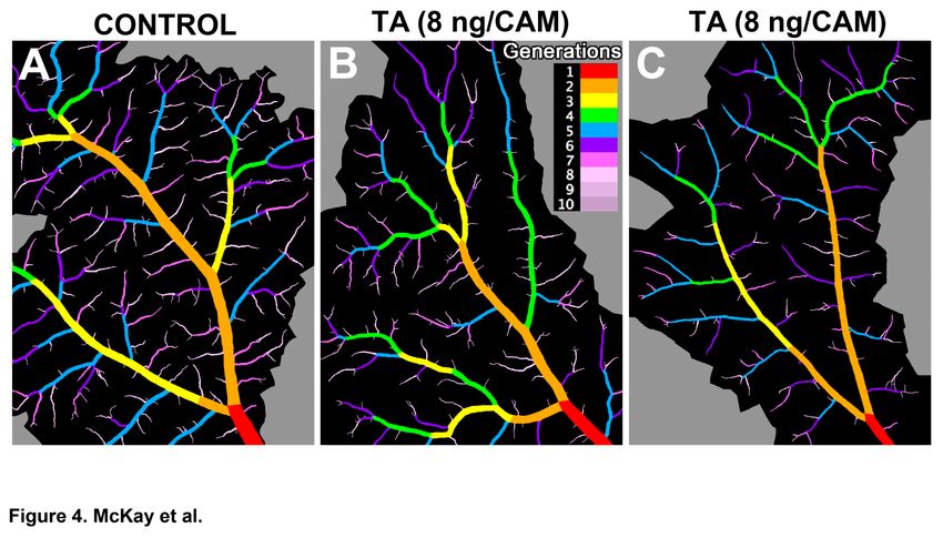
Fig. 4. TA selectively decreases the number of smaller vessels and decreases
vessel diameter throughout the vascular tree.
(A-C) Each representative image displays the major arterial tree with its ROI (in black) as
determined by VESGEN, for which vessel diameter (Dv) and the density of key vascular
parameters that include vessel number density (Nv), vessel branch point density (Brv), vessel
length density (Lv), and vessel area density (Av), were measured. Parameters were specified for
branching generations G1-G10 (see colorized legend) and denoted Dv1, Av1, Lv1, Nv1, etc. Relative
to controls (A), VESGEN results for Av, Lv, Nv and Brv in TA-treated specimens (B-C) show that
vessel density decreased only in the smallest branching generations G7-G10 (Fig. 5). However,
Dv decreased throughout vascular tree (Fig. 6). B above is the VESGEN output image of the
input binary image Fig. 1E.
20McKay et al Steroid Inhibition of Vessel Density and Diameter Page 21
Fig. 5. TA selectively inhibits the angiogenesis of small blood vessels.
Relative to controls, vessel density estimated by vessel number density (Nv) and vessel length
density (Lv) decreased only in the smallest branching generations G7-G10 for higher
concentrations of TA. For clarity, results for various classes of vessels lumped together as G1-2,
G3-4, G5-6 and G≥7 (G7-10). See Figs. 1, 2 and 4 for qualitative confirmation of the quantitative
results. Data are plotted as mean ± S.E. P-values ≤ 0.05 and ≤ 0.01 are indicated by * and **.
21McKay et al Steroid Inhibition of Vessel Density and Diameter Page 22
Fig. 6. TA decreases vessel diameter throughout the vascular tree.
Vessel diameter (Dv) decreased in all vessel branching generations (G1-G10) with increasing
concentration of TA, relative to controls. As for Fig. 5, results were lumped together as G1-2, G3-4,
22McKay et al Steroid Inhibition of Vessel Density and Diameter Page 23
G5-6 and G≥7 (G7-10). For qualitative confirmation of quantitative results, see Figs. 1 and 4. Data
are plotted as mean ± S.E. P-values ≤ 0.05 and ≤ 0.01 are indicated by * and **.
23McKay et al Steroid Inhibition of Vessel Density and Diameter Page 24
REFERENCES
1. Floman N, Zor U: Mechanism of steroid action in ocular inflammation: Inhibition of prostaglandin
production, Invest Ophthalmol Vis Sci 1977, 16:69-73
2. Umland SP, Nahrebne DK, Razac S, Beavis A, Pennline KJ, Egan RW, Billah MM: The
inhibitory effects of topically active glucocorticoids on IL-4, IL-5, and interferon-gamma
production by cultured primary CD4+ T cells, J Allergy Clin Immunol 1997, 100:511-519
3. Kang BS, Chung EY, Yun YP, Lee MK, Lee YR, Lee KS, Min KR, Kim Y: Inhibitory effects of
anti-inflammatory drugs on interleukin-6 bioactivity, Biol Pharm Bull 2001, 24:701-703
4. Sze PY, Iqbal Z: Glucocorticoid action on depolarization-dependent calcium influx in brain
synaptosomes, Neuroendocrinology 1994, 59:457-465
5. Sze PY, Iqbal Z: Glucocorticoid actions on synaptic plasma membranes: modulation of
[125I]calmodulin binding, J Steroid Biochem Mol Biol 1994, 48:179-186
6. Bandi N, Kompella UB: Budesonide reduces vascular endothelial growth factor secretion and
expression in airway (Calu-1) and alveolar (A549) epithelial cells, Eur J Pharmacol 2001,
425:109-116
7. Fischer S, Renz D, Schaper W, Karliczek GF: In vitro effects of dexamethasone on hypoxia-
induced hyperpermeability and expression of vascular endothelial growth factor, Eur J
Pharmacol 2001, 411:231-243
8. Wilson CA, Berkowitz BA, Sato Y, Ando N, Handa JT, de Juan E, Jr.: Treatment with intravitreal
steroid reduces blood-retinal barrier breakdown due to retinal photocoagulation, Arch
Ophthalmol 1992, 110:1155-1159
9. Kaiser PK: Steroids for branch retinal vein occlusion, Am J Ophthalmol 2005, 139:1095-1096
24McKay et al Steroid Inhibition of Vessel Density and Diameter Page 25
10. Margolis R, Singh RP, Kaiser PK: Branch retinal vein occlusion: clinical findings, natural history,
and management, Compr Ophthalmol Update 2006, 7:265-276
11. Brasil OF, Smith SD, Galor A, Lowder CY, Sears JE, Kaiser PK: Predictive factors for short-term
visual outcome after intravitreal triamcinolone acetonide injection for diabetic macular oedema:
an optical coherence tomography study, Br J Ophthalmol 2007, 91:761-765
12. Taban M, Singh RP, Chung JY, Lowder CY, Perez VL, Kaiser PK: Sterile endophthalmitis after
intravitreal triamcinolone: a possible association with uveitis, Am J Ophthalmol 2007, 144:50-54
13. Parsons-Wingerter P, Lwai B, Yang MC, Elliott KE, Milaninia A, Redlitz A, Clark JI, Sage EH: A
novel assay of angiogenesis in the quail chorioallantoic membrane: stimulation by bFGF and
inhibition by angiostatin according to fractal dimension and grid intersection, Microvasc. Res.
1998, 55:201-214
14. Parsons-Wingerter P, Elliott KE, Farr AG, Radhakrishnan K, Clark JI, Sage EH: Generational
analysis reveals that TGF-beta1 inhibits the rate of angiogenesis in vivo by selective decrease
in the number of new vessels, Microvasc. Res. 2000a, 59:221-232
15. Parsons-Wingerter P, Elliott KE, Clark JI, Farr AG: Fibroblast growth factor-2 selectively
stimulates angiogenesis of small vessels in arterial tree, Arterioscler. Thromb. Vasc. Biol.
2000b, 20:1250-1256
16. Parsons-Wingerter P, McKay TL, Leontiev D, Vickerman MB, Condrich TK, Dicorleto PE:
Lymphangiogenesis by blind-ended vessel sprouting is concurrent with hemangiogenesis by
vascular splitting, Anat Rec A Discov Mol Cell Evol Biol 2006a, 288:233-247
17. Parsons-Wingerter P, Chandrasekharan UM, McKay TL, Radhakrishnan K, DiCorleto PE,
Albarran B, Farr AG: A VEGF165-induced phenotypic switch from increased vessel density to
increased vessel diameter and increased endothelial NOS activity, Microvasc Res 2006b,
72:91-100
18. Mandelbrot BB: The Fractal Geometry of Nature. W. H. Freeman, San Francisco, 1983
25McKay et al Steroid Inhibition of Vessel Density and Diameter Page 26
19. Bassingthwaighte JB, Liebovitch LS, West BJ: Fractal Physiology. Oxford University Press, New
York,1994
20. West GB, Brown JH, Enquist BJ: A general model for the origin of allometric scaling laws in x
21. Avakian A, Kalina RE, Sage EH, Rambhia AH, Elliott KE, Chuang EL, Clark JI, Hwang J-N,
Parsons-Wingerter P: Fractal analysis of region-based vascular change in the normal and non-
proliferative diabetic retina, Curr Eye Res 2002, 24:274-280
22. Gan RZ, Tian Y, Yen RT, Kassab GS: Morphometry of the dog pulmonary venous tree, J. Appl.
Physiol. 1993, 75:432-440
23. Kassab GS, Rider CA, Tang NJ, Fung YC: Morphometry of pig coronary arterial trees, Am. J.
Physiol. 1993, 265:H350-365
24. Kassab GS, Lin DH, Fung YC: Morphometry of pig coronary venous system, Am. J. Physiol.
1994, 267:H2100-2113
25. Benjamin LE, Hemo I, Keshet E: A plasticity window for blood vessel remodelling is defined by
pericyte coverage of the preformed endothelial network and is regulated by PDGF-B and VEGF,
Development 1998, 125:1591-1598
26. Gerhardt H, Golding M, Fruttiger M, Ruhrberg C, Lundkvist A, Abramsson A, Jeltsch M, Mitchell
C, Alitalo K, Shima D, Betsholtz C: VEGF guides angiogenic sprouting utilizing endothelial tip
cell filopodia, J. Cell Biol. 2003, 161:1163-1177
26You can also read