Tumorbiology:
Effect of environmental
         factors,
  Cancer immunology
Neogrády Zsuzsa, 2019
←
→
Page content transcription
If your browser does not render page correctly, please read the page content below
Tumorbiology:
Effect of environmental
factors,
Cancer immunology
Neogrády Zsuzsa, 2019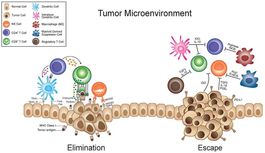
Drs. Yamagiwa and Ichikawa: “Experimental study
 on the pathogenesis of epithelial tumors”, 1915

                                        coal-tar
                                       painting in
                                       rabbit ear:
                                       squamous
                                       carcinom
                                       formation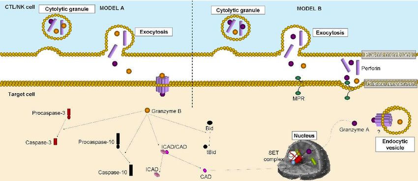
Effect of environmental factors on tumor
                        formation
• General pathomechanism: carcinogen → damaged genetic material
  → tumor formation
   I.) Chemical carcinogens
   II.) Physical carcinogens: radiation, (mechanical effects)
   III.) Biological carcinogens: oncogenic bacteria,
         virus, parasitic worms, (chronic inflammation,
         aging)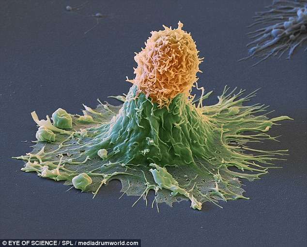
I.) Chemical carcinogens:
mode of action
                                                carcinogen
• Carcinogens can act:
   1. Directly
   2. Indirectly: carcinogenicity develops following structural
   changes in the organism such as by activity of CYP enzymes,
   glutation-S-transferase.
• Carcinogen chemical or its metabolite: electrophile → binds to
  the electron-rich molecules of the cell (mostly DNA) → DNA
  damage → changes in cell division and differentiation, etc →
  tumor formation (One certain carcinogen acts mostly on one
  certain organ! )
I.) Chemical carcinogens
• Carcinogen can be initiator or promoter
   1. Initiator: DNA of the cells will be primarily modified (mutated)
   2. Promoters are the chemicals which cannot cause DNA
   damage but act on cells after application of initiators
• If for some time there is no promoter action, the primarily
  produced DNA modification will be reversed and the tumor
  formation fails following the next promoter action. Human
  carcinogens:       https://www.cancer.org/cancer/cancer-causes/general-info/known-and-probable-human-
  carcinogens.htmlra
I.) Classification of chemical
carcinogens
1. Initiators:
• Alkylating agents (immunsuppressive drugs and cytostatics)
• Polycyclic aromatic hydrocarbons: metabolic activation is needed
  (tobacco, charcoal-broiled meat, smoked meat and fish)
• Aromatic amines and azo dyes: metabolic activation is needed
• Mycotoxin (Aflatoxin B1)
• Nitrosamines and –amides: intestinal microbes produce from nitrate and nitrite
  (source of nitrate and nitrite: preservatives, cured meat, drinking water)
• Asbestos, silicate, talcum, vinylchloride monomer (PVC), metals: chromium,
  nickel, arsenic
2. Promoters:
• Mostly produced in the body (hormones, free radicals, hydrogen peroxide, bile
  acids etc.)
II. Radiation
• The carcinogenic effect of radiation has been proved in each
species both in vivo and in vitro
• Consequence: double and simple DNA strand breaks, damaged bases, covalent
  crosslinks inside and between the DNA strands (pyrimidin dimer), DNA-protein
  crosslinks, protein-protein crosslinks, mutations in tumorsuppressor genes,
  enzyme inactivation, etc.
• Types of radiation:
    1. Sun light UVB (280-320 nm) → mutagenic, UVC (200-280 nm) is mutagenic
    also, but atmospheric osone can protect partly against this radiation.
    Consequence: DNA damage
    2. Ionizing radiation (very low wavelenght): electromagnetic (X-ray, gamma-ray)
    and particle radiation (proton, electron, alpha)
• Ionising radiation sources: radioactive nucleus, (Hiroshima, Csernobil), cosmic
  radiation, X-ray, PET=Positron emission tomograph (x-ray), CT=computed
  tomograph (gamma-ray) Consequence: DNA damage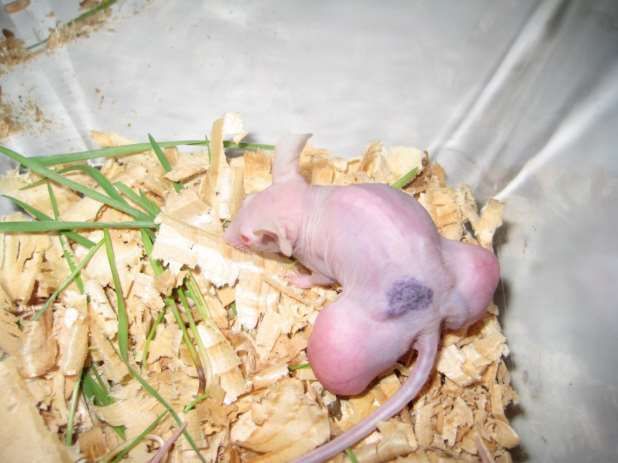
II. Radiation: electromagnetic spectrum → low
wavelenght UV, X-ray, gamma ray → carcinogen
              https://hu.wikipedia.org/wiki/Elektrom%C3%A1gneses_sug%C3%A1rz%C3%A1s
III. Biological carcinogens
            Filtrated chicken sarcoma homogenate contains virus

• Virus: chicken Rous sarcoma virus (1911), hepatitis B, C virus, Bittner
  mouse mammary tumor virus (MMTV), Human papilloma (HPV) virus,
  Epstein-Barr virus, HIV type 1, Human T-cell lymphotrophic virus
• Consequence: virus can be integrated in the genom, changes in gene
  expression: mutations and epigenetic effects
• Bacteria: Helicobacter pylori
• Helminths: Opisthorchis viverrini (bile duct), Clonor-
• chis sinensis (bile duct),
  Spirocerca lupi (canine esophageal carcinom)
                                                    Liver rot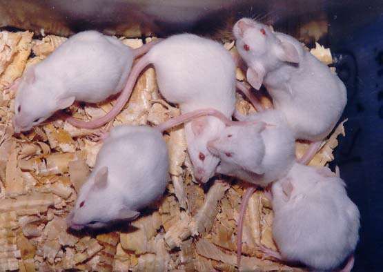
III. Biological carcinogens: role of MMTV= Bittner virus
 in the development of MMT (mouse mammary tumor)

• MMT cells were cultured in vitro
or injected in newborn mouse
(2500 or 7500 cells/animal IP) by us
Bittner virus could be detected both
in the cell culture and in the
developed abdominal tumor
(Gálfi and Neogrády, 1995)

                                       Cell culture   Tumor
Tumor development in the abdomen of 80-day old mice
MMTV: Reverse- inoculated with MMT cells
transcribing RNA viruses
(retroviruses) use the enzyme to
reverse-transcribe their RNA
genomes into DNA, which is
then integrated into the
host genome and replicated
along with it.
(Gálfi and Neogrády, 1995)Tumor development in the abdomen of 80-day old mice
inoculated with MMT cellsCancer immunology
https://www.dailymail.co.uk/health/article-4375120/Cancer-attack-scans-cancer-cell-colours.html
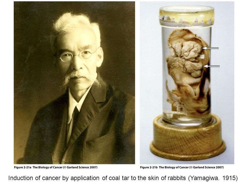
This SEM shows a volcanic-shaped breast
cancer cell (green) being attacked from
above by extracted CAR (chimeric antigen
receptor) linked T-cells (orange) that have
been modified to recognise and attack the
malignant tumourImmunological background 1.: types of immune
response and immune cells
T and B lymphocytes
NK=natural killer cells
http://missinglink.ucsf.edu/lm/immunology_module/prologue/objectives/obj02.htmlImmunological background 2.: adaptive immunity
APC=antigen presenting cells such as dendritic
cells)
MHC=major histocompatibility complex
TCR=T cell receptors
TH=helper T lymphocytes (CD4+)
CTL=cytotoxic T lymphocytes (CD8+)
CD=specific CD (cluster of differentiation) antigens
of T lymphocytes
IL=interleukines
http://www.sinobiological.com/Adaptive-Immunity-a-747.html
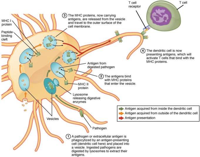
IFN=interferonImmunological background 3.: antigen presentation by
dendritic cells https://courses.lumenlearning.com/suny-ap2/chapter/the-adaptive-immune-response-t-lymphocytes-and-their-functional-types/
MHC=major
histocompatibility
complex
https://courses.lumenlearning.com/suny-ap2/chapter/the-adaptive-immune-
response-t-lymphocytes-and-their-functional-types/Immunological background 4: role of helper T and
cytotoxic T cells
https://courses.lumenlearning.com/suny-ap2/chapter/the-adaptive-immune-response-t-lymphocytes-and-their-functional-types/
(a) CD4 is associated with
helper T cells. An
extracellular pathogen is
processed and presented
in the binding cleft of a
class II MHC molecule, and
this interaction is
strengthened by the CD4
molecule.
(b) CD8 is associated with
cytotoxic T cells. An intra-
cellular pathogen is
presented by a class I MHC
molecule, and CD8
interacts with it.Perforin/granzyme-mediated apoptotic cell death (released by cytotoxic T lymphocytes =(CTL) and
natural killers=NK). Perforin directly forms pores in the plasma membrane of the target cell (Model
A) or internalization occurs via receptor-mediated endocytosis (Model B). Receptor: Mannosa-6-
phosphate receptor (MPR)
IMMUNOLOGICAL
BACKGROUND 5.
perforin
granzyme
Perforin pore
Caspase-dependent
apoptosis
Caspase-independent
apoptosis
SET complex, complex
that contains 3
DNAses: caspase-
Semantic Scholar independent apoptosisImmunological background 6: T-cell independent
activation of B cells
Differentiation
in plasma cells
or cancer cells
Antibody secretionImmunological background 7.
CD=cluster of
differentiation
lymphocytes (CD4: helper,
CD8: cytotoxic T lymphocytes)
lymphocytes
During the immune
function CD
antigens recognize
specific ligands,
activate them and
induce the
https://www.slideshare.net/MUBOSScz/immunology-vi-labcellimmunol
maturation of other
immune cells.Immunological background of tumor formation.
General overview: role of T and B lymphocytes
1. Presentation IV. Stimulated tumor
of dendritic cells I. Inflammatory cytokine production of tumor cells growth
III. Inhibition of apoptosis, genomic
instability, increased cell growth,
tissue expansion, increased
angiogenesis
2. Activation of T and 3. Anti-tumor actions: cell-
B lymphocytes, II. Modified immune mediated cytotoxicity of T
immune response response: such as Treg cells and antibody-
production dependent cytotoxicity of B-
cellsIn order to prove the role of T lymphocytes in tumor development:
congenitally athymic nude mice (nu/nu) are presented as a model for
the study of cell-mediated immunologic deficiencies: tumor
development is quick
https://www.ncbi.nlm.nih.gov/pubmed/1105061Role of dendritic cells in tumor killing 1-2. Apoptosis and necrosis of tumor cells 3-4. Antigen presentation of dendritic cells 5. T- lymphocyte activation 6. Cytotoxic effect of CTL: killing of tumor cells https://www.nature.com/articl es/4402243/figures/1
Modified immune response in cancer 1.: specific
             role of dendritic cells
• Dendritic cells: specialized antigen presenting cells
• To produce active, tumor-killing CTL (cytotoxic T-
  lymphocytes) the followings are needed: interaction of naive
  CD8 cells and matured dendritic cells, further other co-
  stimulating and co-inhibiting stimules
• However in tumors unmatured dendritic cells can be found
  → decreased activity of CTL
Modified immune response in cancer 2.: specific
role of Treg (regulatory T cells)
• Changes of different stimulation markers in tumors → activation of PD-
1 element of T cells by unmatured tumor dendritic cells → T cells →
differentiation in Treg → apoptosis of T cells and inhibition of CTL
(cytotoxic T lymphocytes)
• Further explanation of the decreased activity of CTL in the tumors:
• Unmatured (tumor) dendritic cells → much IL-10 and TGF →
differentiation in Treg
• Treg → further increase of IL-10 and TGF synthesis → decreased
synthesis/function of CTL
• Treg concentration in tumor is high → decreased tumor elimination
→ tumor progressionModified immune response
in cancer 3.: specific role of
Treg cells
Tregs can suppress the
activation, proliferation and
effector function of CD4+ and
CD8+ T cells, natural killer (NK)
cells, dendritic cells (DCs), and
B cells.
FOXP3=transcription factor for
genes involved in regulatory T-cell
function.
https://www.semanticscholar.org/paper/Regulatory-T-cells-in-oral-squamous-cell-carcinoma.-Liu-Liu/68cf83c404041cb0fb795a0c7d981ac10ee4ee36/figure/0Modified immune response in cancer 4.: tumor specific
T-lymphocytess (CD8+ cells) has no cytotoxic action
• In tumors no CTL/CD8 „cell-killing” function (do not produce
interferon, perforin and granzyme )
• The reasons:
• Differentiation of T cells into Treg
• Immunosuppressive mediators
• Existing inhibitory surface receptors on the CD8+ cells
• Few Trp and Arg in tumors: increased activity of IDO (indolamine
2,3 dioxygenase) - Trp-metabolizing enzyme and increased activity
of arginase - Arg-metabolizing enzymeSummary: during elimination phase the effector cells of immune system, CTL and
NK cells recognize (by the help of dendritic and CD4+ T cells) and kill (by the help of
CTL/CD8 +) the tumor cells. Left: Elimination= tumor regression, right: Escape=
inhibition of antitumor activity=tumor progression)
https://hu.wikipedia.org/wiki/Immunonkol%C3%B3gia#/media/File:Tumor_microenvironment.jpg
CTL
Helper
lymphocyte
Immune system destroyes Tumor escapes from immune
tumor controlClassification of tumor antigens • Tumor-Specific Antigens (TSA), which are present only on tumor cells and not on any other cell (specific vaccines) • Tumor-Associated Antigens (TAA), which are present on some tumor cells and also some normal cells (weak immune response against vaccines).
Classification of tumor antigens
1.) Overexpressed or Aberrantly Expressed Cellular Proteins
    Tumor Antigens Produced by Oncogenic Viruses
    Tyrosinase: melanoma
2.) Oncofetal Antigens
    alphafetoprotein: hepatocellular cancer
3.) Altered Cell Surface Glycolipids and Glycoproteins
    Her2=human epidermal growth factor receptor 2: breast cancer,
    Der2=canine epidermal growth factor receptor: mammary cancer
    EGFR=epithelial growth factor receptor
4.) Cell Type-Specific Differentiation Antigens
    PSA=prostate specific antigen: prostate cancer
    1A10 and SB2: canine mammary cancer
Failure of host defenses against tumor antigen
• Many tumors are eliminated by the immune system (and thus are
never detected), others continue to grow.
• Reasons of deficient host response:
• 1.) Suppression of immune response by chemical, physical, or viral
agents, by cytotoxic drugs or radiation
• 2.) Suppression of the immune response by the tumor itself:
• a) No appropriate antigen presentation: unmatured dendritic cells,
no MHC production
• b) Presence of Treg cells
• c) Low concentration of Trp and Arg
• d) High concentration of immunsuppressor mediators (IL-10, TGF)
• e) Low concentration of CD8 (tumor killing mediators (IL- 7,-IL-12,
IL-15)
• f) Immunologically protecting stroma around the tumorAntitumor immunotherapy= immunoncology: „active immunization” • Active immunization: by tumor specific antigens (TSA), tumor associated antigens (TAA) or by the tumor cells • Tumor cells: weak immune response, because they do not express MHC, in the tumor cells can not be found real antigen presenting cells (unmatured dendritic cells), formation of Treg cells, etc • Weak antitumor effect and as side effect: autoimmunity
Antitumor immunotherapy=
immunoncology:
„passive immunization”
• Passive immunization → AST method: in vitro production and injection of
  tumor antigen-specific CD8+ cytotoxic T-cells in patients with cancer
• Consequence: tumor cells carrying the specific antigen will be killed, but
  tumor cells carrying another antigen will survive, so complet recovery will
  not occur
• Lymphodepletion + AST method: systhemic cytostatic treatment, or total
  body irradiation (killing existing T and B lymphocytes, Treg cells), then AST
  → production of high number of injected tumor antigen-specific CD8+ cells
  → followed by lymphotrophic interleukin 2 (IL-2) treatment → production
  of healthy lymphocytes → very good method!
Antitumor vaccines1. : monoclonal antibody
for treatment of B and T cell lymphomas
1. Monoclonal antibody against human B lymphocyte (antibody production
in ovarial cell culture of chinese hamster). Antibody targets and bound to
CD20 cell surface marker of B lymphocyte. Active substance: Rituximab
(artificial monoclonal antibody against B cell lymphoma
2. Rituximab in dog recognizes canine CD20 superficial marker, but will not
bound to it. However, anti-canine CD21 antibody (produced in murine
hybridoma cells) is bound specifically to B lymphocytes of dog.
3. Monoclonal antibody against canine T lymphocyte (T-cell Mab) is used
for treatment of T cell lymphoma (mostly in golden retriever, the boxer,
Australian shepherd, Siberian husky). Each existing T lymphocytes will be
killed, but from the healthy stem cells new, healthy T lymphocytes will
develop.
Antitumor vaccines 2.: monoclonal antibody
for treatment of mammary carcinoma
• 1. Monoclonal antibody targets human epidermal growth factor
  receptor type 2 (HER2) on the surface of breast cancer cells (antibody
  production in ovarial cell culture of chinese hamster), active
  substance: trastuzumab (Herceptin)
• 2. Treatment of canine mammary tumor by herceptin results in
  allergic reaction. Dog epidermal growth factor receptor type2 (DER-
  2) has been isolated from the cell surface of canine mammary tumor,
  antibody targeting DER-2 seems to be a promising step to produce
  appropriate vaccine in the future.
Antitumor vaccines 3.:
for treatment of canine melanoma
Canine melanoma cells have characteristic tyrosinase activity, however this
enzyme can be found in healthy cells also (TAA).
OnceptR canine melanoma vaccine
• Each dose contains plasmid DNA that expresses the gene coding for
  human tyrosinase.
• Upon injection, the DNA is taken up by muscle cells which then express the
  human tyrosinase protein.
• The human tyrosinase protein is different enough from the canine
  tyrosinase protein that it will stimulate an immune response, yet similar
  enough to the canine tyrosinase that the immune response is effective
  against canine melanoma cells which express tyrosinase.
Antitumor vaccines 4.:
for treatment of B cell lymphoma
http://www.morphogenesis-inc.com/wp-content/uploads/2015/06/ImmuneFx-VOS-Brochure.pdf

• ImmuneFx™ is an autologous (personalized) cancer vaccine → made from
  each patient’s own cancer cells.
• The patented ImmuneFx™ priming signal (antigen) is a highly
  immunogenic protein → normally expressed on the surface of a
  streptococcal     bacterium.     Manufacturing       process:    ImmuneFx™
  (=immunogenic protein) → supplied in vitro to the patient’s own tumor
  cells (collected at surgery) → the antigen will be expressed on the tumor
  cells of patient→ the cells are then irradiated → these cells cannot divide
  when returned to the patient as vaccine.
• The irradiated vaccine cells are injected into the dermal layer of the skin →
  large concentration of antigen presenting cells will attack the tumor cells.
  This will be not asked at the exam!
Antitumor vaccines for treatment of canine
T cell lymphoma (lymphosarcoma=LSA) and
feline vaccine-associated fibrosarcoma
1. Canine tumor cells have high telomerase reverse transcriptase
   (TERT) activity, absent in the majority of normal dog tissues.
   Adenovirus expressing TERT injected intradermally induces
   intensive immune response in dogs with LSA.
2. One of the messenger chemicals of your cat’s immune system that
   is important in the fight against cancer and infection is Interleukin
   2 (IL-2). The vaccine contains canary pox virus that has been
   modified to produce feline (IL-2) by this way stimulating the cat’s
   own cancer defense cells (number of cytotoxic T lymphocytes and
   natural killer cells will increase).
    This will be not asked at the exam!
Cancer is a battle that can only be von if everyone
works togetherYou can also read