Unfolded Protein Response-Related Signature Associates With the Immune Microenvironment and Prognostic Prediction in Osteosarcoma
←
→
Page content transcription
If your browser does not render page correctly, please read the page content below
ORIGINAL RESEARCH
                                                                                                                                                 published: 08 June 2022
                                                                                                                                         doi: 10.3389/fgene.2022.911346

                                              Unfolded Protein Response–Related
                                              Signature Associates With the Immune
                                              Microenvironment and Prognostic
                                              Prediction in Osteosarcoma
                                              Zhao Zhang†, Xincheng Liu†, Debin Cheng†, Jingyi Dang, Zhenzhou Mi, Yubo Shi, Lei Wang
                                              and Hongbin Fan *

                                              Division of Musculoskeletal Cancer Service, Department of Orthopaedic Surgery, Xi-jing Hospital, The Fourth Military Medical
                                              University, Xi’an, China

                                              Background: Osteosarcoma is a highly malignant bone tumor commonly occurring in
                                              adolescents with a poor 5-year survival rate. The unfolded protein response (UPR) can
                                              alleviate the accumulation of misfolded proteins to maintain homeostasis under
                                              endoplasmic reticulum stress. The UPR is linked to the occurrence, progression, and
                           Edited by:         drug resistance of tumors. However, the function of UPR-related genes (UPRRGs) in
                      William C. Cho,
          QEH, Hong Kong SAR, China           disease progression and prognosis of osteosarcoma remains unclear.
                      Reviewed by:            Methods: The mRNA expression profiling and corresponding clinical features of
                     Wweibin Zhang,
               Ruijin Hospital, China
                                              osteosarcoma were acquired from TARGET and GEO databases. Consensus
                       Weichun Guo,           clustering was conducted to confirm different UPRRG subtypes. Subsequently, we
Renmin Hospital of Wuhan University,
                                              evaluated the prognosis and immune status of the different subtypes. Functional
                                China
                                              analysis of GO, GSEA, and GSVA was used to reveal the molecular mechanism
                    *Correspondence:
                          Hongbin Fan         between the subtypes. Finally, four genes (STC2, PREB, TSPYL2, and ATP6V0D1)
                   fanhb@fmmu.edu.cn          were screened to construct and validate a risk signature to predict the prognosis of
     †
         These authors have contributed       patients with osteosarcoma.
                    equally to this work
                                              Result: We identified two subtypes according to the UPRRG expression patterns. The
                   Specialty section:         subgroup with higher immune scores, lower tumor purity, and active immune status was
        This article was submitted to
Cancer Genetics and Oncogenomics,             linked to a better prognosis. Meanwhile, functional enrichment revealed that immune-
              a section of the journal        related signaling pathways varied markedly in the two subtypes, suggesting that the UPR
                 Frontiers in Genetics
                                              might influence the prognosis of osteosarcoma via influencing the immune
               Received: 02 April 2022
                                              microenvironment. Moreover, prognostic signature and nomogram models were
               Accepted: 27 April 2022
              Published: 08 June 2022         developed based on UPRRGs, and the results showed that our model has an
                            Citation:         excellent performance in predicting the prognosis of osteosarcoma. qPCR analysis
Zhang Z, Liu X, Cheng D, Dang J, Mi Z,        was also conducted to verify the expression levels of the four genes.
    Shi Y, Wang L and Fan H (2022)
 Unfolded Protein Response–Related            Conclusion: We revealed the crucial contribution of UPRRGs in the immune
Signature Associates With the Immune
    Microenvironment and Prognostic           microenvironment and prognostic prediction of osteosarcoma patients and provided
         Prediction in Osteosarcoma.          new insights for targeted therapy and prognostic assessment of the disease.
            Front. Genet. 13:911346.
    doi: 10.3389/fgene.2022.911346            Keywords: unfolded protein response, prognostic prediction, immune infiltration, nomogram, osteosarcoma

Frontiers in Genetics | www.frontiersin.org                                          1                                            June 2022 | Volume 13 | Article 911346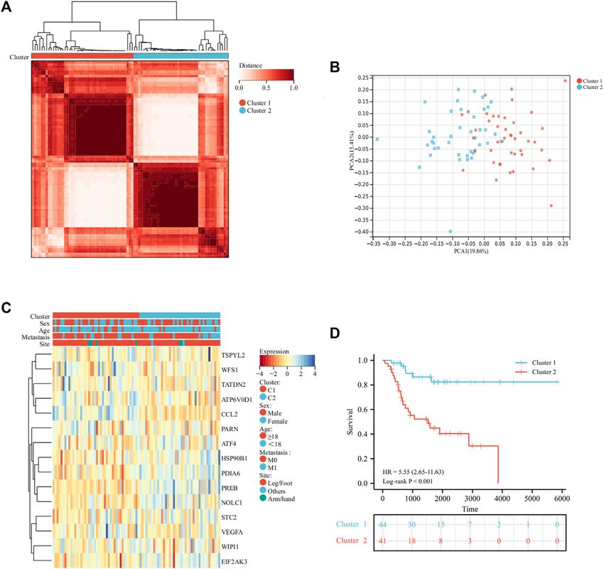
Zhang et al.                                                                                                UPR-Related Signature in Osteosarcoma

INTRODUCTION                                                                a novel prognostic signature for predicting the prognosis of
                                                                            osteosarcoma patients via integrating risk scores and clinical
Osteosarcoma is one of the most frequent primary malignant                  features.
bone tumors in adolescents, often occurring in fast-growing long
bones (Tang et al., 2008; Arndt et al., 2012). With significant
advances in surgical options, neoadjuvant chemotherapy, and                 METHODS AND MATERIALS
diagnostic imaging, the overall survival of patients with
osteosarcoma has been significantly prolonged (Li et al., 2015;              Data Collection
Wakamatsu et al., 2019; Hao et al., 2021). Nevertheless, the                The mRNA expression profiles and corresponding clinical
outcome of metastatic and recurrent patients remains                        features of 85 osteosarcoma patients were acquired from the
unsatisfactory due to its intensely aggressive feature, with a 5-           TARGET database as a training cohort (https://ocg.cancer.gov/
year mortality ratio of below 20% (Ferrari et al., 2005). In                programs/target). UPRRGs were extracted from hallmark gene
addition, high tumor heterogeneity, leading to chemoresistance              sets from the Molecular Signature Database (Zhang et al., 2021).
in some patients, remains challenging clinically (Xiao et al., 2015).       In addition, mRNA expression profiles and relevant clinical
Therefore, there is a need for the discovery of novel targets for           information of 53 osteosarcoma patients from GSE21257 were
targeted treatment and better prognostic markers for                        obtained from the GEO database to serve as an external validation
osteosarcoma.                                                               cohort (https://www.ncbi.nlm.nih.gov/geo/). The clinical
    The unfolded protein response (UPR) is a highly conserved               information for osteosarcoma patients is presented in
homeostatic response in all eukaryotic cells and can help cells             Supplementary Table S1.
mitigate the accumulation of misfolded proteins in the
endoplasmic reticulum (ER) (Hetz et al., 2020). Under ER                    Classification of Molecular Subtypes
stress conditions, such as hypoxia, nutritional deprivation,                To assess the biological functions of UPRRGs in OS, we first
acidosis, and inflammatory stimuli, the UPR can activate three               identified 15 prognosis-related UPRRGs for osteosarcoma based
sensor proteins (IRE1α, PERK, and ATF6) to initiate specific                 on a univariate Cox regression analysis. Subsequently, consensus
signal transduction cascades which regulate the rate of protein             clustering analysis was conducted based on the expression matrix
production for maintaining ER homeostasis (Vanacker et al.,                 of these 15 genes with the R package “ConsensusClusterPlus”. For
2017). Lately, extensive studies have indicated that the UPR acts a         the analysis, 80% of the total samples of the target dataset are
dual function in tumor occurrence and development. In earlier               included in each iteration and reiterated 1,000 times to ensure
stages of tumorigenesis, the UPR is capable of exerting antitumor           cluster stability. The average consistency value and clinical
effects to hinder tumor transformation, while in the established            significance within the clustering group were used together
tumors, tumor cells can induce chronic UPR to relieve ER stress-            with the optimal number of clusters. Principal component
induced apoptosis and develop drug resistance to maintain tumor             analysis (PCA) was performed to confirm the gene expression
survival (Ma and Hendershot, 2004; Yang et al., 2019). Aberrant             patterns in the different clusters. KM curve and log-rank tests
activation of the UPR was found in a wide range of tumors,                  were employed to evaluate the prognosis of different clusters. The
including bladder cancer, cutaneous melanoma, and liver cancer              heat map was used to show the gene expression and clinical
(Houessinon et al., 2016; Wan et al., 2020; Zhu et al., 2021). Apart        characteristics of the different clusters.
from a direct impact on tumor biology, the UPR also has the
ability to remodel the tumor immune microenvironment (TIME)                 TIME Evaluation
to regulate the crosstalk between immune cells, which serves an             The tumor microenvironment scores for individual samples in
essential function in immune surveillance and immune escape                 the different subtypes were assessed by the ESTIMATE algorithm
(Zanetti et al., 2022). Mahadevan et al. demonstrated that the              (Yoshihara et al., 2013). The TIMER algorithm was conducted to
UPR can modulate the phenotype of dendritic cells and CD8+                  comprehensively evaluate the abundance of immune infiltrating
T cells to facilitate tumor growth. Notably, previous studies have          cells in each sample (Li et al., 2020). Moreover, the infiltration
confirmed the UPR to be relevant to the growth, prognosis, and               abundance of 28 immune cell types for an individual sample was
drug resistance of osteosarcoma (Mahadevan et al., 2012; Yan                tested by the single sample gene set enrichment analysis (ssGSEA)
et al., 2015; Shi et al., 2022). However, the role of the UPR-related       algorithm. The expression of immune checkpoint (ICP) genes in
gene (UPRRG) sets on prognostic prediction and immune                       different subtypes was also evaluated.
infiltration in patients with osteosarcoma remains unclear.
    Recently, following the advancement of the high-throughput
sequencing technology for tumor genomics, probing new                       Identification of DEG and Enrichment
molecular patterns through bioinformatics approaches offers                 Analysis
new insights for tumor treatment and prognosis evaluation                   Differentially expressed genes (DEGs) in different subtypes were
(Qian et al., 2021). In the present study, we performed a                   analyzed via the R package “limma”, and log2 (Foldchange) > 1.5
comprehensive analysis of the prognosis and immune                          and FDR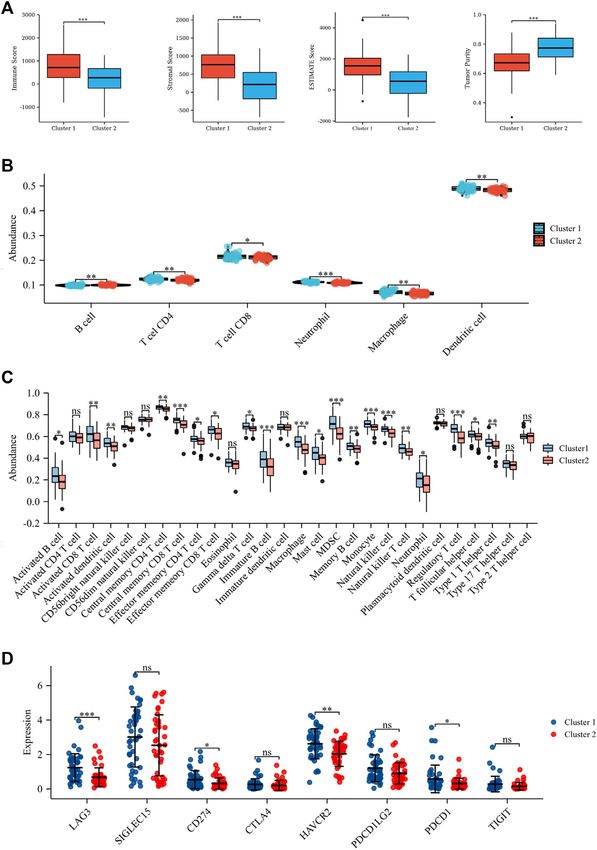
Zhang et al.                                                                                                  UPR-Related Signature in Osteosarcoma

conducted to verify functional pathway variations between the            TABLE 1 | Primer sequences of the candidate genes.
different subtypes according to GO: the biological process (BP).         Gene                                         Sequence (5’ -> 39)
Also, Gene Set Enrichment Analysis (GSEA) was employed to
analyze hallmark gene sets for different subtypes (Subramanian           STC2                               Forward:GGGTGTGGCGTGTTTGAATG
                                                                                                            Reverse:TTTCCAGCGTTGTGCAGAAAA
et al., 2005). |NES| > 1, NOM p-val < 0.05, and FDR<0.25 were
                                                                         TSPYL2                             Forward:ACAGGTGCTGGCCGATATG
taken as the threshold.                                                                                     Reverse:CCGACTCGATGGTAGAATCCC
                                                                         PREB                               Forward:ACGGGCCACCATGAACTTG
                                                                                                            Reverse:GGGTTTCCGCTCCACATTTCT
Establishment and Validation of the                                      ATP6V0D1                           Forward:TTCCCGGAGCTTTACTTTAACG
                                                                                                            Reverse:CAAGTCCTCTAGCGTCTCGC
Prognostic Signature                                                     GAPDH                              Forward:GGAGCGAGATCCCTCCAAAAT
The previously obtained 15 prognosis-related UPRRGs were                                                    Reverse:GGCTGTTGTCATACTTCTCATGG
screened via least absolute shrinkage and selection operator
(LASSO) Cox regression based on the R package “glmnet”, and
the minimum lamba is taken as the optimal value. Then, a
multivariate Cox analysis was conducted to further optimize              Quantitative Real-Time PCR (qRT-PCR)
and establish the prognostic signature. The risk score in the            The TRIzol method was utilized to extract and purify RNA from
training and validation cohorts was calculated with the                  tissues and cells. Then, a cDNA synthesis kit (Takara, China) was
following formula: Risk score = in (Coefi * Xi). In the                 applied to reverse transcribe the RNA. The TB Green Premux Ex
training cohort, all patients were classified into low- and               TaqⅡ (Tli RNaseH Plus) was used for qRT-PCR with the Bio-Rad
high-risk groups by the median risk score, and the overall               CFX96 Real-Time PCR system (Bio-Rad, USA). The internal control
survival (OS) between the two groups was investigated using              was GADPH. The primer sequences of the candidate genes are shown
the KM curve and log-rank tests. In addition, receiver operating         in Table 1, and the analysis was conducted three times for all genes.
characteristic (ROC) curves were applied to measure the
effectiveness of prognostic models. Furthermore, the                     Statistics
abovementioned formula was also used in patients from                    R software (version 4.0.5), SPSS 21.0 software, and GraphPad
GSE23257 to generate a risk model to validate the prognostic             Prism 8 were carried out for all statistical analyses. The t-test was
benefit. Finally, we integrated different clinical characteristics        applied for two groups. One-way ANOVA was applied to three
including gender, age, metastatic status, and disease site to            groups. p < 0.05 was taken as statistically significant. pp < 0.05;
assess if the risk score was an independent prognostic                   ppp < 0.01; and pppp < 0.001.
element in osteosarcoma patients based on univariate and
multivariate Cox regression.
                                                                         RESULTS
Construction and Calibration of the                                      Identification of UPRRG Molecular
Nomogram                                                                 Subtypes via Consensus Analysis
A nomogram model was established to forecast the 3-year and 5-           A total of 113 UPRRGs were retrieved from hallmark gene sets, out
year survival probability of osteosarcoma patients via integrating       of which we identified 15 prognosis-related UPRRGs for
risk scores and clinical profiles. The C-index, ROC curves, and           osteosarcoma, according to univariate COX analysis
calibration plots were employed to survey the predictive                 (Supplementary Table S2, 3). According to the expression
performance of the constructed nomogram in both cohorts.                 profile of these genes, consensus clustering analysis was used to
                                                                         ascertain subgroups of osteosarcoma patients in the training
Patient Sample Collection and Cell Culture                               database. k = 2 is considered the best category number of
A total of six osteosarcoma patient tissues and corresponding            clusters, depending on the average consistency value and clinical
adjacent normal tissues were obtained from the operation and             significance within the clustering group (Figure 1A and
stored in liquid nitrogen banks. The study was approved by the           Supplementary Figure S1). PCA analysis revealed a relatively
institutional review board of Xijing Hospital, the Fourth Military       apparent distinction between the two subtypes (Figure 1B). The
Medical University, and all patients provided signed informed            heat map illustrated the gene expression profile and clinical
consent. Moreover, human osteosarcoma cell lines HOS and MG-             characteristics of the two subtypes (Figure 1C). Moreover, we
63 were procured from Procell Life Science & Technology Co. Ltd          noticed that patients in cluster 2 experienced a dismal prognosis
and cultured in DMEM medium (Gibco, Shanghai, China)                     to that in cluster 1 (p<0.001, Figure 1D). Previous studies have
containing 10% fetal bovine serum (FBS; Gibco, Shanghai,                 proven that the UPR can coordinate the crosstalk between immune
China) and 1% (v/v) penicillin/streptomycin (Gibco, Shanghai,            cells and tumor cells in the TIME to exert immunosurveillance and
China). The osteoblast cell line hFOB 1.19 was provided by Dr.           immunosuppressive functions to influence tumor prognosis
Jianping Bai of Xijing Hospital and cultured in DMEM/F12                 (Vanacker et al., 2017). Thus, we then evaluated the differences
medium with 10% FBS, 0.3 mg/ml G418 (Procell, Wuhan,                     in the TIME across different subtypes. The ESTIMATE algorithm
China), and 1% (v/v) penicillin/streptomycin. At 37 C, 5%                indicated that cluster 1 had higher immune score (p = 8.4e-5),
CO2 environment, all cell lines were incubated.                          stromal score (p = 8.1e-7), ESTIMATE score (p = 9.6e-7), and lower

Frontiers in Genetics | www.frontiersin.org                          3                                         June 2022 | Volume 13 | Article 911346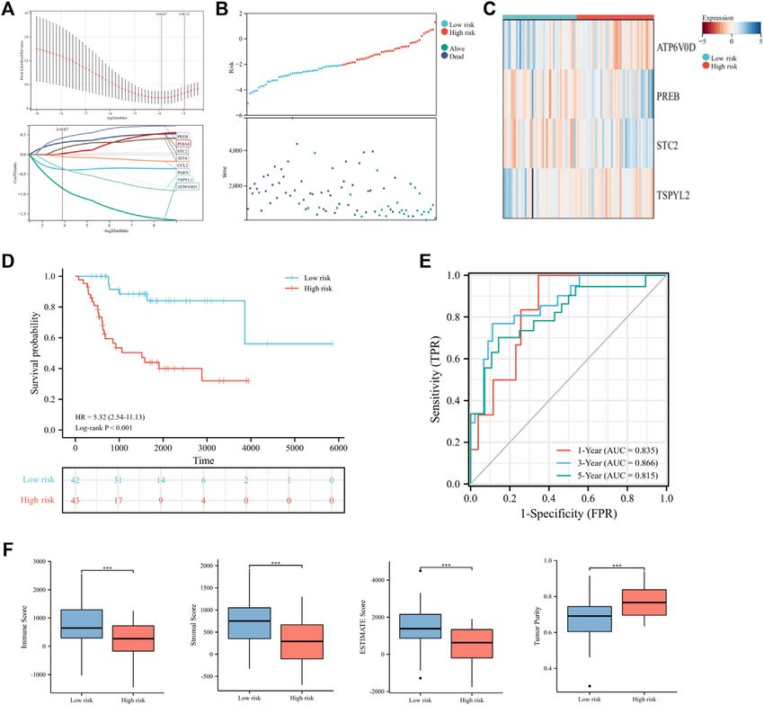
Zhang et al. UPR-Related Signature in Osteosarcoma
FIGURE 1 | Identification of molecular subtypes of UPRRGs by consensus clustering. (A). Clustering heat map at k = 2. (B) PCA plot between the two subtypes. (C)
Heat map of the UPR-related gene expression and clinical features in the two subtypes. (D) Survival curves for the two subgroups.
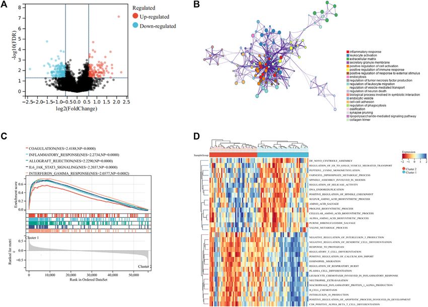
tumor purity (p = 1.1e-6), as compared to cluster 2 (Figure 2A). In clusters. The results indicated that 121 genes were upregulated
addition, the TIMER algorithm discovered that the abundance of and 157 genes were downregulated in cluster 2 to cluster 1
most immune infiltrating cells in cluster1 was significantly increased (Figure 3A). GO analysis implied that these DEGs were
than in cluster 2, including dendritic cells, neutrophil cells, CD4+ primarily associated with inflammatory response, leukocyte
T cells, neutrophil cells, and CD8+ T cells, while B cells showed the activation, and extracellular matrix (Figure 3B). These
opposite result (Figure 2B). As shown in Figure 2C, ssGSEA findings implied that UPR could influence the TIME and
analysis found that the abundance of 20 immune cell types was prognosis of osteosarcoma via modulation of immune-
significantly increased in cluster 1 compared to cluster 2. In addition, associated pathways. We then used GSVA and GSEA analysis
we also observed that CD274, LAG3, HAVCR2, and PDCD1 were to explore the functional differences in the two clusters. GSEA
expressed at an elevated level in cluster 1 than in cluster 2 analysis demonstrated that coagulation, inflammatory response,
(Figure 2D). Our findings suggested that the prognosis of and IL6/JAK/STAT3 signaling were markedly upregulated in
different subtypes may be affected by the TIME. cluster 1 (Figure 3C). GSVA analysis showed that positive
regulation of calcium ion import, regulation of the apoptotic
process involved in the development, and some immune-related
Functions and Pathway Annotations of pathways were significantly upregulated in cluster 1 to cluster 2
DEGs for UPRRG Subtypes (Figure 3D). Therefore, we speculated that the UPR plays an
To reveal the potential mechanisms regulating the TIME between essential role in regulating the immune function, thus
different subtypes, we performed DEGs analysis on the two contributing to the prognosis of osteosarcoma.
Frontiers in Genetics | www.frontiersin.org 4 June 2022 | Volume 13 | Article 911346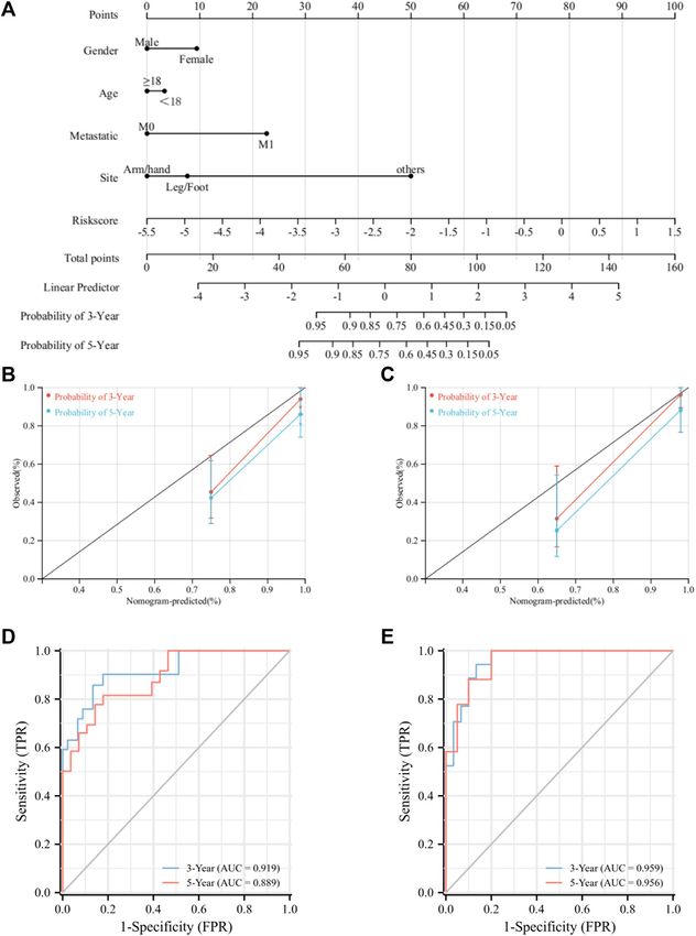
Zhang et al.                                                                                                                UPR-Related Signature in Osteosarcoma

  FIGURE 2 | Tumor microenvironment in the two subtypes. (A) Stromal score, immune score, ESTIMATE score, and tumor purity based on ESTIMATE algorithm.
  (B) Six immune cell abundance assessments by the TIMER algorithm. (C) Twenty-nine immune cell abundance assessments by the ssGSEA algorithm. (D) Differences in
  immune checkpoints between the two subtypes. *p < 0.05; **p < 0.01; ***p < 0.001.

Construction and Validation of the UPRRG                                             ATP6V0D1) were identified to establish a risk model for
Risk Signature                                                                       osteosarcoma via the multivariate Cox regression analysis
To further elucidate the prognostic predictive role of UPRRGs in                     (Figure 4C and Supplementary Table S4). The following
osteosarcoma patients, LASSO regression analysis was applied to                      formula was applied to generate the risk score for each sample:
screen for potential genes, and eight genes were identified by the                    Risk score = -1.523 × ATP6V0D1 + 0.903× PREB +0.586 × STC2
minimal lambo value (Figures 4A,B). Finally, based on the results of                 -0.760 × TSPYL2. All patients in the training cohort were classified
previous screening, four UPRRGs (STC2, PREB, TSPYL2, and                             into high- and low-risk groups by the median risk scores, and

Frontiers in Genetics | www.frontiersin.org                                      5                                           June 2022 | Volume 13 | Article 911346
Zhang et al. UPR-Related Signature in Osteosarcoma
FIGURE 3 | Differentially expressed genes and functional enrichment analyses. (A) Volcano plot showing the DEGs between the two subgroups. (B) Bubble plot
exhibited the functional enrichment of DEGs through GO analysis. (C) GSEA shows the hallmark gene sets in the two subgroups. (D) Heat map depicted the results of the
GSVA analysis.
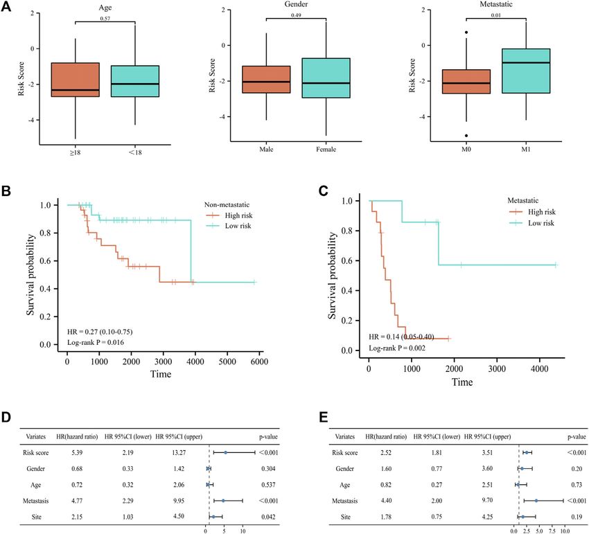
patients in the high-risk group showed shorter survival intervals than multivariate Cox analysis demonstrated that the risk score and
those in the low-risk group (Figure 4D). KM curves demonstrated a metastatic status were independent prognostic factors for
poor prognosis in the high-risk group to the low-risk group osteosarcoma patients, which meant that the UPRRGs risk model
(Figure 4E). Moreover, time-dependent ROC analysis found the was applicable to predict survival in osteosarcoma (Figures 5D,E).
AUC values of 1, 3, and 5 years were 0.84, 0.87, and 0.83, respectively, Moreover, we further tested the prognostic performance of the
which suggested our risk signature showed excellent predictive UPRRG risk signature in a validation cohort. As shown in Figure 6A,
performance (Figure 4F). Notably, we also observed that the the patients in the validation cohort were clearly separated into different
low-risk group saw higher immune scores (p = 3.5e-4), stromal risk groups via the abovementioned formula. The heat map
scores (p = 1.1e-4), ESTIMATE scores (p = 3.3e-5), and lower tumor demonstrated these four genes’ expression profiles in subgroups
purity (p = 2.9e-5) relative to the high-risk group, which suggested (Figure 6B). The KM curve likewise showed that the low-risk
that the TIME might be strongly linked to prognosis in different risk group had a better prognosis (p = 0.04) (Figure 6C). ROC curves
groups (Figure 4G). Then, the relevance between the risk signature suggested the risk signature had better prediction accuracy at 1 and
and clinical features was also evaluated, and the results revealed 3 years (Figure 6D). Similarly, the ESTIMATE algorithm obtained
results consistent with the training cohort, which further confirmed the
metastatic patients had significantly higher risk scores than non-
role of the TIME in the UPRRG risk signature (Figure 6E).
metastatic patients (p = 0.03), while no differences were found in any
other clinical characteristics (Figure 5A). When patients were
reclassified for metastatic status, there was a significantly Construction and Validation of the
improved prognosis for patients in the low-risk group over those Nomogram Prediction Model
in the high-risk group (Figures 5B,C). These findings supported that A nomogram model was constructed to improve the accuracy of
the UPRRG risk signature might be strongly correlated with the predicting the prognosis of osteosarcoma patients at 3 and 5 years
metastasis status in patients with osteosarcoma. Furthermore, by integrating risk scores and clinical characteristics (Figure 7A). Then,
Frontiers in Genetics | www.frontiersin.org 6 June 2022 | Volume 13 | Article 911346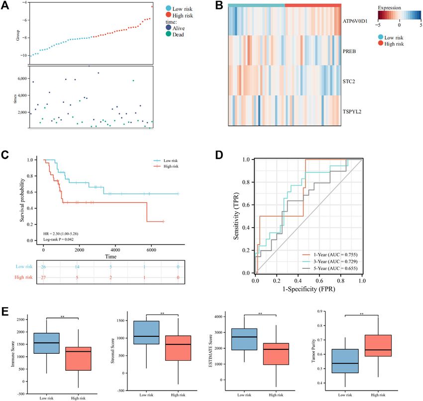
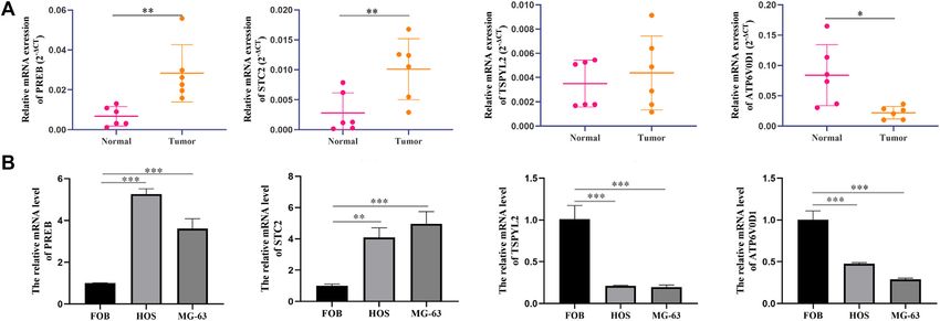
Zhang et al.                                                                                                                      UPR-Related Signature in Osteosarcoma

  FIGURE 4 | Construction of the risk signature based on UPRRGs in the training cohort. (A) Eight optimal UPRRGs filtered by LASSO analysis. (B) Distribution of risk
  scores and patient status in the two risk groups; (C). Heat map showing the expressions of four candidate genes. (D) Survival curves for the two risk groups. (E) Time-
  dependent ROC curve of the risk model. (F) Tumor microenvironment analysis in the two risk groups through the ESTIMATE algorithm. *p < 0.05; **p < 0.01; ***p <
  0.001.

we validated the predictive efficacy of the nomogram model in the two                     Verification of Candidate Genes by
cohorts. The C-index values for the training and validation cohorts were                 qRT-PCR Analysis
0.88 and 0.87, respectively. The ROC curves revealed AUC values of                       To certify the expression levels of these four candidate genes, we
0.93 and 0.90 for the training cohort at 3 and 5 years, respectively, and                performed a qPCR analysis in patients’ tissue and cell lines. The
for the validation cohort, ROC curves also exhibited excellent prediction                results revealed that the expression levels STC2 and PREB were
accuracy (Figures 7B,C). Moreover, the calibration curves for the                        elevated clearly in osteosarcoma tissues than in normal tissues,
training and validation cohort showed that the nomogram model                            whereas the ATP6V0D1 expression level was significantly
has a strong predictive capacity for the prognosis of osteosarcoma                       downregulated in tumor tissues compared with normal tissues
patients at 3 and 5 years (Figures 7D,E). Collectively, the                              (Figure 8A). The results of the cell further identified that the
aforementioned results pointed out that the UPRRG nomogram                               expression levels of PREB and STC2 were higher in osteosarcoma
model has high predictive accuracy and can be applied to predict                         cells than in normal osteoblasts, while TSPYL2 and ATP6V0D1
the prognosis of osteosarcoma patients.                                                  exhibited the opposite results (Figure 8B).

Frontiers in Genetics | www.frontiersin.org                                          7                                             June 2022 | Volume 13 | Article 911346
Zhang et al.                                                                                                                      UPR-Related Signature in Osteosarcoma

  FIGURE 5 | Correlation of the risk signature with clinical features in the training cohort. (A) Differences in risk scores among osteosarcoma patients by age, gender,
  and metastatic status. (B–C) Survival curves for patients with osteosarcoma regrouped by metastatic status. (D–E) Univariate and multivariate cox regression analyses
  for integrating risk characteristics and clinical features.

DISCUSSION                                                                               and built a prognostic model for differentially expressed genes
                                                                                         (Shi et al., 2022). Although previous studies have shown the UPR
The unfolded protein response is an adaptive signaling pathway                           was implicated in the development, progression, and treatment of
that regulates the endoplasmic reticulum homeostasis (Hetz et al.,                       osteosarcoma, the function of UPRRGs on immune infiltration
2020). The UPR has been extensively studied in tumorigenesis,                            and prognostic prediction in patients with osteosarcoma is
and its abnormal activation is involved in various stages of                             not clear.
tumorigenesis and progression (Ma and Hendershot, 2004).                                    In this research, we determined two molecule subtypes of
Chaiyawat et al. revealed that plenty of UPR-related proteins                            osteosarcoma based on prognosis-related UPRRGs and presented
were highly expressed in osteosarcoma patients and closely                               the relevance of different subtypes to clinical profiles. Our study
associated with chemoresistance, suggesting that targeting UPR                           found that different subtypes had different survival intervals and
pathways might be promising for the treatment of osteosarcoma                            TIME. The TIME consists of various components, including
(Chaiyawat et al., 2019). Ji et al. found that PERK could induce                         immune and stromal cells, which together contribute to
autophagy in osteosarcoma by inhibiting the mTORC1 pathway                               tumorigenesis, progression, and prognosis (Zhao et al., 2018).
to counteract ER stress-induced cell apoptosis (Ji et al., 2015). Yan                    The UPR is a critical factor in regulating the balance of immune
et al. demonstrated that the UPR could inhibit cisplatin-mediated                        dynamics in the tumor microenvironment which can directly
apoptosis by triggering the NF-κB pathway, contributing to drug                          affect innate and adaptive immune responses to participate in
resistance in osteosarcoma (Yan et al., 2015). Shi et al. identified                      tumor progression (Martins et al., 2016). Our results indicated
aberrant activation of UPR-related pathways in osteosarcoma                              that cluster 1 has a better prognosis with higher immune and

Frontiers in Genetics | www.frontiersin.org                                          8                                            June 2022 | Volume 13 | Article 911346
Zhang et al. UPR-Related Signature in Osteosarcoma FIGURE 6 | Validation of the constructed risk signature in the verification cohort. (A) Distribution of risk scores and patient status in different risk groups. (B) Heat map displayed the expressions of four candidate genes in the verification cohort. (C) Survival curves of the two risk groups. (D) Time-dependent ROC curve in the verification cohort. (E) Tumor microenvironment analysis by the ESTIMATE algorithm. *p < 0.05; **p < 0.01; ***p < 0.001. stromal scores compared to cluster 2. Previous studies have that as a consequence of a large number of immune infiltration demonstrated that a higher immune score and stromal score cells in cluster 1, the expressions of several ICPs were were connected to a better prognosis in osteosarcoma, which is in correspondingly elevated in cluster 1 compared to Cluster 2, agreement with our findings (Qian et al., 2021). Further analysis implying that patients in cluster 1 might be more sensitive to ICP indicated that the abundance of multiple immune infiltrating cells inhibitors. These findings suggested that the identification of was distinctly greater in cluster 1 than in cluster 2, according to UPRRG subtypes may provide a new clinical strategy for the TIMER, indicating that the active immune status of the prognostic evaluation and targeted therapy of osteosarcoma. UPRRG subtypes might be closely better associated with To reveal the molecular mechanisms underlying regulating osteosarcoma prognosis. ssGSEA analysis found that the TIME between different UPRRG subtypes, we carried functional abundance of a variety of tumor-infiltrating lymphocytes was enrichment analysis of the DEGs between the two subtypes. GO expressed at obviously higher levels in cluster 1 than in cluster 2. analysis suggested that the DEGs were mainly participating in Previous studies have demonstrated that the UPR acts as a critical immune-related pathways including inflammatory response and mediator of tumor immunity as an appropriate UPR can induce leukocyte activation. In addition, GSEA analysis also confirmed immune cells to eliminate tumor cells, whereas the sustained UPR significant enrichment of some immune-related pathways in could induce immune cell apoptosis to enhance tumor cluster 1, including coagulation, inflammatory response, and proliferation and invasion (Vanacker et al., 2017). Hence, we IL6/JAK/STAT3 signaling. The UPR is proven to not only affect believed that the elevated abundance of multiple TILs in cluster 1 the growth and survival of tumor cells but also play an essential role might be due to appropriate UPR, which induced activation of in remodeling the TIME (Zanetti et al., 2022). Batista et al. TILs to deliver anti-tumor immunity. Nevertheless, the sustained identified that the UPR participates in the macrophage UPR led to massive TIL exhaustion, causing a poor prognosis in polarization in the TIME by activating the IRE1α/XBP1 axis, patients with osteosarcoma in Cluster 2. Moreover, we also noted resulting in upregulation of IL-6, IL-23, arginase 1, CD86, and Frontiers in Genetics | www.frontiersin.org 9 June 2022 | Volume 13 | Article 911346
Zhang et al.                                                                                                                          UPR-Related Signature in Osteosarcoma

  FIGURE 7 | Construction and evaluation of the nomogram. (A). Nomogram for predicting the prognosis of patients with osteosarcoma. (B) Calibration for 3-and 5-
  year OS in the training cohort. (C) Calibration for 3-and 5-year OS in the verification cohort. (D) ROC analysis for 3-and 5-year OS in the training cohort. (E) ROC analysis
  for 3-and 5-year OS in the verification cohort.

PD-L1 that lead to local immune dysregulation (Batista et al.,                              from immune-related pathways, apoptosis and calcium
2020). Zanetti et al. showed that tumor cells regulate immune cell                          homeostasis-related pathways were also upregulated in cluster 1.
phenotypes through UPR activation of dendritic cells and T cells to                         These results suggested that the UPR may exert antitumor effects
promote tumor proliferation (Mahadevan et al., 2012). In addition,                          through multiple pathways between UPRRG subtypes. Overall, our
Medel et al. also demonstrated that dendritic cells could enhance                           data showed that patients with osteosarcoma can activate the UPR
CD8+ T-cell–specific responses through activation of the IRE1α/                              in the presence of ER stress to modulate tumorigenesis and
XBP1 axis to exert an anti-tumor effect (Medel et al., 2018). Taken                         progression through multiple pathways and that targeting UPR
together, these findings revealed that the UPR can modulate                                  might be a promising treatment strategy for osteosarcoma.
immune cell infiltration through various immune signaling                                       Next, to assess the role of UPRRGs in predicting the prognosis of
pathways to improve the prognosis of patients with                                          osteosarcoma, we constructed a prognostic signature to predict the
osteosarcoma. Moreover, GSVA analysis revealed that aside                                   survival of osteosarcoma patients via four genes (STC2, PREB,

Frontiers in Genetics | www.frontiersin.org                                            10                                              June 2022 | Volume 13 | Article 911346
Zhang et al.                                                                                                                    UPR-Related Signature in Osteosarcoma

  FIGURE 8 | Candidate gene validation. (A) Expression levels of candidate genes in tumor and normal tissues from osteosarcoma patients. (B) Expression levels of
  candidate genes in different cell lines. *p < 0.05; **p < 0.01; ***p < 0.001.

TSPYL2, and ATP6V0D1). Our results found that high expressions                          and that the low immune status in the high-risk group was strongly
of STC2 and PREB were linked to high risk (risk factors), whereas                       connected with poor prognosis. Moreover, our results identified that
high expressions of TSPYL2 and ATP6V0D1 were linked to low risk                         risk score was an independent prognostic factor for osteosarcoma
(protective factors). STC2 encodes a glycoprotein that performs an                      patients. Subsequently, we performed a nomogram model to better
essential function in the development and invasion of multiple                          predict the prognosis of osteosarcoma patients via risk score and
tumors (Li S. et al., 2021). Previous studies have demonstrated                         clinical characteristics. Previous studies have reported several
endoplasmic reticulum stress could activate PERK-ATF4 to induce                         nomogram models to predict the prognosis of osteosarcoma (Qian
the upregulated expression of STC2 to inhibit cell apoptosis (Ito et al.,               et al., 2021). Li et al. built a nomogram model by autophagy-related
2004). Chen et al. found that STC2 could promote tumor                                  genes with AUC values of 0.735 and 0.726 at 3 and 5 years, respectively
proliferation by activating the AKT-ERK pathway, and increased                          (Li J. et al., 2021). Wen et al. developed a 3-gene nomogram model with
STC2 was strongly correlated with poor prognosis in colorectal                          3-year and 5-year AUC values of 0.853 and 0.818, respectively (Wen
tumors (Chen et al., 2016). PREB can encode transcription factors                       et al., 2020). Wu et al. established a hypoxic nomogram model with an
that bind and activate the basal prolactin promoter activity. Murao                     AUC value of only 0.73 (Wu et al., 2021). In our study, the constructed
et al. showed that PREB could act as a transcription factor for TNF-                    nomogram model had 3-year and 5-year AUC values of 0.93 and 0.90,
αand IL-1β to regulate the expression of monocyte chemoattractant                       respectively, which were consistently superior to other models. We
protein-1, suggesting that PREB plays an active role in immune                          presented that the UPRRG nomogram model could better predict the
responses (Murao et al., 2009). In addition, PREB is a member of a                      prognosis of patients with osteosarcoma than existing models.
eukaryotic family of WD-repeat proteins involved in many                                Collectively, these findings offered new options for personalized
biological activities, including vesicle trafficking, RNA processing,                    treatment and prognostic prediction of osteosarcoma.
and signal transduction (Taylor Clelland et al., 2000). TSPYL2, a                           Despite the many strengths of the current study, there are notable
member of the TSPY-L nucleosome assembly protein-1 superfamily,                         limitations. First, the UPRRG risk signature was constructed based
can exert anti-tumor effects by inhibiting the cell cycle and regulating                on the TARGET and GEO databases, which may be biased due to
DNA damage. Previous studies have found that TSPYL2 maintains                           the limited number of patients. Second, this study lacked some
the G1 checkpoint function by inducing p21 transcription to                             clinical information relevant to the prognosis of osteosarcoma, such
modulate DNA damage and inhibit cellular growth (Tao et al.,                            as tumor pathological grade, which constrained clinical variables that
2011). Liu et al. indicated that TSPYL2 could inhibit SIRT1-mediated                    can be incorporated into the nomogram model. A larger,
FOXO3 deacetylation to reduce gefitinib resistance and inhibit DNA                       multicenter, prospective clinical cohort will be needed to further
damage, implying that TSPYL2 is a promising therapeutic target (Liu                     evaluate the clinical merit of our findings in the future.
et al., 2022). ATP6V0D1 is an encoded protein involved in vacuolar
ATPase formation, which has a crucial role in the modulation of the
acidic microenvironment (Lu et al., 2021). Numerous research                            CONCLUSION
studies have found that dysregulation of V-ATPase is related to
tumor growth and invasion (Whitton et al., 2018). Targeting                             Our study identified two molecular subtypes through consensus
V-ATPase could upregulate ER stress-related markers and inhibit                         clustering based on prognosis-related UPRRGs, and the two
mTOR signaling to exert anticancer effects (Kitazawa et al., 2017).                     subtypes exhibited different survival times and immune
    Survival analysis and ROC curve presented that the UPRRG risk                       statuses. Functional analysis revealed that the UPRRG
signature demonstrated satisfactory predictive accuracy in both cohorts                 subtypes might influence the progression and prognosis of

Frontiers in Genetics | www.frontiersin.org                                        11                                            June 2022 | Volume 13 | Article 911346
Zhang et al. UPR-Related Signature in Osteosarcoma
osteosarcoma patients through immune-related pathways. provided their written informed consent to participate in
Moreover, a novel prognostic model based on UPRRGs was this study.
constructed and validated to better predict the prognosis of
patients with osteosarcoma. We elucidated the important
function of UPRRGs in the development and prognosis of AUTHOR CONTRIBUTIONS
osteosarcoma, shedding light on new insights for targeted
therapy and clinical decision-making in patients with HF and ZZ have designed the study. ZZ and XL have written the
osteosarcoma. manuscript, and DC, JD, ZM, YS, and LW have analyzed the data.
All authors have read and approved it for publication.
DATA AVAILABILITY STATEMENT
FUNDING
The original contributions presented in the study are included in
the article/Supplementary Material; further inquiries can be This work was supported by the National Natural Science
directed to the corresponding author. Foundation of China (No. 31971272).
ETHICS STATEMENT SUPPLEMENTARY MATERIAL
The studies involving human participants were reviewed and The Supplementary Material for this article can be found online at:
approved by the Ethics Committee of Xi-Jing Hospital, The https://www.frontiersin.org/articles/10.3389/fgene.2022.911346/
Fourth Military Medical University. The patients/participants full#supplementary-material
Ji, G.-r., Yu, N.-c., Xue, X., and Li, Z.-g. (2015). PERK-mediated Autophagy in
REFERENCES Osteosarcoma Cells Resists ER Stress-Induced Cell Apoptosis. Int. J. Biol. Sci. 11
(7), 803–812. doi:10.7150/ijbs.11100
Arndt, C. A. S., Rose, P. S., Folpe, A. L., and Laack, N. N. (2012). Common Kitazawa, S., Nishizawa, S., Nakagawa, H., Funata, M., Nishimura, K., Soga, T.,
Musculoskeletal Tumors of Childhood and Adolescence. Mayo Clin. Proc. 87 et al. (2017). Cancer with Low Cathepsin D Levels Is Susceptible to Vacuolar
(5), 475–487. doi:10.1016/j.mayocp.2012.01.015 (H+ )-ATPase Inhibition. Cancer Sci. 108 (6), 1185–1193. doi:10.1111/cas.
Batista, A., Rodvold, J. J., Xian, S., Searles, S. C., Lew, A., Iwawaki, T., et al. (2020). 13240
IRE1α Regulates Macrophage Polarization, PD-L1 Expression, and Tumor Li, J., Guo, Z., Wang, Z., Fan, H., and Fu, J. (2015). Does Microwave Ablation of the
Survival. PLoS Biol. 18 (6), e3000687. doi:10.1371/journal.pbio.3000687 Tumor Edge Allow for Joint-Sparing Surgery in Patients with Osteosarcoma of
Chaiyawat, P., Sungngam, P., Teeyakasem, P., Sirikaew, N., Klangjorhor, J., the Proximal Tibia? Clin. Orthop. Relat. Res. 473 (10), 3204–3211. doi:10.1007/
Settakorn, J., et al. (2019). Protein Profiling of Osteosarcoma Tissue and s11999-015-4447-y
Soft Callus Unveils Activation of the Unfolded Protein Response Pathway. Li, J., Tang, X., Du, Y., Dong, J., Zhao, Z., Hu, H., et al. (2021a). Establishment of an
Int. J. Oncol. 54 (5), 1704–1718. doi:10.3892/ijo.2019.4737 Autophagy-Related Clinical Prognosis Model for Predicting the Overall
Chen, B., Zeng, X., He, Y., Wang, X., Liang, Z., Liu, J., et al. (2016). STC2 Promotes Survival of Osteosarcoma. BioMed Res. Int. 2021, 1–17. doi:10.1155/2021/
the Epithelial-Mesenchymal Transition of Colorectal Cancer Cells through 5428425
AKT-ERK Signaling Pathways. Oncotarget 7 (44), 71400–71416. doi:10.18632/ Li, S., Huang, Q., Li, D., Lv, L., Li, Y., and Wu, Z. (2021b). The Significance of
oncotarget.12147 Stanniocalcin 2 in Malignancies and Mechanisms. Bioengineered 12 (1),
Ferrari, S., Smeland, S., Mercuri, M., Bertoni, F., Longhi, A., Ruggieri, P., et al. 7276–7285. doi:10.1080/21655979.2021.1977551
(2005). Neoadjuvant Chemotherapy with High-Dose Ifosfamide, High-Dose Li, T., Fu, J., Zeng, Z., Cohen, D., Li, J., Chen, Q., et al. (2020). TIMER2.0 for
Methotrexate, Cisplatin, and Doxorubicin for Patients with Localized Analysis of Tumor-Infiltrating Immune Cells. Nucleic Acids Res. 48 (W1),
Osteosarcoma of the Extremity: a Joint Study by the Italian and W509–w514. doi:10.1093/nar/gkaa407
Scandinavian Sarcoma Groups. Jco 23 (34), 8845–8852. doi:10.1200/jco. Liu, Z., Li, C., Yu, C., Chen, Z., Zhao, C., and Ye, L. (2022). TSPYL2 Reduced
2004.00.5785 Gefitinib Resistance and DNA Damage Repair via Suppressing SIRT1-
Hao, Y., An, R., Xue, Y., Li, F., Wang, H., Zheng, J., et al. (2021). Prognostic Value Mediated FOXO3 Deacetylation. Future Med. Chem. 14 (6), 407–419.
of Tumoral and Peritumoral Magnetic Resonance Parameters in Osteosarcoma doi:10.4155/fmc-2021-0136
Patients for Monitoring Chemotherapy Response. Eur. Radiol. 31 (5), Lu, J., Ma, J., Hao, Z., and Li, W. (2021). HPS6 Regulates the Biogenesis of Weibel-
3518–3529. doi:10.1007/s00330-020-07338-y Palade Body in Endothelial Cells through Trafficking V-ATPase to its Limiting
Hetz, C., Zhang, K., and Kaufman, R. J. (2020). Mechanisms, Regulation and Membrane. Front. Cell. Dev. Biol. 9, 743124. doi:10.3389/fcell.2021.743124
Functions of the Unfolded Protein Response. Nat. Rev. Mol. Cell. Biol. 21 (8), Ma, Y., and Hendershot, L. M. (2004). The Role of the Unfolded Protein Response
421–438. doi:10.1038/s41580-020-0250-z in Tumour Development: Friend or Foe? Nat. Rev. Cancer 4 (12), 966–977.
Houessinon, A., Gicquel, A., Bochereau, F., Louandre, C., Nyga, R., Godin, C., doi:10.1038/nrc1505
et al. (2016). Alpha-fetoprotein Is a Biomarker of Unfolded Protein Mahadevan, N. R., Anufreichik, V., Rodvold, J. J., Chiu, K. T., Sepulveda, H., and
Response and Altered Proteostasis in Hepatocellular Carcinoma Cells Zanetti, M. (2012). Cell-Extrinsic Effects of Tumor ER Stress Imprint Myeloid
Exposed to Sorafenib. Cancer Lett. 370 (2), 242–249. doi:10.1016/j. Dendritic Cells and Impair CD8+ T Cell Priming. PLoS One 7 (12), e51845.
canlet.2015.10.032 doi:10.1371/journal.pone.0051845
Ito, D., Walker, J. R., Thompson, C. S., Moroz, I., Lin, W., Veselits, M. L., et al. Martins, A. S., Alves, I., Helguero, L., Domingues, M. R., and Neves, B. M. (2016).
(2004). Characterization of Stanniocalcin 2, a Novel Target of the Mammalian The Unfolded Protein Response in Homeostasis and Modulation of
Unfolded Protein Response with Cytoprotective Properties. Mol. Cell. Biol. 24 Mammalian Immune Cells. Int. Rev. Immunol. 35 (6), 457–476. doi:10.
(21), 9456–9469. doi:10.1128/mcb.24.21.9456-9469.2004 3109/08830185.2015.1110151
Frontiers in Genetics | www.frontiersin.org 12 June 2022 | Volume 13 | Article 911346Zhang et al. UPR-Related Signature in Osteosarcoma Medel, B., Costoya, C., Fernandez, D., Pereda, C., Lladser, A., Sauma, D., et al. Xiao, X., Wang, W., Zhang, H., Gao, P., Fan, B., Huang, C., et al. (2015). (2018). IRE1α Activation in Bone Marrow-Derived Dendritic Cells Modulates Individualized Chemotherapy for Osteosarcoma and Identification of Gene Innate Recognition of Melanoma Cells and Favors CD8+ T Cell Priming. Front. Mutations in Osteosarcoma. Tumor Biol. 36 (4), 2427–2435. doi:10.1007/ Immunol. 9, 3050. doi:10.3389/fimmu.2018.03050 s13277-014-2853-5 Murao, K., Imachi, H., Yu, X., Muraoka, T., Hosami, N., Dobashi, H., et al. (2009). Yan, M., Ni, J., Song, D., Ding, M., and Huang, J. (2015). Activation of Unfolded The Transcriptional Factor PREB Mediates MCP-1 Transcription Induced by Protein Response Protects Osteosarcoma Cells from Cisplatin-Induced Apoptosis Cytokines in Human Vascular Endothelial Cells. Atherosclerosis 207 (1), 45–50. through NF-Κb Pathway. Int. J. Clin. Exp. Pathol. 8 (9), 10204–10215. doi:10.1016/j.atherosclerosis.2009.03.051 Yang, H., Liang, S.-Q., Xu, D., Yang, Z., Marti, T. M., Gao, Y., et al. (2019). HSP90/ Qian, H., Lei, T., Hu, Y., and Lei, P. (2021). Expression of Lipid-Metabolism Genes AXL/eIF4E-regulated Unfolded Protein Response as an Acquired Vulnerability Is Correlated with Immune Microenvironment and Predicts Prognosis in in Drug-Resistant KRAS-Mutant Lung Cancer. Oncogenesis 8 (9), 45. doi:10. Osteosarcoma. Front. Cell. Dev. Biol. 9, 673827. doi:10.3389/fcell.2021.673827 1038/s41389-019-0158-7 Shi, C., Zhao, F., Zhang, T., Xu, D., Hao, Z., Cui, F., et al. (2022). A Novel Yoshihara, K., Shahmoradgoli, M., Martínez, E., Vegesna, R., Kim, H., Torres- Prognostic Signature in Osteosarcoma Characterised from the Perspective of Garcia, W., et al. (2013). Inferring Tumour Purity and Stromal and Immune Unfolded Protein Response. Clin. Transl. Med 12 (3), e750. doi:10.1002/ Cell Admixture from Expression Data. Nat. Commun. 4, 2612. doi:10.1038/ ctm2.750 ncomms3612 Subramanian, A., Tamayo, P., Mootha, V. K., Mukherjee, S., Ebert, B. L., Gillette, Zanetti, M., Xian, S., Dosset, M., and Carter, H. (2022). The Unfolded Protein M. A., et al. (2005). Gene Set Enrichment Analysis: a Knowledge-Based Response at the Tumor-Immune Interface. Front. Immunol. 13, 823157. doi:10. Approach for Interpreting Genome-wide Expression Profiles. Proc. Natl. 3389/fimmu.2022.823157 Acad. Sci. U.S.A. 102 (43), 15545–15550. doi:10.1073/pnas.0506580102 Zhang, F., Feng, D., Wang, X., Gu, Y., Shen, Z., Yang, Y., et al. (2021). An Unfolded Tang, N., Song, W.-X., Luo, J., Haydon, R. C., and He, T.-C. (2008). Osteosarcoma Protein Response Related Signature Could Robustly Predict Survival Outcomes Development and Stem Cell Differentiation. Clin. Orthop. Relat. Res. 466 (9), and Closely Correlate with Response to Immunotherapy and Chemotherapy in 2114–2130. doi:10.1007/s11999-008-0335-z Bladder Cancer. Front. Mol. Biosci. 8, 780329. doi:10.3389/fmolb.2021.780329 Tao, K. P., Fong, S. W., Lu, Z., Ching, Y. P., Chan, K. W., and Chan, S. Y. (2011). Zhao, Y., Shao, Q., Zhu, H., Xu, H., Long, W., Yu, B., et al. (2018). Resveratrol TSPYL2 Is Important for G1 Checkpoint Maintenance upon DNA Damage. Ameliorates Lewis Lung Carcinoma-bearing Mice Development, Decreases PLoS One 6 (6), e21602. doi:10.1371/journal.pone.0021602 Granulocytic Myeloid-derived Suppressor Cell Accumulation and Impairs its Taylor Clelland, C. L., Craciun, L., Bancroft, C., and Lufkin, T. (2000). Mapping Suppressive Ability. Cancer Sci. 109 (9), 2677–2686. doi:10.1111/cas.13720 and Developmental Expression Analysis of the WD-Repeat Gene Preb. Zhou, Y., Zhou, B., Pache, L., Chang, M., Khodabakhshi, A. H., Tanaseichuk, O., Genomics 63 (3), 391–399. doi:10.1006/geno.1999.6089 et al. (2019). Metascape Provides a Biologist-Oriented Resource for the Analysis Vanacker, H., Vetters, J., Moudombi, L., Caux, C., Janssens, S., and Michallet, M.- of Systems-Level Datasets. Nat. Commun. 10 (1), 1523. doi:10.1038/s41467- C. (2017). Emerging Role of the Unfolded Protein Response in Tumor 019-09234-6 Immunosurveillance. Trends Cancer 3 (7), 491–505. doi:10.1016/j.trecan. Zhu, K., Xiaoqiang, L., Deng, W., Wang, G., and Fu, B. (2021). Identification of a 2017.05.005 Novel Signature Based on Unfolded Protein Response-Related Gene for Wakamatsu, T., Kakunaga, S., Takenaka, S., Outani, H., Hamada, K., Imura, Y., Predicting Prognosis in Bladder Cancer. Hum. Genomics 15 (1), 73. doi:10. et al. (2019). Prognostic Implication of Adjuvant/neoadjuvant Chemotherapy 1186/s40246-021-00372-x Consisting of Doxorubicin and Ifosfamide in Patients with Extraskeletal Osteosarcoma. Int. J. Clin. Oncol. 24 (10), 1311–1319. doi:10.1007/s10147- Conflict of Interest: The authors declare that the research was conducted in the 019-01475-1 absence of any commercial or financial relationships that could be construed as a Wan, Q., Jin, L., and Wang, Z. (2020). Comprehensive Analysis of Cancer potential conflict of interest. Hallmarks in Cutaneous Melanoma and Identification of a Novel Unfolded Protein Response as a Prognostic Signature. aging 12 (20), 20684–20701. doi:10. Publisher’s Note: All claims expressed in this article are solely those of the authors 18632/aging.103974 and do not necessarily represent those of their affiliated organizations, or those of Wen, C., Wang, H., Wang, H., Mo, H., Zhong, W., Tang, J., et al. (2020). A Three- the publisher, the editors, and the reviewers. Any product that may be evaluated in Gene Signature Based on Tumour Microenvironment Predicts Overall Survival this article, or claim that may be made by its manufacturer, is not guaranteed or of Osteosarcoma in Adolescents and Young Adults. Aging 13 (1), 619–645. endorsed by the publisher. doi:10.18632/aging.202170 Whitton, B., Okamoto, H., Packham, G., and Crabb, S. J. (2018). Vacuolar ATPase Copyright © 2022 Zhang, Liu, Cheng, Dang, Mi, Shi, Wang and Fan. This is an as a Potential Therapeutic Target and Mediator of Treatment Resistance in open-access article distributed under the terms of the Creative Commons Attribution Cancer. Cancer Med. 7 (8), 3800–3811. doi:10.1002/cam4.1594 License (CC BY). The use, distribution or reproduction in other forums is permitted, Wu, F., Xu, J., Jin, M., Jiang, X., Li, J., Li, X., et al. (2021). Development and provided the original author(s) and the copyright owner(s) are credited and that the Verification of a Hypoxic Gene Signature for Predicting Prognosis, Immune original publication in this journal is cited, in accordance with accepted academic Microenvironment, and Chemosensitivity for Osteosarcoma. Front. Mol. practice. No use, distribution or reproduction is permitted which does not comply Biosci. 8, 705148. doi:10.3389/fmolb.2021.705148 with these terms. Frontiers in Genetics | www.frontiersin.org 13 June 2022 | Volume 13 | Article 911346
You can also read