Unilateral knee effusion in an elderly patient: an unusual presentation of rheumatoid arthritis
←
→
Page content transcription
If your browser does not render page correctly, please read the page content below
CASE REPORT

Unilateral knee effusion in an elderly patient:
an unusual presentation of rheumatoid arthritis
Morteza Khodaee MD, MPH;1,3 Lindsay Ogle MD;2 Cleveland Piggott MD, MPH1
1
  University of Colorado School of Medicine, Department of Family Medicine & Orthopedics, Division of Sports Medicine,
  AF Williams Clinic, Denver, Colorado, USA
2
  University of Colorado School of Medicine, Swedish Family Medicine Residency, Aurora, Colorado, USA
3
  Corresponding author. Email: Morteza.khodaee@cuanschutz.edu

                                                                                                                                       J PRIM HEALTH CARE
                                                                                                                                       2020;12(3).
    ABSTRACT                                                                                                                           doi:10.1071/HC20035
    Unilateral atraumatic knee effusion is a relatively common presenting complaint among geriatric                                    Received 30 April 2020
                                                                                                                                       Accepted 28 August 2020
    patients in primary care and musculoskeletal speciality clinics. Gout, pseudogout, degenerative                                    Published 16 September 2020
    joint diseases and reactive arthritis are the most common causes of the atraumatic knee effusions.
    Rheumatoid arthritis very rarely presents as arthritis of one or two large joints. Arthrocentesis, plain
    radiography and screening blood tests should be performed to help narrow the differential
    diagnosis. In some cases, advanced imaging modalities such as MRI may be indicated. This study
    reports a case of rheumatoid arthritis in a 75-year-old gentleman with oligoarthropathy of two large
    joints as the presenting symptoms.

    KEYWORDS: Synovitis; rheumatoid arthritis; arthrocentesis; oligoarthropathy

Introduction                                                         swelling with inability to bend his knee beyond 908.
                                                                     On further questioning, he also mentions right
Rheumatoid arthritis (RA) is the most common
                                                                     elbow pain and swelling. His past medical history is
systemic inflammatory arthritis, with worldwide
                                                                     significant for well-controlled type 2 diabetes mel-
prevalence of ,0.5–1%.1,2 It is more common
                                                                     litus, hypertension and hyperlipidemia, for which
among women, with the peak incidence between
                                                                     he is taking metformin, lisinopril, and atorvastatin.
age 55 and 65 years.1,2 The hallmark of RA is
                                                                     During physical examination, he is afebrile with
symmetric involvement of small joints. Large joints
                                                                     normal vital signs. He has significant right knee
involvement, particularly as a presenting condition,
                                                                     effusion with mild joint line tenderness and palpa-
is rare. The purpose of this report is to remind
                                                                     ble synovitis (Figure 1a). His right elbow exami-
clinicians to not regard typical presentations of
                                                                     nation also revealed significant effusion and
common conditions as the only possibility in the
                                                                     synovitis (Figure 1b). Plain radiography showed
differential diagnosis. Clinicians should follow basic
                                                                     mild-to-moderate degenerative changes (Figure 2).
and logical approaches to any case, starting with a
thorough history and physical examination and
                                                                     Arthrocentesis of the right knee joint with an
obtaining appropriate imaging and laboratory tests.
                                                                     18-gauge needle (inner diameter of 0.838 mm) pro-
                                                                     duced 47 mL of cloudy synovial fluid with obvious
Case report                                                          synovial tissue (Figure 3). Laboratory investigations
A gentleman aged 75 years presented with bilateral                   revealed unremarkable complete blood cell counts,
(right worse than left) knee pain for years. Previ-                  comprehensive metabolic panel, hemoglobin A1c of
ously, he was told that he had knee osteoarthritis.                  6.1% (reference range [RR] 4.0–6.0%) and uric acid of
Due to personal preference, he has refused any                       6.1 (RR 4.4–7.6 mg/dL). He had a negative antinuclear
intra-articular corticosteroids or viscosupplement                   antibodies (ANA) and elevated C-reactive protein
injections in the past. He complains of right knee                   (CRP) of 25.7 (RR ,10.0 mg/L) and an erythrocyte

CSIRO Publishing
Journal compilation  Royal New Zealand College of General Practitioners 2020
This is an open access article licensed under a Creative Commons Attribution-NonCommercial-NoDerivatives 4.0 International License                               1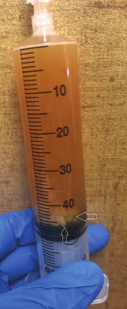
CASE REPORT
CASE REPORT
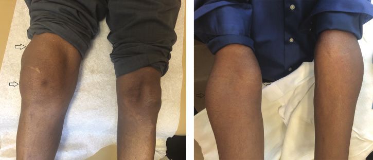
Figure 1. Right knee (a) and right elbow (b) effusion (open arrows).
(a) (b)
Figure 2. Plain radiography reveals moderate (right) and mild (left) tricompartmental Figure 3. Cloudy aspirated synovial fluid with floating
osteoarthritis of the right (a, b) and left (c, d) knees. Radiographs show joint space synovium (open arrows).
narrowing (open arrows), osteophyte formation (arrows), subchondral cyst formation and
sclerosis (arrowheads).
(a) (c)
(b) (d)
.200 U (levels .60 U consistent with strong
positive). His urine chlamydia, gonorrhoea and
sedimentation rate (ESR) of 47 (RR ,10 mm/h). trichomonas nucleic acid amplification tests were
Rheumatoid factor (RF) was 268 (RR 0–14I U/mL) negative. There were no crystals in his synovial fluid,
and anti-cyclic citrullinated peptide (anti-CCP) was with a synovial fluid nucleated cell count of 25,175 per
2 JOURNAL OF PRIMARY HEALTH CARECASE REPORT
CASE REPORT
Figure 4. T2-weighted right knee MRI images (a, b) show significant effusion (*) and synovitis (arrows).
(a) (b)
mm3 (76% segmented neutrophils) and a red (RF, anti-CCP).3 A total of at least six points out of
blood cell count of 13,000 per mm3. maximum 10 defines the diagnosis of RA.3 In this
system, zero points are given to patients who
A MRI revealed severe tricompartmental osteoar- present with mono-arthritis of a large joint
thritis with broad areas of full-thickness cartilage (ie shoulder, elbow, hip, knee and ankle).3
loss, subchondral cystic change and oedema. There
was a large effusion with exuberant synovitis Although RA incidence is four- to five-fold higher
throughout the joint (Figure 4). in females than in males aged ,50 years, the
female-to-male ratio is only two-fold higher in
The patient was counselled about his new diagnosis patients aged .60–70 years.4 Additionally, RA
of RA and therapeutic options during the following should remain on the differential diagnosis of
visit. However, he decided not to start any treat- mono-arthropathy or oligo-arthropathy despite
ment at this point, and wanted to follow up with his rarely presenting initially as swelling of a single
primary care provider in the coming months. large joint.1–3,5–7 This is especially true for patients
who do not have a clear diagnosis and their symp-
toms do not improve after standard treatment for
Discussion
common conditions such as osteoarthritis or
Rheumatoid arthritis typically presents as poly- gout.1,2,6,7 A potential approach to avoid delays in
arthritis of multiple small joints and associated diagnosis and treatment of RA would be to imple-
constitutional symptoms.1,2 Extra-articular mani- ment early plain radiographic imaging in all patient
festations are common. Diagnosis is currently based who present with monoarthritis.1–3,6 This practice
on the 2010 American College of Rheumatology– would close the gap between the 2010 ACR/EULAR
European League Against Rheumatism Classifica- criteria and the previous 1987 ACR criteria, which
tion.3 Criteria include joint involvement (most included radiographic changes.1–3
points given to polyarthritis of small joints),
chronicity (at least 6 weeks), elevated acute-phase In cases with unexplained monoarticular or poly-
reactants (CRP, ESR) and positive serology articular arthritis lasting for .6 weeks, RA should
JOURNAL OF PRIMARY HEALTH CARE 3CASE REPORT
CASE REPORT
be in the differential diagnosis.1–3,7 Other more References
common aetiologies, such as gout, pseudogout and
1. Aletaha D, Smolen JS. Diagnosis and management of rheu-
reactive arthritis (eg chlamydia, gonorrhoea and matoid arthritis: a review. JAMA. 2018;320(13):1360–72.
viral hepatitis) should be ruled out. Obtaining early doi:10.1001/jama.2018.13103
2. Allen A, Carville S, McKenna F, Guideline Development Group.
plain radiographic imaging and arthrocentesis with Diagnosis and management of rheumatoid arthritis in adults:
a large-bore needle is an important diagnostic tool summary of updated NICE guidance. BMJ. 2018;362:k3015.
to help with the diagnosis.6,7 doi:10.1136/bmj.k3015
3. Aletaha D, Neogi T, Silman AJ, et al. 2010 Rheumatoid arthritis
classification criteria: an American College of Rheumatology/
European League Against Rheumatism collaborative initiative.
Competing Interests Arthritis Rheum. 2010;62(9):2569–81. doi:10.1002/art.27584
4. Kvien TK, Uhlig T, Odegard S, Heiberg MS. Epidemiological
The authors declare no competing interests.
aspects of rheumatoid arthritis: the sex ratio. Ann N Y Acad
Sci. 2006;1069:212–22. doi:10.1196/annals.1351.019
5. Devaraj NK. The atypical presentation of rheumatoid arthritis in
Funding an elderly woman: a case report. Ethiop J Health Sci.
2019;29(1):957–8.
This research did not receive any specific funding. 6. Sarazin J, Schiopu E, Namas R. Case series: monoarticular
rheumatoid arthritis. Eur J Rheumatol. 2017;4(4):264–7.
Acknowledgement doi:10.5152/eurjrheum.2017.17011
7. Weissman S, Alsheikh M, Kamar K, et al. Non-insidious large
This work was performed at the University of joint manifestation of severe cachectic rheumatoid arthritis.
Colorado School of Medicine, Colorado, USA. Cureus. 2018;10(9):e3266. doi:10.7759/cureus.3266
4 JOURNAL OF PRIMARY HEALTH CAREYou can also read