Verification of co-existence of BRAF V600E and NRAS Q61 mutations in congenital melanocytic naevi (CMN)
←
→
Page content transcription
If your browser does not render page correctly, please read the page content below
Miss Niamh Mcguire
Verification of co-existence of BRAF V600E and NRAS Q61
mutations in congenital melanocytic naevi (CMN)
(1)
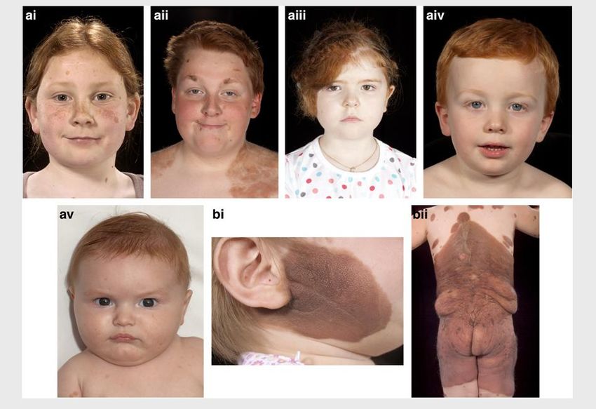
Figure 1: Clinical images of children with congenital melanocytic
naevi (CMN) (1)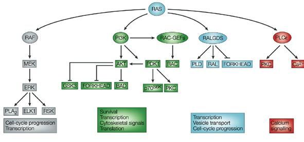
Miss Niamh Mcguire

                                              Introduction

Congenital Melanocytic Naevi (CMN) develop in utero and are therefore present at birth unlike
acquired melanocytic naevi (AMN) which develop post-natally. Small single CMN are common,
occurring in about 1 in 100 babies, but multiple or very large CMN are rare, affecting
approximately 1 in 20,000 infants. Hundreds of well-defined pigmentary birth defects can be
present in an individual with CMN, covering up to 80% of the body’s surface.

CMN is a known risk factor for neurodevelopmental abnormalities and developing melanoma in
postnatal life(2). Increasing size of the naevi has been found to be associated with increased risk
of both conditions(3).

CMN does not follow a Mendelian pattern of inheritance. Phenotypic abnormalities in this
condition are considered to be due to a causative somatic mutation (4, 5).

AMNs frequently harbor oncogenic mutations. BRAF (V-raf murine sarcoma virus oncogene
homolog B1) mutations are present in 59% of melanoma cell lines. It is located on chromosome
7, where the commonest mutation is glutamic acid substitution for valine at codon 600 (V600E).
NRAS (neuroblastoma ras viral oncogene homolog) mutations are present in approximately 15%
of cases with melanoma and the most frequent alterations are codon 61 mutations(6-8). This is a
RAS protein involved in the control of key cell signaling pathways. Transformation from
guanosine diphosphate bound to the active guanosine triphosphate bound state allows RAS to act
as a molecular switch, contributing to the activation of BRAF, one of three closely related Raf
proteins, as well as phosphatidylinositol 3-kinase. This thereby activates the RAF/MEK/ERK
pathway and Akt, respectively (see figure 2) (9, 10) .

                     Figure 2: Diagram to show targeting RAS signaling pathways (10)
Miss Niamh Mcguire
Previous studies have stated that BRAF and NRAS mutations found in melanocytic lesions are
mutually exclusive (8, 11). In recent unpublished data, however, a large cohort of 85 CMN patients
had affected skin samples tested for NRAS and BRAF mutations, using standard Sanger
sequencing and diagnostic laboratory high-resolution melt (HRM) techniques. These results
revealed surprisingly that approximately 70% of the sample cohort carried both mutations
(although one mutation often had a lower variant percentage).
Melanomas which harbor the BRAF V600E mutation are currently treated with BRAF inhibitors
such as Vemurafenib(12). However this mutation specific therapy is contraindicated in individuals
with NRAS mutations due to paradoxical activation of MAPK pathway(13). Therefore this
unpublished data is highly relevant for future treatment, at least for patients with large or
multiple CMN and further laboratory validation is required to identify potential changes that
could be made to the current treatment regimen if BRAF and NRAS mutations coexist in an
individual (14, 15).
Therefore, the aim of the study was to determine the concordance of the first and second BRAF results
and verify the presence of BRAF V600E mutations in confirmed NRAS mutant CMN samples.
Materials and methods
59 anonymised DNA samples extracted from CMN formalin-fixed, paraffin-embedded (FFPE)
tissue samples were genotyped using EntroGen’s B-Raf codon 600 mutation analysis kit, a real
time PCR-based assay that detects V600E in the codon 600 of B-Raf gene. The mutation analysis
was carried out in accordance with the Analysis kit protocol (see below). Alterations (shown in
Table 2) were made to reagent volumes as the protocol has been devised to perform an analysis
on 5 different mutations; V600E, V600K, V600R, V600D and V600M.
4 DNA samples had a concentration of 100ng/πl and were diluted to give a final DNA
concentrations of 20ng/πl.
Reagent Preparation
Table 1
Components Volume
2X PCR reaction mix 15µl
Primer mix 6µl
DNA sample 20ng
Molecular grade water Adjust to make total 30 µlMiss Niamh Mcguire
1. Master mix was prepared for each sample.
Table 2
Reagents Final Volume per sample Volume per sample
concentration for 5 mutations for 1 mutation
(w/10% overage)
2X Reaction Master 1X 82.5 µl 15µl
Mix
DNA, Positive control 20ng 5.5 µl 1µl
mix or Water
Nuclease free water 44 µl 8µl
2. Samples were vortexed and centrifuged for 10 seconds at 2,000rpm.
3. 24µl of master mix were dispensed in 61 wells. Sample well allocation recorded.
4. 6µl of V600E Mutation Detection Primer Mix added to each well.
5. Samples mixed by pipetting up and down several times.
6. The plate was sealed with optical sealing film (ABI plates).
7. The plate was centrifuged for 1,500 rpm for 1 minute.
Instrument setup
The assay was performed using an StepOne PlusTM Real-Time PCR system (Applied
Biosystems®) using StepOne Software.
1. File>New Experiment>Advanced Setup selected
2. Experiment name created and 61 wells selected.
3. Type of experiment: comparative Ct. Reagents: Taqman. Plate setup in left navigation
panel selected.
4. Two targets added: FAM(BRAF) and VIC(Control). NFQ-MGB selected for the
Quencher for both targets.
5. Sample names for the experiment inserted on the right side of the screen.
6. Both targets assigned to all sample wells including PC and NTC.
7. Passive Reference Dye: None
8. ‘Run method’ on left panel selected.
9. Sample volume: 30µl.
10. Cycling parameters set up as shown:Miss Niamh Mcguire
Table 3
Temperature Time Cycles Data Collection
95°C 10 min X1 Off
95°C 15sec X40 Off
95°C 30 sec Off
11. Prepared plate loaded and run started.
12. After one hour the analysis is complete
13. Input of data into SPSS database
Data analysis
1. Criteria used to analyse the data in the real-time PCR instrument software (Applied
Biosystems 7500 Step 1 Plus):
Manual threshold,
FAM (BRAF) threshold: 10,000,
VIC (Control) Threshold: 1,000,
automatic baseline
2. Positive control wells and VIC Ct selected. The threshold was set and the amplification
plots for each sample were checked to assess if each reaction had loaded properly.
The table below explains the interpretation of results using VIC threshold (reproduced from kit
manual).
Table 4
If VIC Ct is: Analysis description Action
Between 26 and 31 Ideal Ct range Continue with step 3
Below 26 Overloaded reaction Dilute DNA and re-test
Above 31 Insufficient DNA, Increase DNA input per
fragmentation, or presence reaction and re-test
of PCR inhibitors in the
DNA
Not present Reaction components or Do not continue with
DNA added incorrectly analysis this sample has
failedMiss Niamh Mcguire
3. Positive control wells and FAM Ct selected. The threshold was set and the amplification
plots for each sample were checked to assess the presence of signal. Higher amounts of
mutant variant produced a lower Ct value.
4. Ct values exported to a file using export function of Step One v.2.3 software. The table
below shows the analysis steps used for the FAM channel (BRAF)
Table 5
If FAM is: Mutation status
37 or lower Positive
Above 37 Re-test sample more DNA (5-10 times). If
Ct value has decreased by 1 cycle, the
sample is positive. If the Ct value stays the
same or is absent, the sample is negative
Absent NegativeMiss Niamh Mcguire
Results
Results were consistent with previous findings obtained from sequencing and high resolution
melt. 2 samples which have previously been identified as a low level variants and the experiment
confirmed them as BRAF mutations. 1 sample was reconfirmed as a BRAF mutant. 1 sample was
identified as a low level variant. BRAF Ct for this sample was 37.37 (above the threshold to be
considered to have a positive mutation status but may be consistent with the low level variant
status) and therefore requires re-testing.
24 out of the total 61 samples did not amplify. According to the protocol this may be due to one
of the reaction components or DNA not being added correctly. These samples should therefore
be re-tested.
Previous methods have displayed a higher number of positive samples. As several samples did
not amplify, 14 results were not obtained for previously identified BRAF mutants.
The table below displays a comparison between previous methods used (HRM and sequencing)
and this experiment:
Table 6
BRAF Mutation Positive Second BRAF Method Results Cross tabulation
Count
Second BRAF Method Results Total
wild type BRAF V600E failed to amplify
mutation
.00 21 0 8 29
BRAF Mutation Positive
1.00 12 4 13 29
Total 33 4 21 58Miss Niamh Mcguire
Conclusions and future work
From the results obtained, the co-existence of BRAF V600E and NRAS Q61 mutations in
congenital melanocytic naevi has been verified by a second diagnostic grade method, in all
samples in which the second test worked.
The results were consistent with previous results obtained using sequencing and High Resolution
Melt. 4 mutations identified as having the NRAS mutation also had the BRAF mutation. A higher
number of mutants may have been identified if all samples had amplified therefore BRAF
mutants that did not amplify could be re-tested in due course to identify other mutations and
validate co-existence further.
These results will need to be replicated in a larger or second cohort of CMN patients, and will
also be checked in a cohort of AMN samples. Functional work will need to be undertaken to
check whether both mutations could be within the same cell, or whether this result implies two
populations of cells within the same naevus.Miss Niamh Mcguire
References
1. Kinsler VA, Abu-Amero S, Budd P, Jackson IJ, Ring SM, Northstone K, et al. Germline
Melanocortin-1-Receptor Genotype Is Associated with Severity of Cutaneous Phenotype in Congenital
Melanocytic Nevi: A Role for MC1R in Human Fetal Development. J Invest Dermatol. 2012;132(8):2026-
32.
2. Kinsler V, Shaw AC, Merks JH, Hennekam RC. The face in congenital melanocytic nevus
syndrome. Am J Med Genet A. 2012 May;158A(5):1014-9.
3. Bittencourt FV, Marghoob AA, Kopf AW, Koenig KL, Bart RS. Large Congenital Melanocytic Nevi
and the Risk for Development of Malignant Melanoma and Neurocutaneous Melanocytosis. Pediatrics.
2000 October 1, 2000;106(4):736-41.
4. Kinsler VA, Thomas AC, Ishida M, Bulstrode NW, Loughlin S, Hing S, et al. Multiple Congenital
Melanocytic Nevi and Neurocutaneous Melanosis Are Caused by Postzygotic Mutations in Codon 61 of
NRAS. J Invest Dermatol. [Commentary]. 2013;133(9):2229-36.
5. Charbel C, Fontaine RH, Malouf GG, Picard A, Kadlub N, El-Murr N, et al. NRAS Mutation Is the
Sole Recurrent Somatic Mutation in Large Congenital Melanocytic Nevi. J Invest Dermatol.
[Commentary]. 2014;134(4):1067-74.
6. Pollock PM, Meltzer PS. A genome-based strategy uncovers frequent BRAF mutations in
melanoma. Cancer Cell. 2002;2(1):5-7.
7. Wu D, Wang M, Wang X, Yin N, Song T, Li H, et al. Lack of BRAFV600E Mutations in Giant
Congenital Melanocytic Nevi in a Chinese Population. The American Journal of Dermatopathology.
2011;33(4):341-4 10.1097/DAD.0b013e3181fb5bc7.
8. Sensi M, Nicolini G, Petti C, Bersani I, Lozupone F, Molla A, et al. Mutually exclusive NRASQ61R
and BRAFV600E mutations at the single-cell level in the same human melanoma. Oncogene.
2006;25(24):3357-64.
9. Saxena N, Lahiri SS, Hambarde S, Tripathi RP. RAS: Target for Cancer Therapy. Cancer
Investigation. 2008;26(9):948-55.
10. Downward J. Targeting RAS signalling pathways in cancer therapy. Nat Rev Cancer.
[10.1038/nrc969]. 2003;3(1):11-22.
11. Colombino M, Capone M, Lissia A, Cossu A, Rubino C, De Giorgi V, et al. BRAF/NRAS Mutation
Frequencies Among Primary Tumors and Metastases in Patients With Melanoma. Journal of Clinical
Oncology. 2012 July 10, 2012;30(20):2522-9.
12. Chapman PB, Hauschild A, Robert C, Haanen JB, Ascierto P, Larkin J, et al. Improved Survival with
Vemurafenib in Melanoma with BRAF V600E Mutation. New England Journal of Medicine.
2011;364(26):2507-16.
13. Wan PTC, Garnett MJ, Roe SM, Lee S, Niculescu-Duvaz D, Good VM, et al. Mechanism of
Activation of the RAF-ERK Signaling Pathway by Oncogenic Mutations of B-RAF. Cell.116(6):855-67.
14. Dumaz N, Hayward R, Martin J, Ogilvie L, Hedley D, Curtin JA, et al. In Melanoma, RAS Mutations
Are Accompanied by Switching Signaling from BRAF to CRAF and Disrupted Cyclic AMP Signaling. Cancer
Research. 2006 October 1, 2006;66(19):9483-91.
15. Kelleher FC, McArthur GA. Targeting NRAS in Melanoma. The Cancer Journal. 2012;18(2):132-6
10.1097/PPO.0b013e31824ba4df.You can also read


