Virtual Course on an Active Surveillance Design Using a 12-point Checklist for Diseases of Aquatic Species - WEEK 1: FAO
←
→
Page content transcription
If your browser does not render page correctly, please read the page content below
TCP/EGY/3705: Enhancing biosecurity governance to support sustainable aquaculture production in Egypt

Virtual Course on an Active Surveillance
 Design Using a 12-point Checklist for
      Diseases of Aquatic Species
                                         WEEK 1:
                               31 August - 2 September 2021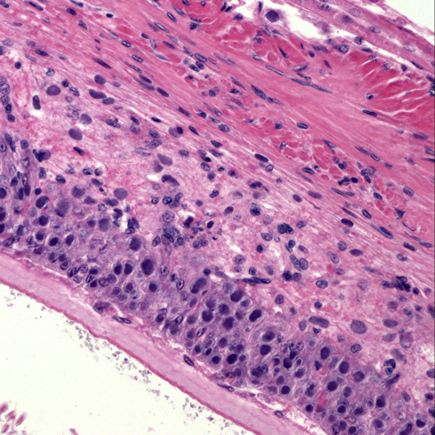
2 September 2021

                 CHECKLIST 6
Diagnostic testing-level I and II:
      Shrimp pathogens
                          Presented by

                     Kathy Tang-Nelson
                  ktangnelson@gmail.com

    TCP/EGY/3705: Enhancing biosecurity governance to support
           sustainable aquaculture production in Egypt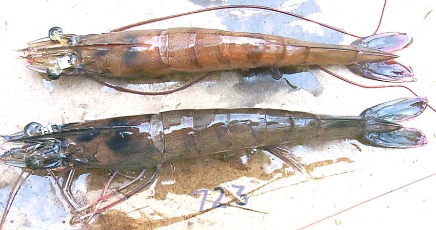
Acute hepatopancreatic necrosis disease (AHPND)
• AHPND is a Vibrio bacterial disease that has caused mass mortalities in farmed
  populations of Penaeus vannamei and P. monodon.
• Mortality from AHPND occurs early, usually within 30-35 days, after stocking
  postlarvae in the ponds. This characteristic led to the disease being initially referred
  as early mortality syndrome (EMS).
• Clinical signs include a pale-to-white atrophied HP, empty stomach and midgut, soft
  carapace.
• In the shrimp, they colonized on the cuticle lining of the stomach, secrete the
  PirABvp toxin, and pass directly into the hepatopancreas (HP), resulting in
  detachment of tubule epithelial cells, and eventually the destruction of HP.
• The histology can be classified by early acute, acute, and terminal phases.
• Tang, K.F.J., Bondad-Reantaso, M.G., Arthur, J.R., MacKinnon, B., Hao, B., Alday-Sanz, V.,
  Liang, Y. & Dong, X. 2020. Shrimp acute hepatopancreatic necrosis disease strategy manual.
  FAO Fisheries and Aquaculture Circular No. 1190. Rome, FAO.

                        TCP/EGY/3705: Enhancing biosecurity governance to support sustainable aquaculture production in Egypt   2 September 2021   3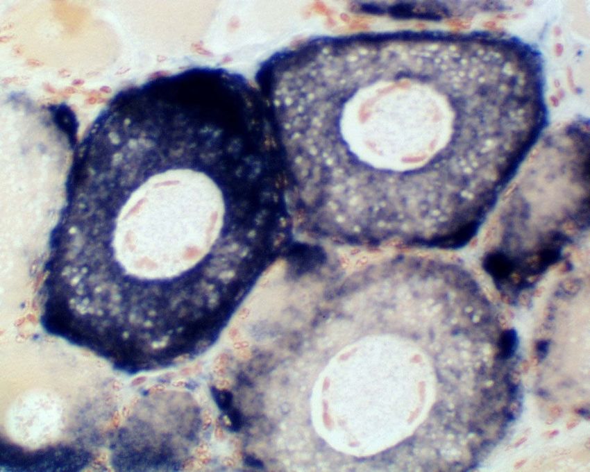
Acute hepatopancreatic necrosis disease (AHPND)

                                         AHPND         Healthy

                                                     Full stomach
                                    Empty stomach
                                     Atrophied HP
                                                     Brownish HP

                                      Empty gut         Full gut

   The occurrence of a mass          Clinical signs of AHPND-
   mortality due to AHPND in         affected P. vannamei versus
   P. vannamei cultured in China.    healthy shrimp.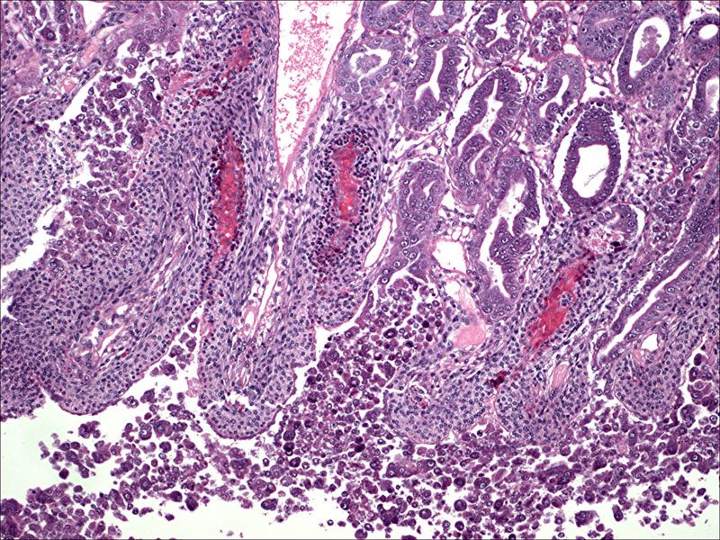
AHPND-early acute phase
                                                   HP                                                                           HP

H&E histology of the HP from an AHPND affected P. vannamei. (A) Shrimp was at early acute phase of
PirABvp toxicosis, arrows indicate the HP epithelial cells starting degenerate, rounding up and slough
into lumen; (B) arrows indicate the appearance of enlarged nuclei (karyomegaly); the inflammatory
response is not evident at this phase
                   TCP/EGY/3705: Enhancing biosecurity governance to support sustainable aquaculture production in Egypt 2 September 2021   5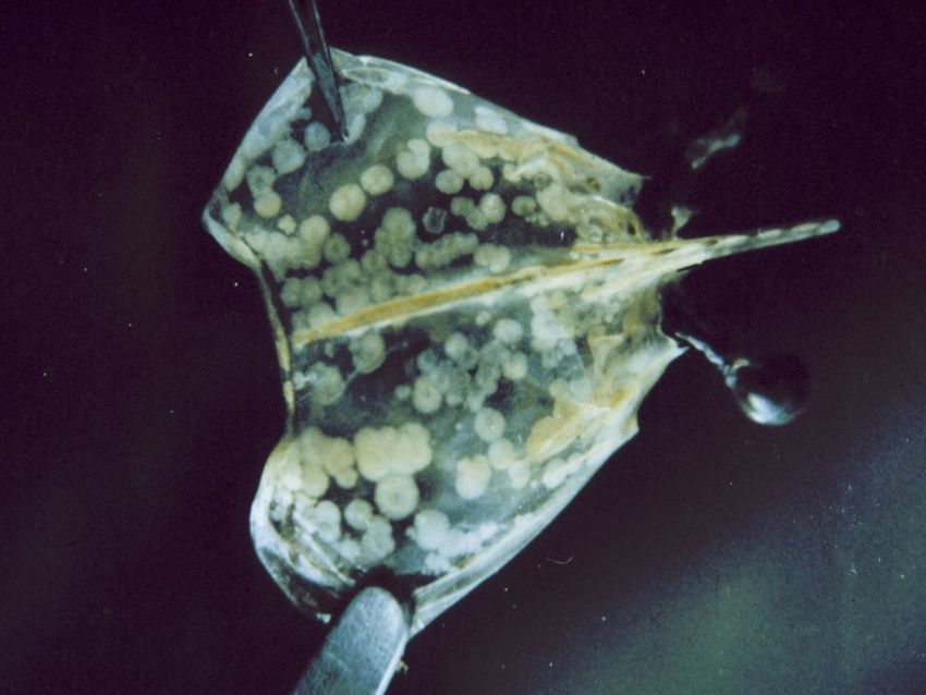
AHPND-acute phase
A                        B                         C                  distal

                                                       proximal

AHPND affected P. vannamei, acute phase, sloughing of tubular epithelial
cells (red arrows); the pathology is characterized by a progressive
degeneration of the HP tubules from proximal to distal; Arrows indicate
significant rounding and massive sloughing of HP epithelial cells.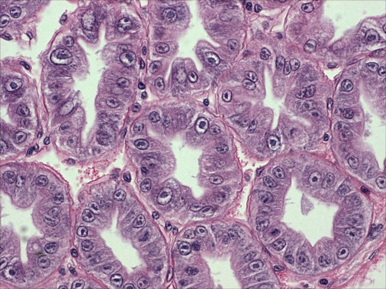
AHPND-terminal phase
A                                  B

AHPND affected Penaeus vannamei; terminal phase. (A) Tubule epithelium
sloughing (black arrows), significant proximal hemocytic inflammation,
most tubules are destroyed, some tubules with putative vibriosis; (B)
showed extensive haemocyte infiltration (red arrows), massive bacterial
colonization in tubule lumens (yellow stars).
White spot disease (WSD)
• White spot syndrome virus (WSSV) has caused high mortality in
  populations of many species of penaeid shrimp and other aquatic
  crustaceans.
• In many penaeids, the clinical signs of WSD are white spots inside the
  carapace and a reddish discoloration of entire body
• Infected shrimp suffer severe mortalities, at times reaching 100%,
  within 3–10 days of the onset of clinical signs.
• WSSV infection is characterized histologically by the presence of
  eosinophilic to pale basophilic intranuclear inclusion bodies in: the
  cuticular epithelial cells, connective tissue cells, and, less frequently, in
  the antennal gland, lymphoid organ sheath cells, hematopoietic tissues,
  and fixed phagocytes of the heart.

                      TCP/EGY/3705: Enhancing biosecurity governance to support sustainable aquaculture production in Egypt   2 September 2021   8
WSSV

A juvenile Penaeus monodon that is displaying the distinctive white
spots of WSD. White spots are especially visible on the carapace and
the rostrum.

         TCP/EGY/3705: Enhancing biosecurity governance to support sustainable aquaculture production in Egypt 2 September 2021   9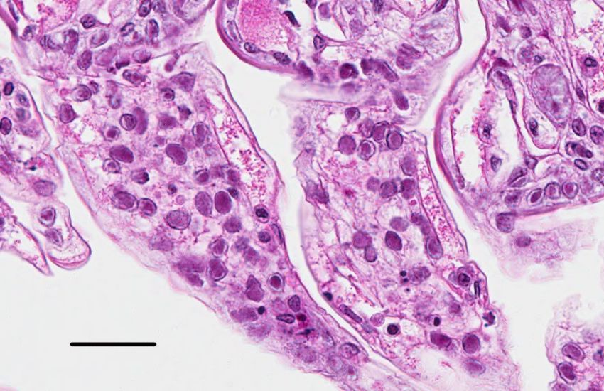
WSSV

                                                WSSV-infected P. vannamei. The
 WSSV-infected P. monodon. The top and
                                                infected shrimp display colour
middle shrimp show a pink to red
                                                variation, with a predominance of
discolouration due to expansion of the
                                                darkened (red-brown or pink-red)
chromatophores. This reddish appearance
                                                body surface and appendages.
may be a gross sign that is more apparent in
the acute phase of the disease. The shrimp on
bottom exhibited white spots that develop
after the acute phase of the disease. Photo
courtesy of Marc LeGroumellec, Madagascar.
WSSV Gill
H&E histology of gills of a WSSV-infected P. vannamei. Arrows indicate
intranuclear inclusion bodies. Red arrowheads indicate the early phase
inclusion bodies (Cowdry-A type) that are centronuclear, eosinophilic, and
separated from the nuclear membrane and marginated chromatin by an
artifactual halo. Scale bar = 30 μm.
TCP/EGY/3705: Enhancing biosecurity governance to support sustainable aquaculture production in Egypt 2 September 2021 11WSSV
Ce
p
sC
n
H&E histology of a WSSV-infected P. indicus. Tissue sections of cuticular
epithelium (Cep) and subcuticular connective (sCn) tissue in a shrimp with
typical WSSV histology. Arrows indicate examples of dark basophilic
intranuclear inclusions.WSSV LO
H&E histology of lymphoid organ (LO) in a WSSV-infected P. indicus.
Arrows indicate examples of WSSV inclusions
TCP/EGY/3705: Enhancing biosecurity governance to support sustainable aquaculture production in Egypt 2 September 2021 13WSSV
H&E wet mount of cuticular epithelium in a WSSV-infected P.
vannamei. Arrows: representative eosinophilic to basophilic WSSV
inclusions.
TCP/EGY/3705: Enhancing biosecurity governance to support sustainable aquaculture production in Egypt 2 September 2021 14Infection with Infectious hypodermal and hematopoietic necrosis
virus (IHHNV)
• Infection with IHHNV had resulted in high mortalities (up to 90%) in infected
populations of Penaeus stylirostris.
• IHHNV infection does not typically cause mortality in stocks of P. vannamei or P.
monodon, it results in a disease called runt-deformity syndrome (RDS).
• IHHNV infection is characterized histologically by the presence of eosinophilic,
intranuclear Cowdry-A type inclusions in tissues of ectodermal and mesodermal
origins, including gills, cuticular epithelium, connective tissues, haematopoietic
tissue, reproductive tissues, antennal gland, and the ventral nerve cord and
associated ganglia.
• As IHHNV infects and replicates in reproductive tissues such as those of the vas
deferens, testes, and ovary, including in oocytes, the findings support the vertical
transmission.
TCP/EGY/3705: Enhancing biosecurity governance to support sustainable aquaculture production in Egypt 2 September 2021 15IHHNV
IHHNV-infected P. vannamei (preserved in Davidson’s fixative). (A) shrimp
gross signs of RDS, bent (to the left or to the right), deformed, rostrums
(arrows) are illustrated; (B) arrows showed cuticular abnormalities in the
sixth abdominal and tail fans.
TCP/EGY/3705: Enhancing biosecurity governance to support sustainable aquaculture production in Egypt 2 September 2021 16IHHNV
Samples of juvenile P. vannamei from a nursery pond population with severe
RDS. A wide size range with numerous "runts" mixed in with shrimp of the
expected size are typical signs of IHHNV infection in juveniles.
TCP/EGY/3705: Enhancing biosecurity governance to support sustainable aquaculture production in Egypt 2 September 2021 17IHHNV Gill
H&E histology of gill in an IHHNV-infected P. stylirostris. Arrows indicate examples of eosinophilic,
intranuclear Cowdry-A type inclusions. The inclusion consists of a large dense center, surrounded by
a clear halo, with marginated chromatin against the nuclear membrane.
TCP/EGY/3705: Enhancing biosecurity governance to support sustainable aquaculture production in Egypt 2 September 2021 18IHHNV
A Heo B Vnc
H&E histology of (A) hemotopoietic organ (Heo) in an IHHNV-infected P.
stylirostris. Arrows indicate examples of Cowdry-A type inclusions; (B) ventral
nerve cord (Vnc).
TCP/EGY/3705: Enhancing biosecurity governance to support sustainable aquaculture production in Egypt 2 September 2021 19IHHNV Testes
H&E histology of testes in an IHHNV-infected P. vannamei. Arrow indicates an
example of Cowdry-A type inclusion. Stars indicate the mitotic figures in the
dividing spermatogonia.
TCP/EGY/3705: Enhancing biosecurity governance to support sustainable aquaculture production in Egypt 2 September 2021 20IHHNV
Ooc
H&E histology (left micrograph) of the ovary in an IHHNV-infected P.
vannamei. Arrows indicate the presence of IHHNV in the maturing
oocytes (Ooc) evidenced by the in situ hybridization positive reactions
(right micrograph) in the oocytes.
TCP/EGY/3705: Enhancing biosecurity governance to support sustainable aquaculture production in Egypt 2 September 2021 21Infection with Enterocytozoon hepatopenaei (EHP)
• The microsporidium Enterocytozoon hepatopenaei (EHP) can infect
penaeid shrimp, including Penaeus monodon, P. stylirostris, P.
vannamei.
• Impacting aquaculture production by severely retarding the growth
of cultured shrimp.
• EHP is an intracellular spore-forming parasite that replicates in the
cytoplasm of epithelial cells of hepatopancreas (HP) tubules and
midgut.
• histological examination of the HP and midgut from the infected
shrimp shows the presence of basophilic inclusions at several
developmental stages of the microsporidian, including the early
sporogonal plasmodia and mature spores.
• The plasmodia are multinucleate; the mature spores are oval shaped,
measuring 0.7–1.1 μm
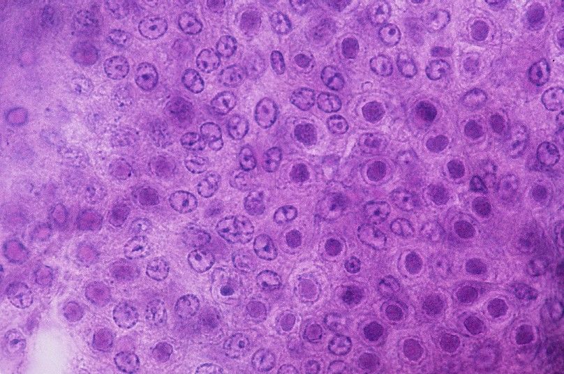
TCP/EGY/3705: Enhancing biosecurity governance to support sustainable aquaculture production in Egypt 2 September 2021 22EHP H&E histology of hepatopancreas (HP) from an EHP-infected P. vannamei. Long arrows indicate mature spores, short arrowheads indicate the plasmodia stage of inclusions.
EHP

      H&E histology of midgut epithelial cells in an EHP-infected P. vannamei,
      arrows indicate EHP inclusions.

            TCP/EGY/3705: Enhancing biosecurity governance to support sustainable aquaculture production in Egypt 2 September 2021   24
EHP
A wet-mount prepared from the HP of an EHP-infected P.
vannamei. Arrows indicate aggregates of spores held
together by remnants of the nuclear membrane are seen.
No stain. Magnification = 1,000X. Photo courtesy of Dr.
Heny Budi Utari, Indonesia.
TCP/EGY/3705: Enhancing biosecurity governance to support sustainable aquaculture production in Egypt 2 September 2021 25EHP
A wet-mount prepared from the HP of an EHP-infected P.
vannamei. Arrows indicate the clusters of spores. The tissue
was fixed in ethanol-formalin, stained with methyl blue.
Magnification = 1,000X. Photo courtesy of Dr. Heny Budi Utari,
Indonesia.
TCP/EGY/3705: Enhancing biosecurity governance to support sustainable aquaculture production in Egypt 2 September 2021 26TCP/EGY/3705: Enhancing biosecurity governance to support sustainable aquaculture production in Egypt
Thank you for
your attention!
Presented by
Kathy Tang-Nelson
ktangnelson@gmail.comYou can also read