White matter microstructure alterations in cortico-striatal networks are associated with parkinsonism in schizophrenia spectrum disorders
←
→
Page content transcription
If your browser does not render page correctly, please read the page content below

European Neuropsychopharmacology 50 (2021) 64–74

                                                 www.elsevier.com/locate/euroneuro

White matter microstructure alterations in
cortico-striatal networks are associated
with parkinsonism in schizophrenia
spectrum disorders
Jakob Wasserthal a,b, Klaus H. Maier-Hein a,c, Peter F. Neher a,b,
Robert C. Wolf d, Georg Northoff e, John L. Waddington f,
Katharina M. Kubera d, Stefan Fritze g, Anais Harneit h,
Lena S. Geiger h, Heike Tost h, Dusan Hirjak g,∗

a
  Division of Medical Imaging Computing (MIC), German Cancer Research Center (DKFZ), Heidelberg,
Germany
b
  Medical Faculty Heidelberg, Heidelberg University, Heidelberg, Germany
c
  Section of Automated Image Analysis, Heidelberg University Hospital, Heidelberg, Germany
d
  Center for Psychosocial Medicine, Department of General Psychiatry, University of Heidelberg,Germany
e
  Mind, Brain Imaging and Neuroethics Research Unit, The Royal’s Institute of Mental Health Research,
University of Ottawa, Ottawa, ON, Canada
f
  School of Pharmacy and Biomolecular Sciences, Royal College of Surgeons in Ireland, Dublin, Ireland
g
  Department of Psychiatry and Psychotherapy, Central Institute of Mental Health, Medical Faculty
Mannheim, Heidelberg University, Mannheim, Germany
h
  Department of Psychiatry and Psychotherapy, Research Group System Neuroscience in Psychiatry,
Central Institute of Mental Health, Medical Faculty Mannheim, Heidelberg University, Mannheim,
Germany

Received 17 September 2020; received in revised form 15 March 2021; accepted 10 April 2021

    KEYWORDS                          Abstract
    White matter;                     The specific role of white matter (WM) microstructure in parkinsonism among patients with
    DTI;                              schizophrenia spectrum disorders (SSD) is largely unknown. To determine whether topographi-
    Parkinsonism;                     cal alterations of WM microstructure contribute to parkinsonism in SSD patients, we examined
                                      healthy controls (HC, n=16) and SSD patients with and without parkinsonism, as defined by

    ∗ Correspondingauthor.
     E-mail address: dusan.hirjak@zi-mannheim.de (D. Hirjak).

https://doi.org/10.1016/j.euroneuro.2021.04.007
0924-977X/© 2021 Elsevier B.V. and ECNP. All rights reserved.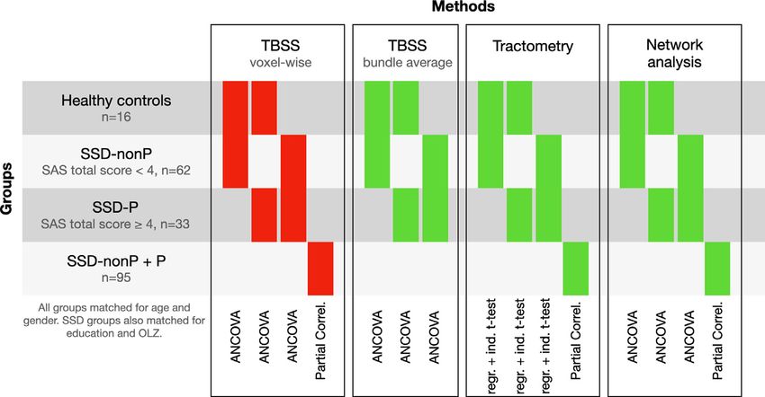
European Neuropsychopharmacology 50 (2021) 64–74

                                   Simpson–Angus Scale total score of ≥4 (SSD-P, n=33) or
J. Wasserthal, K.H. Maier-Hein, P.F. Neher et al.

evaluation of psychopathology, motor assessment and MRI exami-                analysis of the aggregated FA is identical to analysis of the large-
nation was less than 3 days. At the time of examination none of               scale network analysis.
the SSD patients were taking benzodiazepines or anticholinergic                  Tractometry: Along-tract statistics of FA (Tractometry) were
medication and all patients were on stable antipsychotic medica-              computed using TractSeg (Wasserthal et al., 2018, 2019). Tract-
tion for at least two weeks (for details on antipsychotic medication          Seg generates bundle-specific tractograms and then analyzes FA
see Supplementary Information). Daily doses of antipsychotic med-             along 100 points of each bundle (further details are provided in
ication were converted to olanzapine equivalents (OLZ) according              Supplementary Information). Statistics such as T-tests and cor-
to the classical mean dose method (Leucht et al., 2015). For as-              relations can be calculated point-wise along these 100 points.
sessment of parkinsonism we used the Simpson–Angus Scale (SAS)                The permutation-based multiple comparison correction (with
(Simpson and Angus, 1970); for details on SAS domains see Supple-             n=5000 repetitions) published by Nichols and Holmes (Nichols and
mentary Information).                                                         Holmes, 2002; Yeatman et al., 2012) was used to appropriately cor-
   We then excluded 16 SSD patients from the original study sample            rect p values for the 100 tests, given the correlation structure of
(111−16=95) to create two well-balanced (in terms of age, sex,                the data. An uncorrected significance threshold of p < 0.05 was
education and OLZ-equivalent dose) groups of SSD patients with                used, with the corrected p-value being different for each bundle
parkinsonism (SSD-P; SAS total score ≥ 4, n=33) and without parkin-           depending on its correlation structure. Covariates were controlled
sonism (SSD-nonP; SAS
European Neuropsychopharmacology 50 (2021) 64–74

Table 1 Demographics and clinical scores for schizophrenia spectrum disorders patients (n=95) divided into SSD-nonP (SAS total
score
J. Wasserthal, K.H. Maier-Hein, P.F. Neher et al.

Fig. 1 Overview of the different analyses in this study. Colored bars indicate which groups are compared by the respective test. The
color shows if the test gave significant results (p0.05). Using              significant FA difference in CC_7 and right and left ST_FO
partial correlation, we found no significant association be-            between SSD-P or SSD-nonP patients and HC. After ex-
tween SAS score and average FA for CC_7 or left and right              cluding all three covariates (age, sex, and OLZ) and per-
ST_FO in the entire patient sample (p>0.1). After excluding            forming an independent two-sample t-test, we found sig-
all four covariates (age, sex, OLZ and PANSS-N), there was             nificantly lower FA in CC_6 (p=0.041), CC_7 (p=0.009),
still no significant association between SAS scores and FA in           left ST_FO (p=0.005), and right ST_FO (p=0.021) in SSD-
the entire patient sample (p>0.08).                                    P patients when compared to HC. There were no signifi-
   Tractometry: We found significant negative associa-                  cant differences in FA between SSD-nonP patients and HC
tions between SAS total scores and FA in CC_5 (min. p-                 (all p-values>0.066). Thus, these p-values did not survive
value
European Neuropsychopharmacology 50 (2021) 64–74

Fig. 2 Significant differences in FA between SSD patients with (P) and without (nonP) parkinsonism (Tractometry analysis; results
before Bonferroni correction).

old at p=0.002/30=0.00006), CC_7 (threshold at                     feel that it is valuable to discuss them. The specific purpose
p=0.001/30=0.00003), left T_PREM (threshold at                     of this study was to better understand WM microstructure
p=0.002/30=0.00006) and left ST_FO (threshold at                   underlying parkinsonism. Since there are no previous stud-
p=0.003/30=0.0001) survived Bonferroni correction for              ies that have used DTI to address this specific question, the
multiple testing. Finally, only the minimal p-value of the         consequences of a false negative, when using conservative
SSD-nonP vs. HC comparison in left T_PREM (threshold               correction thresholds for multiple comparisons, would be
at p=0.001/30=0.00003) and right ST_FO (threshold at               to miss regional WM variations of heuristic importance in
p=0.003/30=0.0001) survived Bonferroni correction for              relation to parkinsonism. Therefore, as false negatives are
multiple testing.                                                  costly under these conditions, reporting and discussing un-
   Large-scale network analysis: ANCOVA revealed no signif-        corrected results can be valuable in such instances.
icant differences between patient groups and HC (p>0.15)
(Supplementary Table 2). After excluding all three covari-
ates (age, sex, and OLZ) and performing an independent             4.1.   TBSS and tractography
two-sample t-test, we found a significant increase in CCO in
the right SFG in SSD-P patients compared to HC (p=0.038).          Using an aggregated TBSS (similar to a recent study
There was no significant difference in CCO or BC in other           of WM microstructural differences in schizophrenia (SZ)
brain regions between SSD-nonP patients and HC (all p-             Kelly et al., 2018), we found reduced FA in the left CST and
values>0.058).                                                     the ST_FO in SSD-P patients compared to SSD-nonP patients.
                                                                   The FA decrease in CST may relate to decreased input from
                                                                   the thalamus and striatum or altered pallido-thalamic activ-
4.   Discussion                                                    ity (Mole et al., 2016). Since CST carries WM motor tracts to
                                                                   peripheral muscular output (Du et al., 2017), distortion of
The present multiparametric dMRI study investigated, for           structural integrity in CST might lead to aberrant inhibitory
the first time, brain WM microstructure abnormalities un-           signaling and sensorimotor abnormalities. The ST_FO is a
derlying parkinsonism in SSD patients. Three main findings          neural pathway that connects frontal lobe regions with the
emerged: First, we found reduced FA in left CST and ST_FO          basal ganglia (striatum). Impaired fronto-striatal connec-
in SSD-P patients compared to SSD-nonP patients. Second,           tivity in SZ is well documented (de Leeuw et al., 2017;
there were significant positive associations between FA in          Levitt et al., 2017; Quan et al., 2013) and can lead to cog-
CC_5, CST and ST_FO and SAS total scores. Third, there was         nitive impairment (Levitt et al., 2017) as well as sensori-
a significant association between CCO in left OFC and SAS           motor deficits such as parkinsonism (Molina et al., 2018;
total score. Although these findings did not survive conser-        Wolf et al., 2020a). Interestingly, ST_FO reciprocally con-
vative Bonferroni correction for multiple comparisons, we          nects important regions involved in sensorimotor function-

                                                              69
J. Wasserthal, K.H. Maier-Hein, P.F. Neher et al.
ing such as the dorsolateral prefrontal cortex (DLPFC), OFC, from a clinical perspective, there is some overlap between
inferior frontal gyrus, SFG and the striatum (caudate nu- parkinsonism and catatonia in terms of rigidity, immobility
cleus, and putamen) (de Leeuw et al., 2017). More pre- or inhibited movement, psychomotor slowing, mutism, and
cisely, the OFC modulates cognitive control of emotional staring. From a neurobiological perspective, the findings of
experience and is in constant interaction with DLPFC, infe- the present study are in line with two recent MRI studies on
rior frontal gyrus, SFG and striatum (Northoff et al., 2004). catatonia in SSD (Viher et al., 2020; Wasserthal et al., 2020)
The human striatum is mainly responsible for the correct that were able to demonstrate similar WM changes involv-
excitation of goal-directed movement schemas. Recently, ing left-lateralized higher FA in the CST, CC, internal cap-
an MRI study found that iron-loading measures of mainly sule and thalamo-premotor tract. These findings point to-
left-side basal ganglia were associated with extrapyrami- wards some common pathobiology underlying catatonia and
dal motor symptoms and neurological soft signs (NSS) in parkinsonism in SSD.
first-episode psychosis patients (Cuesta et al., 2020). In Taken together, the present FA decrease and CCO in-
accordance with previous findings, axon loss within the crease in different sensorimotor regions suggest a reorgani-
ST_FO reflects aberrant “vertical modulation” of cortico- zation of WM fibers both as selective neurodegeneration and
subcortical relations, which might lead to disturbed reg- a resultant compensatory response in the pathogenesis of
ulation of emotional stimuli and programming and termi- parkinsonism in SSD (Atkinson-Clement et al., 2017). Future
nation of action (Hirjak et al., 2015b; Northoff, 2000), studies should further investigate which of these mecha-
as well as an imbalance between inhibitory and exci- nisms predominates and can be modulated by antipsychotic
tatory processes (Helmich et al., 2012; van der Stouwe drugs.
et al., 2020). Taken together, these conjoint alterations
in bottom-up and top-down modulation might lead to the
development of typical sensorimotor abnormalities such as 4.3. Strengths and limitations
tremor and akinesia, which are characteristic symptoms of
parkinsonism. This study has the following strengths: (i) patient sample
size, (ii) well-matched study groups, and (iii) the use of a
comprehensive set of sophisticated WM microstructural pa-
4.2. Large-scale network analysis rameters. However, the following methodological aspects
limit the generalizability of our results: (i) Antipsychotic
Using graph analytics, we identified significantly higher CCO medication. Although antipsychotic drugs may have promi-
values in the left OFC and left SFG in SSD-P patients com- nent functional effects on the identified WM tracts, our find-
pared to SSD-nonP patients. These findings are relevant for ings do not appear to be confounded by such effects be-
a number of reasons: First, the higher CCO of the OFC cause the two SSD groups were well-matched for dose in
might be interpreted as a compensation mechanism for OLZ-equivalents but showed different levels of parkinson-
disturbed network functionality in the fronto-striatal net- ism. This issue has also been addressed in a recent DTI study
work as represented by reduced FA in both striato-fronto- by Kraguljac and colleagues (Kraguljac et al., 2019), which
orbital tracts (SSDEuropean Neuropsychopharmacology 50 (2021) 64–74
using the SAS as a composite score has proven to better sep- Declaration of Competing Interest
arate specific patient groups (placebo vs. 1 mg haloperidol)
than the six hypokinetic/rigidity items alone. (v) SSD pa- The authors have declared that there are no conflicts of in-
tients in this study may also have had other sensorimotor ab- terest in relation to the subject of this study.
normalities such as NSS or catatonia. However, though this
study concerned both categorical and dimensional assess-
ment of parkinsonism, there can be considerable clinical Acknowledgments
overlap with such other sensorimotor abnormalities. There-
fore, we advocate that sensorimotor assessments combine We are grateful to all the participants and their families for
both clinical ratings scales and instrumental assessments their time and interest in this study.
(Hirjak et al., 2021). This would help to clarify the patho-
physiology of parkinsonism and other sensorimotor abnor- Supplementary material
malities from a broader perspective.
Supplementary material associated with this article can be
found, in the online version, at doi:10.1016/j.euroneuro.
5. Conclusion 2021.04.007.
This study provides a comprehensive, state-of-the-art anal- References
ysis of WM microstructure in SDD patients with and without
parkinsonism that appears to reflect an interaction between Andersson, J.L.R., Sotiropoulos, S.N., 2016. An integrated approach
sensorimotor dysfunction intrinsic to SSD and the effects to correction for off-resonance effects and subject movement in
of antipsychotic drugs; thus, just as movement disorder in- diffusion MR imaging. NeuroImage 125, 1063–1078.
Atkinson-Clement, C., Pinto, S., Eusebio, A., Coulon, O., 2017. Dif-
trinsic to PD can be exacerbated by antipsychotics, parkin-
fusion tensor imaging in Parkinson’s disease: review and meta–
sonism appears to be intrinsic to SSD in a manner that can
analysis. Neuroimage Clin. 16, 98–110.
be exacerbated by these same agents (Waddington, 2020). Bach, M., Laun, F.B., Leemans, A., Tax, C.M.W., Biessels, G.J.,
In this regard, the main finding in the present study is Stieltjes, B., Maier-Hein, K.H., 2014. Methodological consider-
that dysfunction in the topological metrics of small-world ations on tract-based spatial statistics (TBSS). NeuroImage 100,
brain networks in SSD comprise sensorimotor regions such 358–369.
as OFC, SFG and striatum that operate in a dimensional and Boos, H.B., Mandl, R.C., van Haren, N.E., Cahn, W., van Baal, G.C.,
cross-nosological manner. This is indicated by their extents Kahn, R.S., Hulshoff Pol, H.E., 2013. Tract-based diffusion ten-
of expression being related across the triadic psychomo- sor imaging in patients with schizophrenia and their non-psy-
tor abnormalities of parkinsonism, i.e. rigidity, akinesia and chotic siblings. Eur. Neuropsychopharmacol.: J. Eur. Coll. Neu-
ropsychopharmacol. 23, 295–304.
tremor.
Clark, K., Narr, K.L., O’Neill, J., Levitt, J., Siddarth, P., Phillips, O.,
Toga, A., Caplan, R., 2012. White matter integrity, lan-
guage, and childhood onset schizophrenia. Schizophr. Res. 138,
6. Contributors 150–156.
Cuesta, M.J., Lecumberri, P., Moreno-Izco, L., Lopez-Ilundain, J.M.,
DH and RCW designed the study and wrote the protocol. Ribeiro, M., Cabada, T., Lorente-Omenaca, R., de Erausquin, G.,
Garcia-Marti, G., Sanjuan, J., Sanchez-Torres, A.M., Gomez, M.,
DH, SF and LSG performed the motor assessments in all
Peralta, V., 2020. Motor abnormalities and basal ganglia in
study subjects. KMK and AH managed the literature searches
first-episode psychosis (FEP). Psychol. Med. 1–12.
and analyses. JW, KMH, PH and DH undertook the statis- Cuesta, M.J., Sanchez-Torres, A.M., de Jalon, E.G., Campos, M.S.,
tical analysis. DH, RCW, GN, JLW and HT discussed and Ibanez, B., Moreno-Izco, L., Peralta, V., 2014. Spontaneous
interpreted the results. DH wrote the first draft of the parkinsonism is associated with cognitive impairment in antipsy-
manuscript. All authors contributed to and have approved chotic-naive patients with first-episode psychosis: a 6-month
the final manuscript. follow-up study. Schizophr. Bull. 40, 1164–1173.
Dale, A.M., Fischl, B., Sereno, M.I., 1999. Cortical surface-based
analysis. I. Segmentation and surface reconstruction. NeuroIm-
age 9, 179–194.
Role of funding source Dazzan, P., Morgan, K.D., Orr, K.G., Hutchinson, G., Chitnis, X.,
Suckling, J., Fearon, P., Salvo, J., McGuire, P.K., Mallett, R.M.,
This work was supported by the German Research Foun- Jones, P.B., Leff, J., Murray, R.M., 2004. The structural brain
dation (DFG) (grant number DFG HI 1928/2-1 to D.H., WO correlates of neurological soft signs in AESOP first-episode psy-
1883/6-1 to R.C.W., MA 6340/10-1 and MA 6340/12-1 to choses study. Brain 127, 143–153.
K.H.M.-H.) and German Federal Ministry of Education and de Leeuw, M., Bohlken, M.M., Mandl, R.C., Hillegers, M.H.,
Research (BMBF, grant 01GQ1102 to H.T.). The DFG and Kahn, R.S., Vink, M., 2017. Changes in white matter organiza-
tion in adolescent offspring of schizophrenia patients. Neuropsy-
BMBF had no further role in study design; in the collec-
chopharmacology 42, 495–501.
tion, analysis and interpretation of data; in the writing of
Desikan, R.S., Segonne, F., Fischl, B., Quinn, B.T., Dickerson, B.C.,
the report; and in the decision to submit the paper for pub- Blacker, D., Buckner, R.L., Dale, A.M., Maguire, R.P., Hy-
lication. GN is grateful for financial support from PSI and man, B.T., Albert, M.S., Killiany, R.J., 2006. An automated la-
CIHR in Canada. The authors have declared that there are beling system for subdividing the human cerebral cortex on
no conflicts of interest in relation to the subject of this MRI scans into gyral based regions of interest. NeuroImage 31,
study. 968–980.
71J. Wasserthal, K.H. Maier-Hein, P.F. Neher et al.
Du, X., Kochunov, P., Summerfelt, A., Chiappelli, J., Choa, F.S., Wolf, R.C., 2015a. Motor dysfunction within the schizophreni-
Hong, L.E., 2017. The role of white matter microstructure in a-spectrum: a dimensional step towards an underappreciated
inhibitory deficits in patients with schizophrenia. Brain Stimul. domain. Schizophr. Res. 169, 217–233.
10, 283–290. Hirjak, D., Thomann, P.A., Kubera, K.M., Wolf, N.D., Sambataro, F.,
Emsley, R., Asmal, L., du Plessis, S., Chiliza, B., Phahladira, L., Kil- Wolf, R.C., 2015b. Motor dysfunction within the schizophreni-
ian, S., 2017. Brain volume changes over the first year of treat- a-spectrum: a dimensional step towards an underappreciated
ment in schizophrenia: relationships to antipsychotic treatment. domain. Schizophr. Res. 169, 217–233.
Psychol. Med. 47, 2187–2196. Hirjak, D., Wolf, R.C., Kubera, K.M., Stieltjes, B., Thomann, P.A.,
Fischl, B., Dale, A.M., 2000. Measuring the thickness of the human 2016. Multiparametric mapping of neurological soft signs in
cerebral cortex from magnetic resonance images. Proc. Natl. healthy adults. Brain Struct. Funct. 221, 1209–1221.
Acad. Sci. U. S. A. 97, 11050–11055. Jenkinson, M., Beckmann, C.F., Behrens, T.E., Woolrich, M.W.,
Fischl, B., Sereno, M.I., Dale, A.M., 1999. Cortical surface-based Smith, S.M., 2012. FSL. NeuroImage 62, 782–790.
analysis. II: Inflation, flattening, and a surface-based coordinate Jenkinson, M., Smith, S., 2001. A global optimisation method for
system. NeuroImage 9, 195–207. robust affine registration of brain images. Med. Image Anal. 5,
Fischl, B., van der Kouwe, A., Destrieux, C., Halgren, E., 143–156.
Segonne, F., Salat, D.H., Busa, E., Seidman, L.J., Goldstein, J., Kellner, E., Dhital, B., Kiselev, V.G., Reisert, M., 2016. Gibbs-ringing
Kennedy, D., Caviness, V., Makris, N., Rosen, B., Dale, A.M., artifact removal based on local subvoxel-shifts. Magn. Reson.
2004. Automatically parcellating the human cerebral cortex. Med.: Off. J. Soc. Magn. Reson. Med./Soc. Magn. Reson. Med.
Cereb. Cortex 14, 11–22. 76, 1574–1581.
Freeman, L.C., 1977. A set of measures of centrality based on be- Kelly, S., Jahanshad, N., Zalesky, A., Kochunov, P., Agartz, I., Al-
tweenness. Sociometry 40, 31–40. loza, C., Andreassen, O.A., Arango, C., Banaj, N., Bouix, S.,
Fritze, Stefan, Harneit, Anais, Waddington L., John, Ku- Bousman, C.A., Brouwer, R.M., Bruggemann, J., Bustillo, J.,
bera M., Katharina, Schmitgen M., Mike, Otte, Marie-Luise, Cahn, W., Calhoun, V., Cannon, D., Carr, V., Catts, S.,
Geiger S., Lena, Tost, Heike, Meyer-Lindenberg, Andreas, Chen, J., Chen, J.X., Chen, X., Chiapponi, C., Cho, K.K.,
Wolf C., Robert, Hirjak, Dusan, 2021. Structural alterations in Ciullo, V., Corvin, A.S., Crespo-Facorro, B., Cropley, V., De
brainstem, basal ganglia and thalamus associated with parkin- Rossi, P., Diaz-Caneja, C.M., Dickie, E.W., Ehrlich, S., Fan, F.M.,
sonism in schizophrenia spectrum disorders. European Archives Faskowitz, J., Fatouros-Bergman, H., Flyckt, L., Ford, J.M.,
of Psychiatry and Clinical Neuroscience In press. Fouche, J.P., Fukunaga, M., Gill, M., Glahn, D.C., Gollub, R.,
Goch, C.J., Stieltjes, B., Henze, R., Hering, J., Poustka, L., Goudzwaard, E.D., Guo, H., Gur, R.E., Gur, R.C., Gurholt, T.P.,
Meinzer, H.P., Maier-Hein, K.H., 2014. Quantification of changes Hashimoto, R., Hatton, S.N., Henskens, F.A., Hibar, D.P.,
in language-related brain areas in autism spectrum disorders us- Hickie, I.B., Hong, L.E., Horacek, J., Howells, F.M., Hulshoff, P.,
ing large-scale network analysis. Int. J. Comput. Assist. Radiol. Hyde, H.E., Isaev, C.L., Jablensky, D., Jansen, A., Janssen, P.R.,
Surg. 9 (3), 357–365. doi:10.1007/s11548- 014- 0977- 0. Jonsson, J., Jung, E.G., Kahn, L.A., Kikinis, R.S., Liu, Z.,
Helmich, R.C., Hallett, M., Deuschl, G., Toni, I., Bloem, B.R., Klauser, K., Knochel, P., Kubicki, C., Lagopoulos, M., Langen, J.,
2012. Cerebral causes and consequences of parkinsonian rest- Lawrie, C., Lenroot, S., Lim, R.K., Lopez-Jaramillo, K.O.,
ing tremor: a tale of two circuits? Brain 135, 3206–3226. Lyall, C., Magnotta, A., Mandl, V., Mathalon, R.C.W., McCar-
Hirjak, D., Kubera, K.M., Northoff, G., Fritze, S., Bertolino, A.L., ley, D.H., McCarthy-Jones, R.W., McDonald, S., McEwen, C.,
Topor, C.E., Schmitgen, M.M., Wolf, R.C., 2019. Cortical contri- McIntosh, S., Melicher, A., Mesholam-Gately, T., Michie, R.I.,
butions to distinct symptom dimensions of catatonia. Schizophr. Mowry, P.T., Mueller, B., Newell, B.A., O’Donnell, D.T., Oertel–
Bull. 45 (6), 1184–1194. doi:10.1093/schbul/sby192. Knochel, P., Oestreich, V., Paciga, L., Pantelis, S.A., Paster-
Hirjak, D., Kubera, K.M., Thomann, P.A., Wolf, R.C., 2018a. Motor nak, C., Pearlson, O., Pellicano, G., Pereira, G.R., Pineda Zap-
dysfunction as an intermediate phenotype across schizophre- ata, A., Piras, J., Potkin, F., Preda, S.G., Rasser, A., Roalf, P.E.,
nia and other psychotic disorders: progress and perspectives. Roiz, D.R., Roos, R., Rotenberg, A., Satterthwaite, D., Savad-
Schizophr. Res. 200, 26–34. jiev, T.D., Schall, P., Scott, U., Seal, R.J., Seidman, M.L., Shan-
Hirjak, D., Kubera, K.M., Wolf, R.C., Northoff, G., 2020. Going non, L.J., Weickert, ., Whelan, C., Shenton, C.D., Kwon, M.E.,
back to Kahlbaum’s psychomotor (and GABAergic) origins: is Spalletta, J.S., Spaniel, G., Sprooten, F., Stablein, E., Stein, M.,
catatonia more than just a motor and dopaminergic syndrome? Sundram, D.J., Tan, S., Tan, Y., Tang, S., Temmingh, S., West-
Schizophr Bull. 46 (2), 272–285. doi:10.1093/schbul/sbz042. lye, H.S., Tonnesen, L.T., Tordesillas-Gutierrez, S., Doan, D.,
Hirjak, D., Meyer-Lindenberg, A., Fritze, S., Sambataro, F., Ku- Vaidya, N.T., van Haren, J., Vargas, N.E.M, Vecchio, C.D., Ve-
bera, K.M., Wolf, R.C., 2018b. Motor dysfunction as re- lakoulis, D., Voineskos, D., Voyvodic, A., Wang, J.Q., Wan, Z.,
search domain across bipolar, obsessive-compulsive and neu- Wei, P., Weickert, D., Whalley, T.W., White, H., Whitford, T.,
rodevelopmental disorders. Neurosci. Biobehav. Rev. 95, Wojcik, T.J., Xiang, J.D., Xie, H., Yamamori, Z., Yang, H.,
315–335. Yao, F., Zhang, N., Zhao, G., van Erp, J., Turner, T.G.M., Thomp-
Hirjak, D., Meyer-Lindenberg, A., Kubera, K.M., Thomann, P.A., son, J., Donohoe, P.M., 2018. Widespread white matter mi-
Wolf, R.C., 2018c. Motor dysfunction as research domain in the crostructural differences in schizophrenia across 4322 individ-
period preceding manifest schizophrenia: a systematic review. uals: results from the ENIGMA Schizophrenia DTI Working Group.
Neurosci. Biobehav. Rev. 87, 87–105. Mol. Psychiatry 23, 1261–1269.
Hirjak, Dusan, Meyer-Lindenberg, Andreas, Sambataro, Fabio, Kraguljac, N.V., Anthony, T., Skidmore, F.M., Marstrander, J., Mor-
Wolf C., Robert, 2021. Sensorimotor Neuroscience in Mental gan, C.J., Reid, M.A., White, D.M., Jindal, R.D., Melas Ske-
Disorders: Progress, Perspectives and Challenges. Schizophrenia fos, N.H., Lahti, A.C., 2019. Micro- and macrostructural white
Bulletin. In press. matter integrity in never-treated and currently unmedicated
Hirjak, D., Rashidi, M., Kubera, K.M., Northoff, G., Fritze, S., patients with schizophrenia and effects of short-term antipsy-
Schmitgen, M.M., Sambataro, F., Calhoun, V.D., Wolf, R.C., chotic treatment. Biol. Psychiatry Cognit. Neurosci. Neuroimag-
2020. Multimodal magnetic resonance imaging data fusion re- ing 4, 462–471.
veals distinct patterns of abnormal brain structure and func- Leucht, S., Samara, M., Heres, S., Patel, M.X., Furukawa, T., Cipri-
tion in catatonia. Schizophr. Bull. 46 (1). doi:10.1093/schbul/ ani, A., Geddes, J., Davis, J.M., 2015. Dose equivalents for
sbz042. second-generation antipsychotic drugs: the classical mean dose
Hirjak, D., Thomann, P.A., Kubera, K.M., Wolf, N.D., Sambataro, F., method. Schizophr. Bull. 41, 1397–1402.
72European Neuropsychopharmacology 50 (2021) 64–74
Levitt, J.J., Nestor, P.G., Levin, L., Pelavin, P., Lin, P., Ku- ley, R.W., Shenton, M.E., Levitt, J.J., 2013. White matter
bicki, M., McCarley, R.W., Shenton, M.E., Rathi, Y., 2017. Re- tract abnormalities between rostral middle frontal gyrus, in-
duced structural connectivity in frontostriatal white matter ferior frontal gyrus and striatum in first-episode schizophrenia.
tracts in the associative loop in schizophrenia. Am. J. Psychi- Schizophr. Res. 145, 1–10.
atry 174, 1102–1111. Quispe Escudero, D., Herold, C.J., Kong, L., Schroder, J., 2020.
Li, C.T., Chou, K.H., Su, T.P., Huang, C.C., Chen, M.H., Bai, Y.M., Neurological soft signs (NSS) and gray matter volume (GMV) in
Lin, C.P., 2013. Gray matter abnormalities in schizophrenia pa- first-episode psychosis: an analysis of NSS motor subscores. Psy-
tients with tardive dyskinesia: a magnetic resonance imaging chiatry Res. Neuroimaging 300, 111067.
voxel-based morphometry study. PLoS One 8, e71034. Rubinov, M., Bullmore, E., 2013. Fledgling pathoconnectomics of
Liemburg, E.J., Sibeijn-Kuiper, A., Knegtering, H., Aleman, A., psychiatric disorders. Trends Cognit. Sci. 17, 641–647.
2018. The effect of aripiprazole versus risperidone on prefrontal Sass, H., Wittchen, H.U., Zaudig, M., I., H., 2003. Diagnostisches
brain metabolite levels and brain volume in psychotic disorders: und Statistisches Manual Psychischer Störungen DSM-IV-TR: Tex-
an exploratory study. Neuropsychiatry 8, 176–185. trevision, 1. Hogrefe Verlag Auflage(1. Januar 2003).
Loonen, A.J., van Praag, H.M., 2007. Measuring movement disor- Segonne, F., Dale, A.M., Busa, E., Glessner, M., Salat, D.,
ders in antipsychotic drug trials: the need to define a new stan- Hahn, H.K., Fischl, B., 2004. A hybrid approach to the skull strip-
dard. J. Clin. Psychopharmacol. 27, 423–430. ping problem in MRI. NeuroImage 22, 1060–1075.
Martinez-Horta, S., Sampedro, F., Pagonabarraga, J., Fernan- Simpson, G.M., Angus, J.W., 1970. A rating scale for extrapyramidal
dez-Bobadilla, R., Marin-Lahoz, J., Riba, J., Kulisevsky, J., 2017. side effects. Acta Psychiatr. Scand. Suppl. 212, 11–19.
Non-demented Parkinson’s disease patients with apathy show Smeland, O.B., Shadrin, A., Bahrami, S., Broce, I., Tesli, M.,
decreased grey matter volume in key executive and reward-re- Frei, O., Wirgenes, K.V., O’Connell, K.S., Krull, F., Bettella, F.,
lated nodes. Brain Imaging Behav. 11, 1334–1342. Steen, N.E., Sugrue, L., Wang, Y., Svenningsson, P., Sharma, M.,
Mittal, V.A., Bernard, J.A., Northoff, G., 2017. What can differ- Pihlstrom, L., Toft, M., O’Donovan, M., Djurovic, S., Desikan, R.,
ent motor circuits tell us about psychosis? An RDoC perspective. Dale, A.M., Andreassen, O.A., 2021. Genome-wide association
Schizophr. Bull. 43, 949–955. analysis of Parkinson’s disease and schizophrenia reveals shared
Mole, J.P., Subramanian, L., Bracht, T., Morris, H., Metzler-Badde- genetic architecture and identifies novel risk loci. Biol. Psychia-
ley, C., Linden, D.E., 2016. Increased fractional anisotropy in try 89 (3), 227–235. doi:10.1016/j.biopsych.2020.01.026.
the motor tracts of Parkinson’s disease suggests compensatory Smith, R.E., Tournier, J.D., Calamante, F., Connelly, A., 2015. The
neuroplasticity or selective neurodegeneration. Eur. Radiol. 26, effects of SIFT on the reproducibility and biological accuracy of
3327–3335. the structural connectome. NeuroImage 104, 253–265.
Molina, V., Lubeiro, A., Blanco, J., Blanco, J.A., Rodriguez, M., Ro- Smith, S.M., 2002. Fast robust automated brain extraction. Hum.
driguez-Campos, A., de Luis-Garcia, R., 2018. Parkinsonism is Brain Mapp. 17, 143–155.
associated to fronto-caudate disconnectivity and cognition in Smith, S.M., Jenkinson, M., Johansen-Berg, H., Rueckert, D.,
schizophrenia. Psychiatry Res. Neuroimaging 277, 1–6. Nichols, T.E., Mackay, C.E., Watkins, K.E., Ciccarelli, O.,
Neher, P.F., Descoteaux, M., Houde, J.C., Stieltjes, B., Cader, M.Z., Matthews, P.M., Behrens, T.E., 2006. Tract-based
Maier-Hein, K.H., 2015. Strengths and weaknesses of state spatial statistics: voxelwise analysis of multi-subject diffusion
of the art fiber tractography pipelines – a comprehensive in-vivo data. NeuroImage 31, 1487–1505.
and phantom evaluation study using tractometer. Med. Image Thomann, P.A., Hirjak, D., Kubera, K.M., Stieltjes, B., Wolf, R.C.,
Anal. 26, 287–305. 2015. Neural network activity and neurological soft signs in
Nichols, T.E., Holmes, A.P., 2002. Nonparametric permutation tests healthy adults. Behav. Brain Res. 278, 514–519.
for functional neuroimaging: a primer with examples. Hum. Tustison, N.J., Avants, B.B., Cook, P.A., Zheng, Y., Egan, A., Yushke-
Brain Mapp. 15, 1–25. vich, P.A., Gee, J.C., 2010. N4ITK: improved N3 bias correction.
Northoff, G., 2000. Brain imaging in catatonia: current findings and IEEE Trans. Med. Imaging 29, 1310–1320.
a pathophysiologic model. CNS Spectr. 5, 34–46. van der Stouwe, M., Nieuwhof, F., Helmich, R.C., 2020. Tremor
Northoff, G., 2002. What catatonia can tell us about "top-down pathophysiology: lessons from neuroimaging. Curr. Opin. Neu-
modulation": a neuropsychiatric hypothesis. Behav. Brain Sci. rol. 33 (4), 474–481. doi:10.1097/WCO.0000000000000829.
25, 555–577 discussion 578-604. Veraart, J., Novikov, D.S., Christiaens, D., Ades-Aron, B., Si-
Northoff, G., Hirjak, D., Wolf, R.C., Magioncalda, P., Martino, M., jbers, J., Fieremans, E., 2016. Denoising of diffusion MRI using
2021. All roads lead to the motor cortex: psychomotor mecha- random matrix theory. NeuroImage 142, 394–406.
nisms and their biochemical modulation in psychiatric disorders. Viher, P.V., Stegmayer, K., Federspiel, A., Bohlhalter, S., Wiest, R.,
Mol. Psychiatry 26 (1), 92–102. doi:10.1038/s41380- 020- 0814- 5. Walther, S., 2020. Altered diffusion in motor white matter
Northoff, G., Kotter, R., Baumgart, F., Danos, P., Boeker, H., tracts in psychosis patients with catatonia. Schizophr. Res. 220,
Kaulisch, T., Schlagenhauf, F., Walter, H., Heinzel, A., Witzel, T., 210–217.
Bogerts, B., 2004. Orbitofrontal cortical dysfunction in aki- Waddington, J.L., 2020. Psychosis in Parkinson’s disease and parkin-
netic catatonia: a functional magnetic resonance imaging study sonism in antipsychotic-naive schizophrenia spectrum psychosis:
during negative emotional stimulation. Schizophr. Bull. 30, clinical, nosological and pathobiological challenges. Acta Phar-
405–427. macol. Sin. 41, 464–470.
Oldfield, R.C., 1971. The assessment and analysis of handedness: Walther, S., Schappi, L., Federspiel, A., Bohlhalter, S., Wiest, R.,
the Edinburgh inventory. Neuropsychologia 9, 97–113. Strik, W., Stegmayer, K., 2017. Resting-state hyperperfusion of
Pappa, S., Dazzan, P., 2009. Spontaneous movement disorders in the supplementary motor area in catatonia. Schizophr. Bull. 43,
antipsychotic-naive patients with first-episode psychoses: a sys- 972–981.
tematic review. Psychol. Med. 39, 1065–1076. Walther, S., van Harten, P.N., Waddington, J.L., Cuesta, M.J., Per-
Peralta, V., Basterra, V., Campos, M.S., de Jalon, E.G., alta, V., Dupin, L., Foucher, J.R., Sambataro, F., Morrens, M.,
Moreno-Izco, L., Cuesta, M.J., 2012. Characterization of spon- Kubera, K.M., Pieters, L.E., Stegmayer, K., Strik, W., Wolf, R.C.,
taneous Parkinsonism in drug-naive patients with nonaffective Hirjak, D., 2020. Movement disorder and sensorimotor abnor-
psychotic disorders. Eur. Arch. Psychiatry Clin. Neurosci. 262, malities in schizophrenia and other psychoses – European con-
131–138. sensus on assessment and perspectives. Eur. Neuropsychophar-
Quan, M., Lee, S.H., Kubicki, M., Kikinis, Z., Rathi, Y., Sei- macol.: J. Eur. Coll. Neuropsychopharmacol.
dman, L.J., Mesholam-Gately, R.I., Goldstein, J.M., McCar-
73J. Wasserthal, K.H. Maier-Hein, P.F. Neher et al.
Wassermann, D., Rathi, Y., Bouix, S., Kubicki, M., Kikinis, R., Shen- Wolf, R.C., Rashidi, M., Fritze, S., Kubera, K.M., Northoff, G., Sam-
ton, M., Westin, C.F., 2011. White matter bundle registration bataro, F., Calhoun, V.D., Geiger, L.S., Tost, H., Hirjak, D.,
and population analysis based on Gaussian processes. Inf. Pro- 2020a. A neural signature of parkinsonism in patients with
cess. .Med Imaging 22, 320–332. schizophrenia spectrum disorders: a multimodal MRI study us-
Wasserthal, J., Maier-Hein, K.H., Neher, P.F., Northoff, G., Ku- ing parallel ICA. Schizophr. Bull. 46, 999–1008.
bera, K.M., Fritze, S., Harneit, A., Geiger, L.S., Tost, H., Wolf, R.C., Rashidi, M., Fritze, S., Kubera, K.M., Northoff, G., Sam-
Wolf, R.C., Hirjak, D., 2020. Multiparametric mapping of white bataro, F., Calhoun, V.D., Geiger, L.S., Tost, H., Hirjak, D.,
matter microstructure in catatonia. Neuropsychopharmacology 2020b. A neural signature of parkinsonism in patients with
45 (10), 1750–1757. doi:10.1038/s41386- 020- 0691- 2. schizophrenia spectrum disorders: a multimodal MRI study us-
Wasserthal, J., Neher, P., Maier-Hein, K.H., 2018. TractSeg - Fast ing parallel ICA. Schizophr. Bull 46 (4), 999–1008. doi:10.1093/
and accurate white matter tract segmentation. NeuroImage schbul/sbaa007.
183, 239–253. Woolrich, M.W., Jbabdi, S., Patenaude, B., Chappell, M., Makni, S.,
Wasserthal, J., Neher, P.F., Hirjak, D., Maier-Hein, K.H., 2019. Behrens, T., Beckmann, C., Jenkinson, M., Smith, S.M., 2009.
Combined tract segmentation and orientation mapping for Bayesian analysis of neuroimaging data in FSL. NeuroImage 45,
bundle-specific tractography. Med. Image Anal. 58, 101559. S173–S186.
Whitty, P.F., Owoeye, O., Waddington, J.L., 2009. Neurological signs Yeatman, J.D., Dougherty, R.F., Myall, N.J., Wandell, B.A.,
and involuntary movements in schizophrenia: intrinsic to and in- Feldman, H.M., 2012. Tract profiles of white matter prop-
formative on systems pathobiology. Schizophr. Bull. 35, 415–424. erties: automating fiber-tract quantification. PLoS One
Wolf, R.C., Hose, A., Frasch, K., Walter, H., Vasic, N., 2008. Vol- 7, e49790.
umetric abnormalities associated with cognitive deficits in pa- Yeatman, J.D., Dougherty, R.F., Rykhlevskaia, E., Sherbondy, A.J.,
tients with schizophrenia. Eur. Psychiatry 23, 541–548. Deutsch, G.K., Wandell, B.A., Ben-Shachar, M., 2011. Anatomi-
Wolf C., Robert, Kubera M., Katharina, Waddington L., John, cal properties of the arcuate fasciculus predict phonological and
Schmitgen M., Mike, Fritze, Stefan, Rashidi, Mahmoud, reading skills in children. J. Cognit. Neurosci. 23, 3304–3317.
Thieme E., Cristina, Sambataro, Fabio, Geiger S., Lena,
Tost, Heike, Hirjak, Dusan, 2021. A neurodevelopmental signa-
ture of parkinsonism in schizophrenia.. Schizophr Res. 54–60.
doi:10.1016/j.schres.2021.03.004.
74You can also read