Xanthogranulomatous pyelonephritis with calculus migration into the psoas abscess: an unusual complication - SciELO
←
→
Page content transcription
If your browser does not render page correctly, please read the page content below
Clinical Case Report
Xanthogranulomatous pyelonephritis with calculus migration into
the psoas abscess: an unusual complication
Manpreet Singh1 , Dillibabu Ethiraj1 , Venkatraman Indiran1 ,
Niranjan Dhanaji Kanase1 , Prabakaran Maduraimuthu1
How to cite: Singh M, Ethiraj D, Indiran V, Kanase ND, Maduraimuthu P. Xanthogranulomatous pyelonephritis
with calculus migration into the psoas abscess: an unusual complication. Autops Case Rep [Internet]. 2021;
11:e2020200. https://doi.org/10.4322/acr.2020.200
ABSTRACT
Xanthogranulomatous pyelonephritis (XGP) is a rare variant of chronic pyelonephritis. It is characterized by progressive
parenchymal destruction caused by chronic renal obstruction due to calculus, stricture, or rarely tumor, resulting in kidney
function loss. Herein, we describe the case of a 36-year-old female who presented with left loin pain, left lower limb
pain, and dysuria. On contrast-enhanced computed tomography (CECT), multiple abscesses and an obstructive staghorn
calculus were depicted in the left kidney with the classical appearance of “Bear Paw Sign.” An abscess with calculi was
also present within the left psoas muscle. Though psoas muscle abscess in association with XGP was described, a ureteric
fistula and calculi within the psoas muscle have not yet been reported in the literature. Left nephrostomy was performed,
which came out to be positive for E. coli on culture. The patient underwent left nephrectomy, and the histopathological
report of the surgical specimen confirmed XGP.
Keywords
Pyelonephritis, Xanthogranulomatous; Urinary Tract Infections; Escherichia coli
INTRODUCTION
Xanthogranulomatous pyelonephritis (XGP) is a of imaging modalities, surgical therapy, followed by
chronic granulomatous inflammatory disease of renal histopathological correlation.
parenchyma, which is associated with urinary tract
Case Report
infection and obstruction. It is more frequently seen
in middle-age females, and elderly.1,2 However, it has A 36-year-old non-diabetic female presented to
also been reported to occur in neonates. Patients our hospital, complaining of left loin pain over the last
usually present with abdominal pain, pyrexia, burning 3 months, followed by dysuria and left lower limb pain.
micturition, hematuria. 3 In XGP, the normal renal On examination, the patient was afebrile with pulse of
parenchyma is infiltrated by xanthomatous histiocytes, 80 beats per minute, and blood pressure 130/80 mmHg.
lymphocytes, neutrophils, and multinucleated giant She exhibited a painful flexion of the left thigh and
cells.1,3 Herein, we report a case of XGP associated left loin tenderness. Laboratory parameters revealed
with fistula of the ureter to the psoas muscle with leukocytosis WBC ~22.5 x 10 3 /µI (normal range:
multiple calculi. The patient went through a series 5000‑11000 /µL) and anemia - Hemoglobin of 8.2 g/dL
1
Sree Balaji Medical College and Hospital, Department of Radio-diagnosis, Chennai, Tamilnadu, India
Copyright: © 2020 The Authors. This is an Open Access article distributed under the terms of the Creative
Commons Attribution License, which permits unrestricted use, distribution, and reproduction in any medium,
provided the original work is properly cited.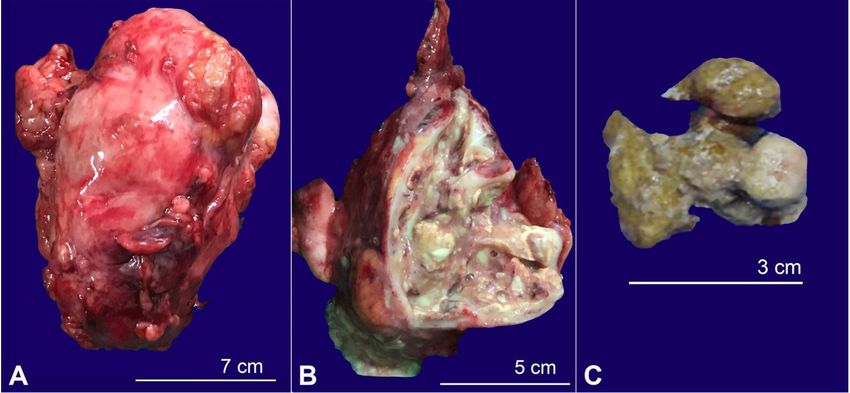
Xanthogranulomatous pyelonephritis with calculus migration into the psoas abscess: an unusual complication

(normal range: 11.5-16.5 g/dL). Renal Function test                     ~ 3.0 cm with a twinkling artifact and posterior
and serum electrolytes were within normal limits.                       acoustic shadowing (Figure 2A and 2B). Also, the
The urinalysis revealed 10-15 leukocytes/HPF (normally                  proximal left ureter was dilated, measuring 1.76 cm
< 5 leukocytes/HPF). On a plain radiograph, there was                   and was seen draining into a large collection measuring
a large radio-opaque shadow in the left renal fossa                     11.2 × 4.1 cm within the left psoas region (Figure 2C).
(Figure 1).                                                                   The CECT depicted an enlarged left kidney
    The abdominal ultrasound (USG) showed a large                       with gross hydroureteronephrosis with thinning of
calculus, probably a staghorn calculus measuring                        the renal cortex and a staghorn calculus measuring
                                                                        ~ 3.8 × 2.4 cm within the renal pelvis. The renal pelvis
                                                                        appeared contracted, whereas calyces were dilated,
                                                                        giving the appearance of a “Paw of a Bear,” referred to
                                                                        as “Bear Paw Sign” (Figure 3A). Also, a large collection
                                                                        measuring 13.0 × 4.0 cm was found along the left
                                                                        iliopsoas muscle harboring multiple images consistent
                                                                        with calculi, the largest measuring ~1.6 × 1.4 cm
                                                                        (Figure 3B and 3C).
                                                                              The working diagnosis based on the imaging exams
                                                                        was left XGP with psoas abscess. Left nephrostomy was
                                                                        performed, and purulent fluid was drained, from which
                                                                        Escherichia coli (E. coli) was isolated on culture. After
                                                                        5 days, a left-sided nephrectomy was performed, and
                                                                        the specimen was sent for histopathology examination.
                                                                        The surgical specimen weighed around 150 g (average
                                                                        124g for female’s left kidney). The excised kidney
                                                                        measured 10.0 × 7.0 × 4.0 cm and was partially
                                                                        covered with perinephric fat. The attached ureter
                                                                        measured ~ 4.0 cm in length. The cut section of
                                                                        the kidney showed a staghorn calculus filling the
                                                                        pelvicalyceal system measuring 3.5 × 1.0 cm. Also, a
                                                                        few areas of yellowish discoloration were noted within
                                                                        the pelvicalyceal system, probably due to xanthoma
                                                                        cells (Figure 4A, 4B and 4C).
Figure 1. Abdominal plain radiograph shows a large
radio-opaque shadow in the left renal fossa and multiple                     Microscopically, the kidney sections showed
small radio-opaque shadows in the left pelvic region.                   hyalinized glomeruli and atrophic tubules lined by
These findings are consistent with multiple calculi.                    flattened cells, and the surrounding blood vessels

Figure 2. Abdominal USG showing a calculus and left psoas abscess. A and B – A large calculus probably a staghorn
calculus (white arrow) with a twinkling artifact and posterior acoustic shadowing; C – Dilated proximal left ureter
(white arrow) in close contact with a large collection in the left psoas region (black arrow).

2-6                                                                                         Autops Case Rep (São Paulo). 2021;11:e2020200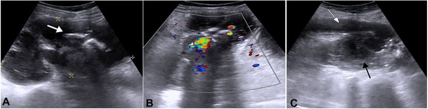
Singh M, Ethiraj D, Indiran V, Kanase ND, Maduraimuthu P

Figure 3. Abdominal CECT axial and coronal section shows XGP of the left kidney. A – Corticomedullary phase of
CECT shows an enlarged left kidney with gross hydroureteronephrosis with thinning of renal cortex and a staghorn
calculus within the renal pelvis, which is contracted, whereas calyces are dilated giving the appearance of a “Paw
of A Bear” (white arrow); B – Excretory phase of CECT shows a staghorn calculus measuring 3.8 × 2.4 cm (black
arrow) within the renal pelvis. Multiple calculi, the largest measuring 1.6 × 1.4 cm (arrowheads), were noted in the
ureter and the collection; C – Large collection within the left iliopsoas muscle consistent with left psoas abscess
(white arrow).

Figure 4. Gross findings of the excised left kidney. A – Gross view of the left kidney and attached ureter partially
covered by perinephric fat; B – The cut surface of the left kidney. Note the presence of multiple calculi and necrotic
cheesy material replacing the renal parenchyma with loss of cortico-medullary differentiation; C – The staghorn
calculus.

were thickly walled. Interstitial fibrosis with a dense     DISCUSSION
collection of inflammatory cells composed of sheets of
lymphocytes, plasma cells, neutrophils, histiocytes, and          XGP is a suppurative granulomatous infection
multinucleated giant cells were seen. Islands of foamy      characterized by progressive parenchymal destruction
macrophages, foci of calcification, lymphoid follicles,     caused by chronic renal obstruction due to calculus or
and thickened capsule were also seen. At some places,       stricture or rarely tumor, resulting in a non‑functioning
the inflammatory cells and the proliferating capillaries    kidney. XGP accounts for 0.6% to 1% of all
were overlined by a transitional epithelium with            pyelonephritis cases, globally. The common organisms
ulceration. (Figure 5A-D).                                  on urine culture are Proteus mirabilis, E coli, or

Autops Case Rep (São Paulo). 2021;11:e2020200                                                                           3-6
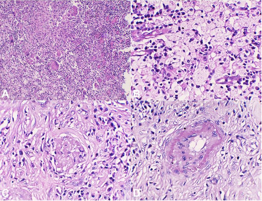
Xanthogranulomatous pyelonephritis with calculus migration into the psoas abscess: an unusual complication

Figure 5. Photomicrographs of the left kidney. A – Microscopic section showing replacement of the renal
parenchyma by sheets of acute and chronic inflammatory cells composed of neutrophils, lymphocytes, and plasma
cells, scattered foreign body giant cells (H&E10X); B and C – Section shows sheets of polygonal histiocytes having
abundant pale eosinophilic and foamy cytoplasm with a round nucleus and annexed with focal aggregates of
lymphocytes (H&E 40X); D – Section shows the hyalinised glomeruli and thick-walled blood vessel (H&E 40X).

Staphylococcus aureus. The annual incidence of XGP is                   enlarged kidney with dilated calyces and loss of
1.4 per 100,000. XGP is a rare variant of pyelonephritis                cortico‑medullary differentiation (CMD). In more than
and is seen more commonly among females. 1,4                            70% of the XGP cases, renal calculus is present, which
The most common presenting symptoms are flank or                        on USG appears as an amorphous echogenic structure
abdominal pain, fever, pyuria, weight loss, hematuria.                  and generally shows a posterior acoustic shadowing
There are three types of XGP (i) diffuse, which is the                  and twinkling artifact.3 In our case, the patient showed
most common type; (ii) segmental; and (iii) focal, which                a large radiopaque shadow in the left renal fossa on
is limited to the cortex.5-7 Diffuse XGP is associated                  the radiograph. The abdominal USG revealed large
with massive renal enlargement, peri-pelvic fibrosis,                   hyperechoic foci in the pelvicalyceal system of the
hydronephrosis, lithiasis, and lobulated mass replacing                 left kidney with posterior acoustic shadowing and
the renal parenchyma. Preoperatively, it is difficult to                twinkling artifact, which is suggestive of calculus.
differentiate, based on clinical and radiological features                   CT is the gold standard diagnostic method for
alone, XGP from other entities like tuberculosis and                    XGP because it demonstrates the specific intrarenal
renal cell carcinoma (RCC). Hence, histopathological                    findings and shows the extrarenal extension, which is
and immunohistochemistry correlation is required.3                      useful for surgical scheduling.8,9 In our case, the CECT
The ultrasound of XGP typically demonstrates an                         showed an enlarged kidney with low-density areas

4-6                                                                                         Autops Case Rep (São Paulo). 2021;11:e2020200
Singh M, Ethiraj D, Indiran V, Kanase ND, Maduraimuthu P

within the renal parenchyma and a large obstructive           junction by soft yellow nodules, along with filling of
staghorn calculus causing hydroureteronephrosis.              the calyces with pus and debris. On the histological
The low-density areas were suggestive of an abscess           examination, there is diffuse infiltration of the renal
or fluid. “Bear paw sign” is better appreciated on the        parenchyma with plasma cells, histiocytes, lymphocytes,
corticomedullary phase of CECT.4,5 Also, a collection         neutrophils, multinucleated giant cells, and lipid-laden
containing multiple calculi was noted within the left         macrophages (xanthoma cells). The xanthomatous
iliopsoas muscle. This is an unusual finding in XGP           cell of XGP sometimes resembles the clear cells of
presenting with psoas abscess and, according to our           RCC. Unlike that of xanthoma cells, the cytoplasm of
knowledge, has not yet been reported in the literature.       tumoral cells is clearer, which has a foamier cytoplasm.
Due to the presence of ureteric calculus, chronic             Therefore, Immunohistochemistry (IHC) study may be
obstruction with concomitant infection is likely to           helpful in certain cases.3,6
have caused significant damage to the ureteric wall
and resultant fistula, and urinoma formation within
the psoas muscle. Subsequent secondary infection of           CONCLUSION
the urinoma could have resulted in the psoas abscess
                                                                    XGP is an uncommon entity associated with
formation. The drained abscess was positive for E coli,
                                                              urinary tract obstruction and infection. Also, its
which is the most frequent bacterial cause of urinary
                                                              association with calculus migration into psoas abscess
tract infection.10
                                                              is an unusual complication. It is difficult to differentiate
     The patient was prescribed broad-spectrum
                                                              it from renal cell carcinoma on imaging, and hence
antibiotics for 5 days, followed by total left nephrectomy.
                                                              histopathological correlation is required. Imaging
Usually, on MRI, XGP appears as a hyper signal on
                                                              features are not enough to confirm the diagnosis
T1- weighted image due to the lipid-laden foamy
                                                              of XGP, but in this case, demonstration of “Bear
macrophages and shows a signal void if calculi are
                                                              paw sign” helped in ruling out other uncommon
present within the collecting system. However, this MRI
                                                              differentials. In this case, the correlation of the clinical
sign is not always reliable if the lesion lacks a certain
                                                              findings with radiologic features, followed by proper
amount of xanthoma cells.
                                                              treatment and histopathological correlation not only
     Medical and surgical management plays an
                                                              helped in confirming the disease but also improved
important role in the treatment of XGP. In cases of
                                                              patient recovery.
unilateral diffuse XGP, nephrectomy is the usual
therapeutic option. While in cases of bilateral XGP,
when surgery is not feasible or in patients with              REFERENCES
focal XGP, the broad-spectrum antibiotic regimen is
indicated.3 However, Leoni et al. observed a mortality        1. F r i e d l A , T u e r k C , S c h i m a W , B r o e s s n e r C .
                                                                 Xanthogranulomatous pyelonephritis with staghorn
rate of 10% in a series of 10 patients, despite the
                                                                 calculus, acute gangrenous appendicitis and enterocolitis:
nephrectomy.11 Since our case could not be managed               a multidisciplinary challenge of kidney-preserving
conservatively with only on antibiotics regimen,                 conservative therapy. Curr Urol. 2015;8(3):162-5. http://
the abscess drainage and a left-sided nephrectomy                dx.doi.org/10.1159/000365709. PMid:26889137.
were necessary to prevent the development of a                2. Lintong PM, Durry M. Xanthogranulomatous
septic shock. In cases of absence of the typical signs           pyelonephritis. MOJ Clin Med Case Rep. 2015;2(2):36-8.
like staghorn calculus, the differential diagnosis
                                                              3. Li L, Parwani AV. Xanthogranulomatous pyelonephritis.
of XGP, based strictly on imaging, includes renal                Arch Pathol Lab Med. 2011;135(5):671-4. PMid:21526966.
tuberculosis, renal cell carcinoma, renal abscess, and
                                                              4. Lee JH, Kim SS, Kim DS. Xanthogranulomatous
renal angiomyolipoma. Hence, in these cases, the
                                                                 pyelonephritis: “bear’s paw sign. J Belg Soc Radiol.
histopathological examination is necessary. A partial            2019;103(1):31. http://dx.doi.org/10.5334/jbsr.1807.
or total nephrectomy should be performed with the                PMid:31139769.
histopathological diagnostic confirmation, in the cases
                                                              5. C h o w J , K a b a n i R , L i t h g o w K , S a r n a M A .
where the biopsy is not feasible.3,12 XGP, generally             Xanthogranulomatous pyelonephritis presenting as acute
presents with replacement of the cortico‑medullary               pleuritic chest pain: a case report. J Med Case Reports.

Autops Case Rep (São Paulo). 2021;11:e2020200                                                                                 5-6
Xanthogranulomatous pyelonephritis with calculus migration into the psoas abscess: an unusual complication
2017;11(1):101. http://dx.doi.org/10.1186/s13256-017- 9. Craig WD, Wagner BJ, Travis MD. Pyelonephritis: radiologic-
1277-4. PMid:28399929. pathologic review. Radiographics. 2008;28(1):255-77.
http://dx.doi.org/10.1148/rg.281075171. PMid:18203942.
6. Kundu R, Baliyan A, Dhingra H, Bhalla V, Punia RS.
Clinicopathological spectrum of xanthogranulomatous 10. Kempegowda P, Eshwarappa M, Dosegowda R, Aprameya
pyelonephritis. Indian J Nephrol. 2019;29(2):111-5. IV, Khan MW, Kumar PS. Clinico-microbiological profile
PMid:30983751. of urinary tract infection in south India. Indian J Nephrol.
2011;21(1):30-6. http://dx.doi.org/10.4103/0971-
7. Chandanwale SS. Xanthogranulomatous pyelonephritis: 4065.75226. PMid:21655167.
Unusual clinical presentation: a case report with literature
11. Leoni FA, Kinleiner P, Revol M, Zaya A, Odicio A.
review. J Family Med Prim Care. 2013;2(4):396-
Xanthogranulomatous pyelonephritis: review of 10
8. http://dx.doi.org/10.4103/2249-4863.123942. cases. Arch Esp Urol. 2009;62(4):259-71. http://
PMid:26664851. dx.doi.org/10.4321/S0004-06142009000400001.
8. Barral M, Sánchez Crespo JM, Pérez Herrera JC, PMid:19717876.
Ortega Garcia JL, Hidalgo Ramos FJ, Porcuna Cazalla 12. Ghoz HM, Williams M, Perepletchikov A, James N, Babeir
G. Xanthogranulomatous pyelonephritis: radiologic review. AA. An unusual presentation of xanthogranulomatous
In: ECR 2014 Congress; 2014; Vienna. Vienna: European pyelonephritis: psoas abscess with reno-colic fistula.
Society of Radiology; 2014. http://dx.doi.org/10.1594/ Oxf Med Case Rep. 2016;(7):150-3. http://dx.doi.
ecr2014/C-0557. org/10.1093/omcr/omw063. PMid:27471599.
Study was carried out the Department of Radiodaignosis, Sree Balaji Medical College and Hospital, Chennai,
Tamil Nadu, India.
Authors’ contributions: Manpreet Singh, Dillibabu Ethiraj, Venkatraman Indiran, Niranjan Dhanaji Kanase and
Madhuraimuthu Prabakaran equally contributed to the conception or design of the work; acquisition, analysis,
and interpretation of data. Similarly, the drafting, revising, and critics for relevant content were equally granted
by all authors, which collectively proofread the final version and approved it for publication.
Ethics statement: The current work is in accordance with the ethical standards of the institutional and/or
national research committee and with the 1964 Helsinki Declaration and its later amendments or comparable
ethical standards. The authors retain informed consent signed by the patient authorizing the data publication.
Conflict of interest: None
Financial support: None
Submitted on: April 29th, 2020
Accepted on: June 19th, 2020
Correspondence
Venkatraman Indiran
Sree Balaji Medical College and Hospital, Department of Radio-diagnosis
7 Works Road, Chromepet, 600044, Chennai, Tamilnadu, India
Phone: +91 967 708 4438
ivraman31@gmail.com
6-6 Autops Case Rep (São Paulo). 2021;11:e2020200You can also read