2022 Wellcome Centre for Cell Biology

Page created by Ron Ramsey
 
CONTINUE READING
2022 Wellcome Centre for Cell Biology
2022
2022 Wellcome Centre for Cell Biology
Reliable identification of protein-protein interactions by crosslinking mass spectrometry.
Nat. Commun. 12, 3564.

5) Title of Research Programme:

    Content
Cellular Tomography

6) Images:

                                                          A                                                             C           SU

                                                                                                                              X
                                                                                                                                     P
                                                                                                                                   (SU
                                                                                                                                  E3-Li

                                                                                                                           heterochro

                                                          B            apical-heterochromatin

                                                           wildtype
    021:
Figure                          16                            30                                43
    Director's Introduction     Atlanta Cook               Hiro Ohkura                          International Scientific
Retention time data complements substantially the currently exclusively
    04                                                                  used mass
                                                              basal-euchromatin                 Advisory Board
                               18                           32
spectrometric
    About theevidence
              Wellcomefor the identification of crosslinks between proteins.
                                Owen Davies                   Juri Rappsilber                   44
                                                           PIAS RNAi

    Centre for Cell Biology
Left: The combined retention information
                                20         of crosslinked peptides
                                                              34     from three different Public Engagement
    06
chromatography  modes suffices Bill
                                to effectively
                                    Earnshaw separate plausible    identifications
                                                              Kenneth  E Sawin     (green)46
    Facilities                                                     E. coli lysate. Middle:PIAS
from modelled noise (all other colours) in a crosslink analysis ofDAPI                    iCM PhD Programme Histone H3
Crosslink network from  the     22
                            Fanconi  anemia   complex         36
                                                       analysis, shown   in the circular
    08
view. Unique residue pairs fromPatrick
    Robin Allshire
                                 xiSCOREHeun(gray), after rescoring (green), and shared 47
                                                              David Tollervey
                                                                                                iCM Summer Internship
                                24                              Figure 2: The SUMO     E3 Ligase PIAS is requ
                                                                                   Programme
    10                                                        38
                                Georg Kustatscher               formation. A) Embryonic cycle 1-14 (image W
                                                              Julie Welburn
    A. Jeyaprakash
    Arulanandam
                                                                cycle 13 embryos48 showing ⍺PIAS and
                                26                            40⍺H3K9me3 in apical List of Groups
                                Adele Marston
                                                                                      heterochromatin in wildt
    12                                                        Marcus D Wilson      54
                                                                Proposed role of PIAS in chromosome organisa
    Adrian Bird                 28                                                              Centre Publications
                                                              42
                                Dónal O'Carroll               Emeritus Centre
    14                                                                                          61
    Dhanya Cheerambathur                                      Members                           Sustainability at WCB
2022 Wellcome Centre for Cell Biology
Director's Introduction
    In November 2021, our former Director David Tollervey stepped down after over 10 years of outstanding leadership of the         We are extremely fortunate to have access to world-class technology platforms supporting our research. I would like to
    Wellcome Centre for Cell Biology. On behalf of the whole Centre, I would like to express our extreme gratitude to David for     thank the technology platform managers Dave Kelly, Shaun Webb, Christos Spanos, Martin Singleton, and Martin Wear for
    all he has done to support and develop our inspiring research community. It is an honour to build on his legacy and that of     maintaining exceptionally high standards of service and support for our research through challenging times. I would also
    the inaugural Director, Prof Adrian Bird.                                                                                       like to extend a welcome to Martin Singleton, who joined us this year to replace Maarten Tuijel as the Cryo-EM Platform
                                                                                                                                    Manager.
    This year, the Centre bounced back following the period of reduced laboratory access and lack of face-to-face interactions
    that were enforced by the pandemic. It is uplifting to feel the buzz in the laboratories and corridors again. Those impromptu   Despite on-going restrictions for much of the year, the WCB Public Engagement team led by Sarah-Jane Judge has
    conversations in hallways are so important and often the start of new cross-disciplinary collaborations upon which our          delivered a busy and diverse programme of events. WCB have also been awarded two ScotPEN Wellcome Engagement
    Centre thrives.                                                                                                                 Awards (SWEA). David Tollervey’s SWEA will focus on engagement with Prader-Willi syndrome patients and families. Julie
                                                                                                                                    Welburn, Atlanta Cook, Alison Pidoux and Tony Ly (Dundee) will use their SWEA to create fabric with science patterns
    This brochure presents a brief overview of our world-class discovery science from individual research groups and provides       for public engagement projects. My thanks to all who participated in the design, organisation and delivery of our public
    snapshots of our broader research community. This year, we are delighted to welcome two new groups to WCB. Owen                 engagement programme.
    Davies is a Wellcome Senior Fellow whose research focuses on the structural biology of meiosis. Georg Kustascher is an
    MRC Career Development Fellow who investigates how cells regulate protein levels, and how this is disrupted in disease.         Finally, I would like to finish by thanking and congratulating our entire WCB community for their resilience and excellent
                                                                                                                                    work this year. WCB is known for its collaborative ethos and landmark scientific discoveries, both of which are the product
    I congratulate several WCB groups on their success in attracting major grant funding this year. David Tollervey and Robin       of the collective efforts of its many talented and dedicated individuals.
    Allshire both renewed their Wellcome Principal Research Fellowships for 5 years, Adrian Bird was awarded a Wellcome
    Investigator Award and Patrick Heun was awarded a BBSRC response-mode grant. Gerard Pieper in the Marston group
    was awarded a Sir Henry Wellcome Fellowship.

    It was also wonderful to see Julie Welburn recognised as an early Career Researcher for her exceptional achievements in
    life sciences with the award of the Patrick Neill Medal from the Royal Society of Edinburgh. SBS achievement awards were
    made to Fiona Cullen in the Ohkura group for her long service and to Tania Auchnynnikava in the Allshire group for her
    contribution to the PhD student experience.

    Professor Malcolm Walkinshaw has wound down his research group, but I am delighted that he will maintain his
    association with WCB as Emeritus Centre Member. We look forward to continuing to benefit from his influence and wisdom.
                                                                                                                                                                                                                                                 Adele Marston
    I congratulate WCB alumni who moved on to prestigious new positions this year and wish them every success in their
    future careers. Philipp Voigt moved to a new position in the Babraham Institute, Cambridge and Tomasz Turowski (Tollervey
    group) obtained an independent PI position in Warsaw at the Institute of Biochemistry and Biophysics, Polish Academy of
    Science. Tania Auchnynnikava (Allshire group) has taken a senior laboratory research scientist position in proteomics at
    the Francis Crick Institute.

    We were also sorry to say goodbye to WCB staff who took retirement this year. As Centre Manager and Administrator of
    the Wellcome PhD programme for over 10 years, Karen Trail kept WCB running smoothly and offered support to many
    generations of students. Sarah Keer-Keer, Public Engagement Manager, established a thriving and prominent public
    engagement vision for WCB. John Connelly, a long-standing member of the Bird group, retired after 28 years. I thank them
    all for their exceptional service to WCB and wish them all the best in their future endeavours.

2                                                                                                                                                                                                                                                                 3
2022 Wellcome Centre for Cell Biology
About the Wellcome Centre for Cell Biology

    The Wellcome Centre for Cell Biology has a mission to discover the fundamental molecular
    mechanisms that determine cell function in health and disease.

    Our vision is to explore      Our collective expertise        Our environment is that       Public engagement is           Our history began in 1992         Wellcome Trust Centre for
    and understand how cell       straddles discipline            of a cutting-edge research    integrated into our research   with the vision to expand         Cell Biology was founded
    states are established and    boundaries, catalyses high      institute embedded within     vision and reaches into        research in cell biology,         in October 2001. Professor
    maintained in contexts        quality research and is alert   a globally influential        diverse communities, with      developed by Professor Sir        Adrian Bird served as
    that include infection,       to translation, with ultimate   University. The Wellcome      a particular emphasis          Kenneth Murray (Biogen            inaugural Director and
    development, aging and        benefits for human health       Centre for Cell Biology       on targeting those that        Professor) and the Institute      successfully renewed
    disease.                      and wellbeing.                  benefits from access to a     have few opportunities for     of Cell and Molecular             Wellcome Centre status in
                                                                  thriving student population   scientific discourse.          Biology. A seed contribution      2006. He was succeeded
    Our culture nurtures ideas,   Our research themes are         and enjoys strong                                            of £2.5 million from the          by Professor David
    disseminates knowledge        intersecting and synergistic:   interdisciplinary links and                                  Darwin Trust leveraged            Tollervey in 2011 who led
    and fosters a collaborative                                   collaborations with other                                    financial support from the        the Centre through a further
                                  • Gametogenesis,
    environment.                                                  University departments                                       Wolfson Foundation, the           renewal in 2016. Our current
                                    inheritance and fertility.
                                                                  including engineering,                                       University of Edinburgh           Director, Professor Adele
                                  • Cell cycle, differentiation   physics, informatics,                                        and the Wellcome Trust,           Marston took over in 2021.
                                    and genetic disease.          medicine and chemistry.                                      allowing construction of the
                                  • Adaptation, gene                                                                           Michael Swann building.
                                    expression and drug                                                                        The majority of the research
                                    resistance                                                                                 space was earmarked for
                                                                                                                               Wellcome Trust-funded
                                                                                                                               research. Recruitment,
                                                                                                                               based on research
                                                                                                                               excellence at all levels in the
                                                                                                                               area of cell biology, began
                                                                                                                               in earnest in 1993. This was
                                                                                                                               mostly, but not exclusively,
                                                                                                                               through the award of
                                                                                                                               Research Fellowships from
                                                                                                                               the Wellcome Trust. The
                                                                                                                               Michael Swann building
                                                                                                                               was first occupied in
                                                                                                                               January 1996 and the

4                                                                                                                                                                                               5
2022 Wellcome Centre for Cell Biology
Facilities

    The Centre Optical                         Media Prep                                 Cryo-Electron                             Edinburgh Protein                        Proteomics Platform                        Bioinformatics Core
    Instrumentation                                                                       Microscopy Platform                       Production Platform                                                                 Platform
    Laboratory

    The Centre Optical Instrumentation         The Media Prep and Wash Up provide         The CryoEM facility offers electron       Rapid solutions to the production        In our Proteomics Facility we use a        The bioinformatics core facility
    Laboratory (COIL) staff provide            the Wellcome Centre with high              microscopy support and training           of proteins and the biophysical          wide range of techniques to address        supports research by providing
    technical support for a wide range         volumes of Buffers, Growth Media,          for analysing a variety of biological     characterisation of their ligands        important biological questions. We         data analysis expertise and high-
    of imaging technologies and image          Agar plates and Fly food. They collect     samples. We are primarily focussed        underpins many of the questions          are equipped with four state-of-the-       performance compute infrastructure.
    analysis software. As well as user         glassware and equipment daily, for         on single-particle approaches but are     in structural, translational and cell    art mass spectrometers, which are          We collaborate on research projects
    training the facility staff are able       washing, sterilization and reuse as well   also interested in electron diffraction   biology today.                           employed to accurately identify,           from inception through to publication,
    to help with experimental design           as safely decontaminating lab waste.       and electron-tomography techniques.                                                quantify, provide structural information   by offering advice on experimental
                                                                                                                                    Located in labs in the Michael Swann
    and provide image analysis advice.         The team provide many hundreds of                                                                                             and demonstrate interactions of            design, managing large amounts of
                                                                                          Our 200 kV Tecnai F20 microscope          Building, The Wellcome Trust, and
    Bespoke ImageJ plugins for analysis        litres of Media, Agars and Buffers per                                                                                        proteins even in the most complex          data and performing computational
                                                                                          has been recently upgraded with a         University of Edinburgh funded Protein
    pipelines or to extend the functionality   week and over 35,000 fly vials per                                                                                            biological samples. We are currently       analysis. We have a large focus
                                                                                          direct electron detector. We have         Production Facility (EPPF) provides
    of ImageJ are written on request.          year. Despite the demanding workload                                                                                          moving into large scale high               on high throughput sequencing
                                                                                          also installed an automated data          researchers with access to state-of-
                                               the team are keen to promote and                                                                                              throughput proteomic analyses.             experiments, including ChIP-seq, HiC,
    Researchers have access to both laser                                                 collection system, complete with          the-art equipment and excellent end-
                                               improve sustainability and have been                                                                                                                                     RNA-seq and long read sequencing,
    scanning and spinning disk confocals,                                                 online processing pipeline. This allows   user core facilities to address these    We provide in-person training
                                               working closely with the labs to tackle                                                                                                                                  and we develop workflows,
    a TIRF microscope, several widefield                                                  the microscope to be used both for        questions.                               to the researchers on proteomic
                                               single use plastic waste and various                                                                                                                                     visualisations and interactive
    microscopes and a flow cytometer.                                                     sample screening prior to submission                                               applications (experimental design,
                                               other environmental issues.                                                          The facility is operated by a team of                                               applications for the processing and
    All microscopes have environmental                                                    to external high-end facilities such                                               sample preparation and data analysis)
                                                                                                                                    three highly skilled experimentalists                                               interrogation of these datasets.
    chambers to maintain temperature                                                      as eBIC, as well as in-house data                                                  and we offer an annual proteomics
                                                                                                                                    who not only ensure that the                                                        The core facility takes a lead role in
    and CO2 for live cell imaging. The                                                    collection.                                                                        course on applications, experimental
                                                                                                                                    equipment is well maintained, but                                                   encouraging researchers to develop
    equipment is bookable online from a                                                                                                                                      approaches and data interpretation.
                                                                                          We have equipment for room-               also provide training, project advice                                               their own skills in bioinformatics by
    central booking site.
                                                                                          temperature and cryogenic sample          and will help design and implement                                                  offering regular training courses
                                                                                          preparation including a vitrification     your experiments to obtain the best                                                 and networking events as well as
                                                                                          robot and work closely with the SBS       possible results from the equipment.                                                promoting the tenets of reproducible
                                                                                          EM facility to accommodate a wide                                                                                             research.
                                                                                          range of sample types.
6                                                                                                                                                                                                                                                                77
2022 Wellcome Centre for Cell Biology
0.8                                         epe1у                                                                                                                                      3

                                                                                                                                                                                                                                                                                                                                                                                                              Epe1-GFP enric
                                                                                                                                                                                                                        H3K9me enric

                                                                                                                                                                                                                                                                                                                                  Epe1 protein lev
                                                                                                                                                                                                                                            0.6                                                                                                                                                                                    2
                                                                                                                                                                                                                                                                                                                                                      0.5
                                                                                                                                                                                                                                            0.4
                                                                                                                                                                                                                                                                                                                                                                                                                                   1
                        Robin Allshire                                                                                                                                                                                              Epigenetic
                                                                                                                                                                                                                                     0.2
                                                                                                                                                                                                                                               mechanisms mediating antifungal resistance
                        Co-workers: Tatsiana Auchynnikava, Roberta Carloni, Andreas Fellas, Elisabeth Gaberdiel, Nitobe London,                                                                                                                  0                                                                                                       0                                                                         0
                                                                                                                                                                                                                                                 0      2     4          8                                                                                          -      low                                                         -    low med
                        Alison Pidoux, Severina Pociunaite, Desislava Staneva, Manu Shukla, Sharon White, Weifang Wu,                                                                                                                          Hours after TetR-Clr4* release                                                                                           CAF                                                                 CAF
                        Imtiyaz Yaseen, Rebecca Yeboah
                                                                                                                                                                                                                                                                                                                                                                                                                               16 cen-IRC
                                                                                                                                                                                  A e                                                                                                                                                                                                                                                  D
                                                                                                                                                                                   A                                                        Wild-type                                  Dead cells                                                                                                                              12          D    Cup1         LYR
                                                                                                                                                                                                                                              cells                Lethal                                                                                 Insult
    Antifungal resistance is increasing in prevalence, raising fungal-borne disease frequencies in humans and crops important                                                                                                                                                                                      Survivors
                                                                                                                                                                                                                                                                   insult                                                                                removal                       Mutant
                                                                                                                                                                                                                                                                                                                                                                                                                                   8                        L73G
    for human well-being. The survival of fungi in harsh environments involves stress-sensing pathways that reprogram their                                                                                                                                                                                                                                                           phenotype
    proteomes. New environmental conditions, including global heating, can push opportunistic fungi to colonise novel                                                                                                                                                                                                                                                                                                                                    − antifungal         + antifungal
                                                                                                                                                                                                                                                                                                                                                                                                                                   4
                                                                                                                              Figure 2                                                                                                                                                                                                                                                                                                            Torres-Garcia
                                                                                                                                                                                                                                                                                                                                                                                                                                                   wt           et al.
    niches, thus increasing their potential to become harmful pathogens. Effective antifungal treatments are limited in number                                                                                                                                                                          Resistant                                              Wild-type
    precisely because fungi are adept at resisting challenges.                                                                                                     a ChIP-seq: H3K9me2                                                                              Resistant                           epimutant                                              phenotype                                                           0
                                                                                                                                                                                                      tel1L
                                                                                                                                                                                                                                                              Chr I
                                                                                                                                                                                                                                                                      cells
                                                                                                                                                                                                                                                                       cen1                        tel1R tel2L                   Resistant
                                                                                                                                                                                                                                                                                                                                 cen2
                                                                                                                                                                                                                                                                                                                                            Chr II
                                                                                                                                                                                                                                                                                                                                                                               tel2R tel3L
                                                                                                                                                                                                                                                                                                                                                                                                 Chr III
                                                                                                                                                                                                                                                                                                                                                                                                 cen3             tel3R
                                                                                                                                                                                                                                                                                                                                                                                                                                       -     lowhba1
                                                                                                                                                                                                                                                                                                                                                                                                                                                 medD
                                                                                                                                                                                                                                                                                                                                   mutant                                                                                      125          CAF
    Resistance to fungicides/antifungal compounds can result from genetic mutations, however, it was unknown if                                     Figure 2                                     wt                                                                                                                                                                                                                                        cup1-L73G
                                                                                                                                                                                                                                                                                                                                                                                                                                                  Torres-Garcia et al.
                                                                                                                                                                                B                B
                                                                                                                                                                                       ChIP-seq: H3K9me2
                                                                                                                                                                                                                                                                                                                                         UR-1
                                                                                                                                                                                                                                                                                                                                                                                                                               0

                                                                                                                                                                            H3K9me2 enrichment
                                                                                                                                                                                 UR-1
    resistance might also arise from heritable epigenetic changes mediated by post-translational modifications carried on                                                       a ChIP-seq:
                                                                                                                                                                                       UR-2
                                                                                                                                                                                            H3K9me2 Chr I                                                                                                                                       ChrUR-2
                                                                                                                                                                                                                                                                                                                                                    II                                           Chr III
                                                                                                                                                                                                                   tel1L                                                       cen1                  tel1R tel2L                  cen2                                         tel2R tel3L       cen3             tel3R
    histones in chromatin. Using the model fission yeast (Schizosaccharomyces pombe) fungal system, we discovered that                                                                 UR-3                                                                        UR-3
                                                                                                                                                                                                                                                                                                                                                                                                                               125
                                                                                                                                                                                                                                                                                                              UR-4
                                                                                                                                                                                       UR-4 wt
    heterochromatin island-mediated ‘epimutations’ confer resistance following exposure to external insults (Torres-Garcia et                                                                                                                                                           UR-5                                                UR-1
                                                                                                                                                                                                                                                                                                                                                                                                                               0

                                                                                                                                                               H3K9me2 enrichment
                                                                                                                                                                                         UR-1
                                                                                                                                                                                       UR-5
    al. 2020; Figure A). Heterochromatin islands are formed by addition of methyl groups to lysine 9 of histone H3 (H3K9me)                                                              UR-2
                                                                                                                                                                                       UR-6
                                                                                                                                                                                                                                                                                                                                                        UR-2UR-6
                                                                                                                                                                                                                                                                                                                                                                                                                                       E
    over regions of chromatin, resulting in reduced expression of underlying genes (Figure B). For example, epimutation-
                                                                                                                                                                                                   0 kb
                                                                                                                                                                                                 UR-3                                                                 UR-3                    5570 kb 0 kb                                                               4530 kb 0 kb                        2450 kb                       E    Epe1          JmjC                  Myc
                                                                                                                                                                   b                         ChIP-seq:
                                                                                                                                                                                              UR-4     H3K9me2
                                                                                                                                                                                                                                                                                                                 UR-4

    mediated repression of the cup1+ gene encoding a mitochondrial LYR protein confers resistance through mitochondrial                                                                          UR-5
                                                                                                                                                                                                                                                                                            UR-5

                                                                                                                                                                                                 UR-6
                                                                                                                                                                                                                                20        hba1 locus                                               UR-1                 30       cup1 locus locus
                                                                                                                                                                                                                                                                                                                                 ncRNA.394    UR-6                                                    UR-2                                                               + antifungal
    dysfunction (Figure C, D).                                                                                                                                                                                                                                                                        wt                                                                                                wt

                                                                                                                                                                                                               H3K9me2 enrichment
                                                                                                                                                                                                                   0 kb                                                                         5570 kb 0 kb                                                              4530 kb 0 kb                       2450 kb
                                                                                                                                                                                b                ChIP-seq: H3K9me2                                                                                                                                                                                                                                      +     JmjC                  Myc
    Transient ectopic H3K9me-dependent heterochromatin is normally rapidly erased by the counteracting H3K9 JmjC-domain                                                                                                         10
                                                                                                                                                                                                                                  20
                                                                                                                                                                                                                                                                                                                        15
                                                                                                                                                                                                                                            hba1 locus                                                 UR-1              30      ncRNA.394 locus                                                      UR-2
    Epe1 demethylase. Surprisingly, external insults such as antifungal compounds (e.g. caffeine, fluconazole) induce cleavage                                                                                                                                                                           wt                                                                                             wt

                                                                                                                                                                                                  H3K9me2 enrichment
    of Epe1 allowing heterochromatin islands to persist and confer resistance in selected lineages (Figure E). Unlike genetic                                                                                                       0                                                                                    0
                                                                                                                                                                                                                                                                                                                                                     SPBC17G9.13c
                                                                                                                                                                                                                                                                                                                                                          cup1     eno101
                                                                                                                                                                                                                                     10                                                                                  15
                                                                                                                                                                                                                                          pmt3 kin17 arp5 grp2                hba1          alp4                           rpl1102                         ncRNA.394    cut2                 tim44 fma2
    mutations, such epimutations are unstable - causative heterochromatin islands, associated gene repression and resistance                                                                                                                                          ish1        uge1                                                   pyr1                                            prl68
    are lost in the absence of antifungal selection. Thus, epigenetic processes promote phenotypic plasticity so that wild-type                                                                                                         tmf1
                                                                                                                                                                                                                                               der1
                                                                                                                                                                                                                                                    ncRNA.1523
                                                                                                                                                                                                                                                                                            ncRNA.1524
                                                                                                                                                                                                                                                                                               ncRNA.135
                                                                                                                                                                                                                                                                                                                                         SPBC17G9.12c
                                                                                                                                                                                                                                                                                                                                                  ncRNA.393
                                                                                                                                                                                                                                      0                                                                                   0
    cells adapt to unfavourable environments without irreversible genetic alterations.                                                                                                                                              Chr II   2,510
                                                                                                                                                                                                                                           pmt3 kin17 arp5 grp2
                                                                                                                                                                                                                                                                2,520
                                                                                                                                                                                                                                                                   hba1                       alp4
                                                                                                                                                                                                                                                                                                            kb          Chr
                                                                                                                                                                                                                                                                                                                        chr II
                                                                                                                                                                                                                                                                                                                            rpl1102
                                                                                                                                                                                                                                                                                                                                                       2,195     2,200eno101
                                                                                                                                                                                                                                                                                                                                                      SPBC17G9.13c
                                                                                                                                                                                                                                                                                                                                                             ncRNA.394     cut2               tim44 fma2
                                                                                                                                                                                                                                                                                                                                                                                                        kb

                                                                                                                                                                                                                                                                       ish1          uge1                                              pyr1                                              prl68
                                                                                                                                                                                                                                60        ppr4 locus                                               UR-3                 80       grt1 locus                                                           UR-4
    We are exploiting fission yeast to define the mechanisms of epigenetic regulation that govern adaptation to challenging
                                                                                                                                                                                  CC
                                                                                                                                                                                                                                                     der1                                     ncRNA.1524
                                                                                                                                                                                                                                                                                                     wt                                      SPBC17G9.12c                                               wt

                                                                                                                                                                                                              H3K9me2 enrichment
                                                                                                                                                                                                                                            tmf1             ncRNA.1523                          ncRNA.135                                            ncRNA.393
                                                                                                                                                                                                                                    Cup1-GFP                                             Mito-mCherry                                                        Merge
    environments. The resulting findings will drive our investigations of processes governing the frequent emergence of                                                                                                               Chr II       2,510                       2,520                          kb         Chr
                                                                                                                                                                                                                                                                                                                         chr II                          2,195            2,200                              kb
                                                                                                                                                                                                                                30                                                                                      40
    antifungal resistance in divergent human (Cryptococcus neoformans) and plant (wheat; Zymoseptoria tritici) pathogens to                                                                                                       60        ppr4 locus                                                 UR-3
                                                                                                                                                                                                                                                                                                         wt
                                                                                                                                                                                                                                                                                                                         80       grt1 locus                                                          UR-4
                                                                                                                                                                                                                                                                                                                                                                                                        wt

                                                                                                                                                                                                  H3K9me2 enrichment
    identify and understand similarities and differences in the underlying processes.
                                                                                                                                                                                                                                    0                                                                                    0
                                                                                                                                                                                                                                     30                                                                                  40
    Key questions:                                                                                                                                                                                                                            vps32         cgs1       dtd1          rps5                                  SPBC1348.05                        ght7 fah1 eno102               SPBPB10D8.03
                                                                                                                                                                                                                                          get3     fhl1     ncRNA.925            fyv7 rrp14          mrs3                                                                      fex2
    1. How are heterochromatin-dependent epimutations formed and maintained?                                                                                                                                                          0               SPAC8C9.04 ppr4 mug129 fra2                                            0                                          grt1
                                                                                                                                                               A. Model:ChrResistant
                                                                                                                                                                             I      vps32 3,645  isolates    arise
                                                                                                                                                                                                     dtd1 3,655          in fission
                                                                                                                                                                                                                               kb Chryeast
                                                                                                                                                                                                                                      II      after40insult     exposure.
                                                                                                                                                                                                                                                                60            Resistance can be mediated by changes in DNA (resistant
                                                                                                                                                                                                                                                                              kb
    2. What features allow specific loci and individual cells to acquire epimutations and survive insults?                                                                                   cgs1             rps5                    SPBC1348.05  ght7 fah1 eno102   SPBPB10D8.03

                                                                                                                                                               mutants)30chror reversible,          heterochromatin-based
                                                                                                                                                                                  get3 fhl1 ncRNA.925      fyv7 rrp14     mrs3            epimutations       fex2(resistant epimutants). Upon withdrawal of insult, epimutants lose
                                                                                                                                                                             fio1 locus                                   UR-5         mbx2 locus                         UR-6
    3. Do related epigenetic mechanisms mediate antifungal resistance in divergent pathogenic fungi?                                                           heterochromatin islands,                 gene
                                                                                                                                                                                           SPAC8C9.04 ppr4  mug129 repression
                                                                                                                                                                                                                      fra2 wt      and resistance,grt1reverting towtwild-type (sensitive phenotype). In contrast, genetic
                                                                                                                                                                                                                                  40

                                                                                                                                                                                                             H3K9me2 enrichment
                                                                                                                                                               mutants continue
                                                                                                                                                                            Chr I
                                                                                                                                                                                            to3,645
                                                                                                                                                                                                  exhibit the
                                                                                                                                                                                                            3,655
                                                                                                                                                                                                                   mutantkbresistant
                                                                                                                                                                                                                                   Chr II
                                                                                                                                                                                                                                            phenotype.
                                                                                                                                                                                                                                                    40           60            kb
                                                                                                                                                                                                                                      chr
                                                                                                                                                                                                                                15
                                                                                                                                                               B. Unstable   resistant epimutants UR-1
                                                                                                                                                                         30 fio1 locus                UR-5 and
                                                                                                                                                                                                             20
                                                                                                                                                                                                                UR-2  exhibit novel H3K9me-dependent
                                                                                                                                                                                                                 mbx2 locus             UR-6         heterochromatin islands compared to wild-
    Selected Publications                                                                                                                                                                               wt    40                          wt

                                                                                                                                                                                                  H3K9me2 enrichment
                                                                                                                                                               type cells (wt). Repression of hba1+ and cup1+ genes confer caffeine or antifungal resistance in UR-1 and UR-2, respectively.
    Fitz-James, M.H., Tong, P., Pidoux, A.L., Ozadam, H., Yang, L., White, S.A., Dekker, J., Allshire, R.C. (2020). Large domains of                           C. GFP-tagged
                                                                                                                                                                        0
                                                                                                                                                                         15        Cup1 protein (cup1+ gene, 020
                                                                                                                                                                                                              UR-2) localises to mitochondria.
                                                                                                                                                                                                                                                 hsp3105      snoR54b dal2              rpl801 aca1 tif224                                    ncRNA.1624                              pop7
    heterochromatin direct the formation of short mitotic chromosome loops. Elife 9, e57212. doi: 10.7554/eLife.57212.
                                                                                                                                                               D. Mutationcdc22
                                                                                                                                                                           of a fip1
                                                                                                                                                                                conserved
                                                                                                                                                                                     fio1      leucine
                                                                                                                                                                                          SPAC1F7.11c pcr1 residue (L73G)    in thencRNA.1626
                                                                                                                                                                                                                     pcn1 ncRNA.1625 Cup1 LYR       domain confers antifungal resistance.
                                                                                                                                                                                                                                              rng9 mug45
    Torres-Garcia, S., Yaseen, I., Shukla, M., Audergon, P.N.C.B., White, S.A., Pidoux, A.L., Allshire, R.C. (2020). Epigenetic gene silencing by                                                                                                                  rRNA.20             yak3          cyp8                                 tad2               mbx2        SPBP8B7.32          rpl402
    heterochromatin primes fungal resistance. Nature 585, 453-458. doi: 10.1038/s41586-020-2706-x.                                                             E. Exposure   0 of  fissionSPAC1F7.10
                                                                                                                                                                                ncRNA.243         yeast to clinical (FLC,0 Fluconazole) ncRNA.426           or agriculturalnce103 (TEB, Tebuconazole; ENL, Enilconazole) antifungals, or
                                                                                                                                                                                  hsp3105 snoR54b dal2        rpl801 aca1 tif224             ncRNA.1624           pop7

    Staneva, D.P., Carloni, R., Auchynnikava, T., Tong, P., Rappsilber, J., Jeyaprakash A.A., Matthews K.R., Allshire, R.C. (2021) A systematic                caffeine (CAF)
                                                                                                                                                                         chr
                                                                                                                                                                         Chr I     results
                                                                                                                                                                               cdc22
                                                                                                                                                                                                  in
                                                                                                                                                                                               4,240
                                                                                                                                                                                       fip1 fio1
                                                                                                                                                                                                     cleavage
                                                                                                                                                                                                           4,250
                                                                                                                                                                                                     SPAC1F7.11c pcr1
                                                                                                                                                                                                                      of Epe1kb   promoting
                                                                                                                                                                                                                                 chr
                                                                                                                                                                                                                                 Chr II     3,620    heterochromatin
                                                                                                                                                                                                                                                           3,630
                                                                                                                                                                                                                                         pcn1 ncRNA.1625 ncRNA.1626 rng9 mug45
                                                                                                                                                                                                                                                                               island
                                                                                                                                                                                                                                                                                kb    and resistant epimutation formation (Yaseen, White
    analysis of Trypanosoma brucei chromatin factors identifies novel protein interaction networks associated with sites of transcription                      et al, BioRxiv doi.org/10.1101/2021.12.20.473483
                                                                                                                                                                                                 rRNA.20    yak3       cyp8                  ). mbx2 SPBP8B7.32 rpl402
                                                                                                                                                                                                                                           tad2
                                                                                                                                                                                                                                                  ncRNA.243            SPAC1F7.10                                                     ncRNA.426                                                       nce103
    initiation and termination. Genome Research 31:2138. doi: 10.1101/gr.275368.121.                                                                                                                                                  chr
                                                                                                                                                                                                                                      Chr I                           4,240              4,250                kb
                                                                                                                                                                                                                                                                                                                          chr
                                                                                                                                                                                                                                                                                                                         Chr II                      3,620                 3,630                             kb
8                                                                                                                                                                                                                                                                                                                                                                                                                                                                                            99
2022 Wellcome Centre for Cell Biology
A. Jeyaprakash Arulanandam                                                                                                                                       Structural Biology of Cell Division
                        Co-workers: Bethan Medina-Pritchard, Maria Alba Abad Fernandaz, Carla Chiodi, Pragya Srivastava,
                        Paula Sotelo Parrilla, Lorenza Di Pompeo*, Thomas Davies** and Anjitha Gireesh
                        (* joint iCM PhD student with Prof Bill Earnshaw; ** joint iCM PhD student with Prof. Kevin Hardwick)

     Accurate distribution of chromosomes to the daughter cells during cell division requires selective stabilisation of
     chromosome-microtubule attachments, capable of supporting chromosome bi-orientation (where sister chromatids are
     attached to microtubules emanating from opposite spindle poles) and maintaining sister-chromatid cohesion until all sister-                               A
     chromatids achieve bipolar attachment. Two chromosomal sites work at the heart of these processes: the centromere,                                   A                Haspin-H3T3p pathway                              Bub1-H2AT120p pathway                               Cross-talk between
     defined by the enrichment of CENP-A (a Histone H3 variant) nucleosomes, and the inner centromere, which lies between                                                                                                                                                    Haspin- and Bub1 pathways?
                                                                                                                                                                                                  Borealin
     the two sister-chromatids. The centromere acts as an assembly site for the kinetochore, where microtubules attach. Unlike                                                       Haspin                                                                                                  Haspin
                                                                                                                                                                                                  Survivin
     canonical chromatin, CENP-A nucleosome undergo DNA replication-mediated dilution due to the distribution of existing                                                                         INCENP                                            CPC
     CENP-A to the newly made DNA strand during each round of the cell cycle. To preserve centromere identity and hence                                                                                                                                        Bub1                                     Bub1
                                                                                                                                                                                               CPC
     to maintain the microtubule attachment site at the right place, CENP-A levels must be replenished during each cell cycle                                                                                                                                                          CPC
                                                                                                                                                                                                                                                                         H3T3p
     round. The inner centromere acts as a signalling/regulatory hub, recruiting factors that regulate kinetochore-microtubule                                                 P                                                                           ?                     P
     attachments and control timely sister-chromatid separation.                                                                                                          H3T3p
                                                                                                                                                                                                                                                               P H2AT120p                          P H2AT120p
                                                                                                                                                                                                                                                    Sgo1                                   Sgo1
     We have a good understanding of the mechanisms controlling the assembly and function of the kinetochore. However,                                                   (Aurora B not shown for clarity)
     structural and molecular bases for the mechanisms underlying the maintenance of centromere identity and the
     establishment of the centromere-associated regulatory interaction network are just emerging. The overarching goal of our
     current work is to obtain high-resolution, mechanistic understanding of centromere/inner centromere assembly and their
                                                                                                                                                               B
     function in ensuring accurate segregation of chromosomes during cell division. This is crucial as defective chromosome
                                                                                                                                                          B        Borealin                Sgo1
                                                                                                                                                                                                      1                    120                                 313 353               466     527
     segregation often results in aneuploidy, a chromosomal numerical aberration implicated in miscarriages, infertility, birth                                    Survivin                           PP2A binding                                         Cohesin binding       H2AT120p binding
                                                                                                                                                                                                                                                                                                                  ****
     defects and several human cancers.                                                                                                                            INCENP
                                                                                                                                                                   Aurora B                                                                          DAPI       Sgo1-GFP Borealin                 ACA

                                                                                                                                                                                                                       Sgo1 siRNA+Sgo1-GFP
     Exploiting our experience in integrating structure-function approaches (X-ray crystallography, cryo electron microscopy,                                                                             H2
                                                                                                                                                                                                            AT
     Crosslinking/Mass Spectrometry, biochemical/biophysical methods with human cell-line based functional assays) to study

                                                                                                                                                                                                                                             WT
                                                                                                                                                                                                               120
                                                                                                                                                                                                                   p
     chromosome segregation, we currently aim to address three important questions:
                                                                                                                                                                                                 Sgo1
     1. How is the inner centromere signalling/regulatory platform established?                                                                                          CPC

                                                                                                                                                                                                                                             Nmut
     2. How does the inner centromere recruit enzymatic activities to ensure accurate chromosome segregation?                                                                              multiple
                                                                                                                                                                                           contacts
     3. How is the centromere identity preserved through generations of cell division?
                                                                                                                                                                         Sgo1 Nt
                                                                                                                                                                      interacts with                                                                                                                            WT Nmut
     Recently, we discovered that the Chromosomal Passenger Complex (CPC), which is a major centromere associated                                                  Survivin-BIR domain
     regulator of chromosome segregation has an intrinsic nucleosome binding activity essential for its chromosome
     association and function (Abad et al., 2019, J Cell Biol). We have also characterised the molecular basis for how CPC
     interacts with Sgo1, a key regulator of sister-chromatid cohesion (Abad et al., 2021, bioRXiv).
     Our ongoing and future work will provide unprecedented details of centromere-mediated control of chromosome                                    A. Overview of proposed pathways responsible for the centromere localization of the Chromosomal Passenger Complex (CPC;
                                                                                                                                                    Borealin, Survivin, INCENP and Aurora B), a master regulator of chromosome segregation. Two histone phosphorylations, Histone
     segregation and allow us to build a comprehensive mechanistic model for error-free chromosome segregation, a process
                                                                                                                                                    H3 Thr3 (H3T3p) and Histone H2A Thr120 (H2AT20p), mediated by Haspin and Bub1 kinases respectively, recruit CPC to the inner
     that has been fascinating researchers for more than a century.
                                                                                                                                                    centromere. CPC binds H3T3p directly via Survivin and H2AT120p indirectly via Sgo1.

     Selected Publications                                                                                                                          B. Molecular basis for CPC-Sgo1 interaction: CPC-Sgo1 binding requires physical recognition of Histone H3 like N-terminal tail of
     Abad, M. A*., Gupta, T*., Hadders, M, A., Meppelink, A., Wopken, J. P., Blackburn, E., Zou, J., Buzuk, L., Kelly, D, A., McHugh, T.,           Sgo1 by Survivin. Disrupting this interaction perturbs CPC centromere association and leads to chromosome missegregation.
     Rappsilber, J., Lens, S. M. A and Jeyaprakash, A. A. (2021) Molecular Basis for CPC-Sgo1 Interaction: Implications for Centromere
     Localisation and Function of the CPC. bioRxiv Doi: https://doi.org/10.1101/2021.08.27.457910 (*equal contribution)
     Medina-Pritchard, B., Lazou, V., Zou, J., Byron, O., Abad, M. A., Rappsilber, J., Heun, P and Jeyaprakash, A. A. (2020) Structural Basis for
     Centromere Maintenance by Drosophila CENP-A Chaperone Cal1. EMBO J e103234. Doi:10.15252/embj.2019103234
     Abad, M. A., Ruppert, J. G*., Buzuk, L*., Wear, M. A., Zou, J., Webb, K. M., Kelly, D. A., Voigt, P., Rappsilber, J., Earnshaw, W. C and
10   Jeyaprakash, A. A. (2019) Direct Nucleosome Binding of Borealin Secures Chromosome Association and Function of the CPC. J Cell Biol                                                                                                                                                                                  11
                                                                                                                                                                                                                                                                                                                           11
     218, 3912-3925. (*equal contribution)
2022 Wellcome Centre for Cell Biology
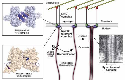
Adrian Bird                                                                                                                            Understanding proteins that stabilise cell identity
                        Co-workers: Beatrice Alexander-Howden, Megan Brown, Kashyap Chhatbar, Sara Giuliani, Jacky Guy,
                        Matthew Lyst, Baisakhi Mondal, Raphael Pantier, Katie Paton, Christine Struthers

     MeCP2 is highly expressed in mature neurons and MeCP2-deficiency causes the profound neurological disorder Rett
     syndrome (RTT), in which neurons show morphological and electrophysiological defects. We previously showed that the
     mouse provides a convincing model of this disorder and found, remarkably, that the severe phenotypes are reversed if the                    A                                                               B
     protein is restored in adulthood. Thus, MeCP2 is dispensable for neurodevelopment, but essential for maintenance of the
     mature neuronal state.
     We have made significant recent progress in elucidating the molecular mechanism underlying MeCP2 function. We showed
     previously that DNA binding by MeCP2 depends on 5-methycytosine in a mCG context. Work by others showed that mCA
     also bound MeCP2 and this was subsequently narrowed down by our demonstration that the trinucleotide mCAC is the
     overwhelmingly prefered non-CG DNA binding motif. Coincidentally, CAC is the preferred non-CG target for the DNA
     methyltransferase DNMT3A and is highly methylated in mature neurons. To determine the biological importance of mCAC
     binding, we replaced the MeCP2 DNA binding domain with that of the related protein MBD2. The MBD2 domain specifically
     binds mCG but does not detectably interact with mCAC in vitro or in vivo. The results showed that mice expressing only the
     domain-swap protein displayed Rett syndrome like phenotypes, indicating that mCAC is an essential MeCP2 target.
     Comparative transcriptomics indicates that MeCP2 functions to restrain expression of large numbers of genes in a DNA
     methylation-dependent manner. Assuming that transcriptional disturbance leads to the neuronal dysfunction that underlies
     RTT, two extreme hypotheses are: 1) RTT is the aggregate outcome of slightly perturbed expression of very many genes;
     2) RTT strongly depends on dysregulation of a few key genes. Our recent work highlights shared dysregulated genes in
     different mouse models with RTT-like phenotypes, allowing a test the second possibility. Specifically, mice expressing
     a chimaeric MeCP2 that is unable to bind mCAC and Mecp2-KO mice both up-regulate genes causally implicated in
     autism-related disorders, including AUTS2, CNTN4, MEF2C, GRIN2A, raising the possibility that their abnormal expression
     contributes disproportionately to RTT. Interestingly, these genes are among the most methylated and highly affected by
     MeCP2 deficiency. Such “convergence” of pathways involved in different intellectual disability syndromes could have
     therapeutic relevance for neurodevelopmental disorders generally.
     A second study published during 2021 involves SALL4 (Figure 1), a multi-zinc-finger protein that plays an important role in
     development and disease (e.g. SALL4 is highly expressed in many cancers with poor prognosis). We identified this protein
     in a screen for proteins that might interpret DNA base composition by recognising AT-rich DNA. Zinc finger cluster 4 of
     SALL4 specifically targets short A/T-rich motifs and recruits a partner corepressor. Inactivation of ZFC4 in embryonic stem                A. A cartoon showing loss of preferential repression of AT-rich genes by SALL4 when the AT binding domain ZFC4 is mutated,
                                                                                                                                                leading to precocious differentiation towards a neuronal fate.
     cells leads to precocious differentiation and up-regulates AT-rich genes that are normally silenced in embryonic stem cells,
     thereby destabilising the pluripotent state. Our SALL4 study provides the first evidence that base composition can be read                 B.Microscopy of mouse embryonic stem cell nuclei showing co-localisation of wildtype SALL4 (WT) with heterochromatic
     as a biological signal to regulate gene expression.                                                                                        foci containing AT-rich DNA (stained with DAPI). When zinc finger cluster 4 is mutated (ZFC4mut), SALL4 becomes dispersed
                                                                                                                                                throughout the nucleus. As a control, we show that staining with the SALL4 antibody is absent when the SALL4 gene is deleted
                                                                                                                                                (S4KO).
     Selected Publications
     Pantier R, Chhatbar K, Quante T, Skourti-Stathaki K, Cholewa-Waclaw J, Alston G, Alexander-Howden B, Lee HY, Cook AG, Spruijt CG,
     Vermeulen M, Selfridge J, Bird A. SALL4 controls cell fate in response to DNA base composition. Mol Cell. 2021 Feb 18;81(4):845-858.e8.
     doi: 10.1016/j.molcel.2020.11.046. Epub 2021 Jan 5. PMID: 33406384; PMCID: PMC7895904.
     Tillotson R, Cholewa-Waclaw J, Chhatbar K, Connelly JC, Kirschner SA, Webb S, Koerner MV, Selfridge J, Kelly DA, De Sousa D, Brown K,
     Lyst MJ, Kriaucionis S, Bird A. Neuronal non-CG methylation is an essential target for MeCP2 function. Mol Cell. 2021 Mar 18;81(6):1260-
     1275.e12. doi: 10.1016/j.molcel.2021.01.011. Epub 2021 Feb 8. PMID: 33561390; PMCID: PMC7980222.
12   Bird A. The Selfishness of Law-Abiding Genes. Trends Genet. 2020 Jan;36(1):8-13. doi: 10.1016/j.tig.2019.10.002. Epub 2019 Oct 29. PMID:                                                                                                                                  13
                                                                                                                                                                                                                                                                                13
     31662191.
2022 Wellcome Centre for Cell Biology
Dhanya Cheerambathur                                                                                                                       Role of microtubule cytoskeleton in building and
                        Co-workers: Mattie Green, Cameron Finlayson, Lana Buzuk, Vasilis Ouzounidis, Emmanuel Fiagbedzi,
                        Henrique Alves Domingos                                                                                                                          regenerating the neural connectome

                                                                                                                                                           A                                                        B                             C
     The central nervous system is a complex network of neurons and supporting cells that form the information relaying unit of
     an organism. During neural development, pioneer neurons extend axons in response to guidance cues from other neurons
     and non-neuronal cells to establish the framework that build the neural circuits. The assembly of this circuit is a highly
     orchestrated event that involves neurite outgrowth, fasciculation (axon bundling) and synapse formation to generate a
     functional nervous system. How these organizational features emerge during development is poorly understood
     Microtubules are critical for neuron formation and function. As neurons develop, microtubules are organized and sculpted
     by the cell machinery to form the axons, dendrites and the neural network. Several human neurodevelopmental disorders
     are linked to mutations in microtubule cytoskeleton-related proteins. Despite the central role of the microtubule, little is
     known about how the microtubule cytoskeleton contributes to the assembly of the neural circuit. We aim to understand
     how the microtubule cytoskeleton uses distinct molecular machinery to build and regenerate 3 dimensional neuronal
     circuits using the simple multicellular organism C. elegans as a model.
                                                                                                                                                           D                                                        E
     During my post-doc, I discovered an unexpected role for kinetochore, the chromosome segregation machinery, in
     developing neurons of C. elegans. Our work showed that the evolutionarily conserved 10 subunit KMN (Knl1-Mis12-
     Ndc80) network, the microtubule coupler within the kinetochore, acts post-mitotically in developing neurons. A similar
     function for kinetochores proteins has also been described in Drosophila and rat hippocampal cultures. KMN proteins are
     enriched in the dendritic and axonal outgrowth during neurodevelopment. Removal of KMN components post-mitotically
     from developing neurons resulted in a disorganized nerve ring, a network of 181 axons and synapses, considered as the
     “brain” of C. elegans. We hypothesize that the kinetochore proteins facilitate nerve ring assembly by promoting the proper
     formation of axon bundles.
     Starting from this unique angle, we aim to understand how the microtubule cytoskeleton integrates distinct molecular
     machinery to build and regenerate 3 dimensional neuronal circuits in C. elegans. Our goal is to 1) define the function of
     the kinetochore proteins in building the nerve ring; 2) build a functional map of microtubule cytoskeleton during nerve ring
     assembly by addressing the function of non-kinetochore microtubule factors; 3) investigate how kinetochore proteins build
     and maintain neuronal network by addressing its role in dendritic branching and regeneration.
                                                                                                                                                   Nerve ring assembly in C.elegans
                                                                                                                                                   A. The C.elegans head nervous system in L1 larvae (PH marks the membrane and histone the cell body). The axon bundle in the
                                                                                                                                                   nerve ring is between white arrowheads (scale 10 mm).
                                                                                                                                                   B. Schematic of KMN network: Mis-12 interface (red) with the centromere, Ndc80 (purple) binds the microtubule and Knl1 (blue)
                                                                                                                                                   functions as a scaffold.
     Selected Publications                                                                                                                         C. Structure of C.elegans nerve ring in control and after post-mitotic degradation of KNL-1 in the neurons. Axon defasciculation
     Cheerambathur, D.K., Prevo, B., Chow, T.-L., Hattersley, N., Wang, S., Zhao, Z., Kim, T., Gerson-Gurwitz, A., Oegema, K., Green, R., et al.   defect (white arrowhead, scale 5 mm).
     (2019). The Kinetochore-Microtubule Coupling Machinery Is Repurposed in Sensory Nervous System Morphogenesis. Dev. Cell.
                                                                                                                                                   D. Fluorescence image and cartoon of the developing nerve ring in C.elegans embryo (pioneer neurons (PN) in purple, amphid
     Cheerambathur, D.K., Prevo, B., Hattersley, N., Lewellyn, L., Corbett, K.D., Oegema, K., and Desai, A. (2017). Dephosphorylation of the
     Ndc80 Tail Stabilizes Kinetochore-Microtubule Attachments via the Ska Complex. Dev. Cell 41, 424–437.e424.
                                                                                                                                                   sensory neurons (ASN) in blue). Note that the ASNs have already extended their dendrites (scale 2.5 mm).
     Cheerambathur, D.K., Gassmann, R., Cook, B., Oegema, K., and Desai, A. (2013). Crosstalk between microtubule attachment complexes             E. Schematic representing the initial stages of nerve ring formation. Insets show the extension and bridging of bilaterally
     ensures accurate chromosome segregation. Science 342, 1239–1242.                                                                              symmetrical PN axons (scale 1 mm).

14                                                                                                                                                                                                                                                                                    15
                                                                                                                                                                                                                                                                                       15
2022 Wellcome Centre for Cell Biology
Atlanta Cook                                                                                                                    Structural biology of macromolecular complexes in RNA
                        Co-workers: Uma Jayachandran, Ola Kasprowicz, Alexander Will, Mickey Oliver, James Le Cornu (iCM student),
                        Atika Al Haisani, Laura Croenen                                                                                                         metabolism and transcriptional silencing

                                                                                                                                                 A
     The expression of individual genes is controlled at the levels of mRNA transcription and also post-transcriptionally, by
     processes such as splicing, localization, modification or editing, and degradation. To gain a mechanistic understanding of
     these processes it is important to understand the interactions between the individual players, including both protein and
     nucleic acid components, at the molecular level. We have used structural approaches to tackle mechanistic questions
     about how protein-RNA interactions can control RNA maturation and RNA editing and how transcriptional repressors are
     recruited to methylated DNA. By combining structural studies with biochemical, biophysical and cell-based functional
     assays we can gain powerful insights into these molecular processes.
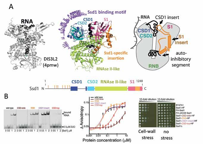
     Recently, we solved a crystal structure of a yeast RNA binding protein, Ssd1, that is important in cell wall biogenesis. It
     is thought that Ssd1 functions by repressing translation of cognate transcripts. Using CRAC, we found that Ssd1 binds
     to specific sequences in the 5’UTRs of a small set of transcripts, several of which encode proteins required for cell wall
     biogenesis. This suggests that Ssd1 functions by blocking ribosome scanning along 5’UTRs. The structure of Ssd1
     shows that it has a classical fold of an RNase II family nuclease. However, RNA degradation activity has been lost by two
     mechanisms. First, the catalytic residues have been altered during evolution. Second, a channel that, in active enzymes,
     allows RNA substrates to funnel into the active site has been blocked. We propose that Ssd1 has evolved a new RNA
     interacting surface.

                                                                                                                                                 B

                                                                                                                                               A. The structure of Ssd1 (middle) compared with the structure of DIS3L2 (left), where RNA is bound, shows the different RNA
     Selected Publications                                                                                                                     binding sites. Domains of Ssd1 are marked in blue (cold shock domain 1, CSD1), cyan (CSD2), green (RNase II-like) and
     Bayne R.A., Jayachandran U., Kasprowicz A., Bresson S., Tollervey D., Wallace E.W.J., Cook A.G. (2021) Yeast Ssd1 is a non-enzymatic      pink (S1). The Ssd1-specific insert is shown in the domain overview (below) and structure in orange. The yellow lollipops are
     member of the RNase II family with an alternative RNA recognition interface. Nucleic Acids Research, DOI: 10.1093/nar/gkab615.            phosphorylation sites. RNA travels down the central channel of DIS3L2 while Ssd1 binds a sequence-specific motif (purple) on
     Pantier R., Chhatbar K., Quante T., Skourti-Stathaki K., Cholewa-Waclaw J., Alston G., Alexander-Howden B., Lee H.Y., Cook A.G., C        the outside of the CSD domains. Two segments of the Ssd1 structure are shown in black – these block the active site funnel. A
     Spruijt C.G., Vermeulen M., Selfridge J., and Bird A. (2021) SALL4 controls cell fate in response to DNA base composition. Mol Cell       cartoon overview of the Ssd1-specific structures is shown on the right.
     81:845-858.e8.                                                                                                                            B. Four sets of point mutations were tested for RNA binding by electrophoretic mobility shift assay (left). Mutations to the side
     Ballou E.R., Cook A.G. and Wallace E.W.J. (2020) Repeated evolution of inactive pseudonucleases in a fungal branch of the Dis3/RNase II   and top of the CSDs block binding to RNA. This is further demonstrated by fluorescence anisotropy assays (middle). Phenotypic
     family of nucleases. Mol. Biol. Evol. doi:10.1093/molbev/msaa324                                                                          assays in yeast show that mutations that prevent RNA binding have a cell wall stress phenotype.

16                                                                                                                                                                                                                                                                                 17
                                                                                                                                                                                                                                                                                    17
Owen Davies                                                                                                                                          Structural biology of meiosis
                       Co-workers: Eleanor Casey, Simona Debilio, Gurusaran Manickam

     How is the chromosome number halved during meiosis to create haploid spermatozoa and oocytes that form
     healthy diploid zygotes upon fertilisation?
     Meiosis involves a unique chromosome choreography in which chromosomes search throughout the cell to find their
     homologous partners, with which they synapse, exchange genetic material by crossing over, and then segregate upon
     cell division. This is achieved by the combined actions of several molecular machines. Firstly, double-strand breaks
     are induced across the genome, triggering recombination searches, which result in the formation of recombination
     intermediates that physically connect matching sequences of homologous chromosome pairs. This is process facilitated
     by rapid chromosomal movements, in which microtubule forces are transmitted via the LINC complex to chromosome
     telomere ends that are tethered to the nuclear envelope by the meiotic telomere complex. Once established, the discrete
     physical connections of recombination are converted into continuous synapsis between homologous chromosomes by
     assembly of the synaptonemal complex, a supramolecular protein structure that ‘zips’ together homologous chromosome
     pairs along their entire length. The assembled synaptonemal complex then facilitates the resolution of recombination
     intermediates, with the formation of crossovers in which diversity is enhanced by the exchange of genetic material between
     homologous chromosome partners that subsequently segregate into daughter cells.
     Our research aims to uncover the structural basis of how the synaptonemal complex, recombination machinery, meiotic
     telomere complex and meiotic LINC complex perform their critical functions in meiosis, and how they operate together as
     an integrated molecular machine. Our main research questions are:
     1. What is the structure, function and assembly mechanism of the synaptonemal complex?
     2. How is meiotic recombination regulated within the synaptonemal complex?
     3. How are meiotic chromosome telomere-ends anchored to the nuclear envelope?
     4. How are cytoskeletal forces transmitted to chromosomes by the meiotic LINC complex?
     We adopt a structural biology approach in which we integrate solution biophysics, high-resolution structure determination
     by X-ray crystallography and Cryo-EM, with EM-based imaging of macromolecular assemblies formed by recombinant
     proteins and within heterologous cellular systems. We translate our structural findings to a functional understanding of
     meiosis through the structure-directed design of separation-of-function mutations that are tested in vivo, in mouse and
     lower organism systems, by our collaborators.
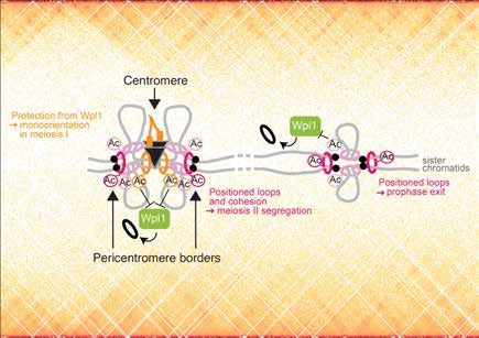
                                                                                                                                           Schematic of recombination and chromosome synapsis in meiosis, highlighting the meiotic LINC complex formed of SUN1-
     Ultimately, we aim to achieve a complete molecular understanding of how the integrated machineries of the synaptonemal                KASH5 (Gurusaran and Davies, 2021), the meiotic telomere complex containing MAJIN-TERB2 (Dunce et al, 2018), and the
     complex, recombination, telomere complex and LINC complex perform the chromosome choreography of meiosis.                             synaptonemal complex.

     Selected Publications
     Dunce, J.M., Salmon, L.J., and Davies, O.R. (2021). Structural basis of meiotic chromosome synaptic elongation through hierarchical
     fibrous assembly of SYCE2-TEX12. Nat Struct Mol Biol 28, 681-693.
     Gurusaran, M., and Davies, O.R. (2021). A molecular mechanism for LINC complex branching by structurally diverse SUN-KASH 6:6
     assemblies. eLife 10.
     Sanchez-Saez, F., Gomez, H.L., Dunne, O.M., Gallego-Paramo, C., Felipe-Medina, N., Sanchez-Martin, M., Llano, E., Pendas, A.M., and
     Davies, O.R. (2020). Meiotic chromosome synapsis depends on multivalent SYCE1-SIX6OS1 interactions that are disrupted in cases of
18   human infertility. Science advances 6.                                                                                                                                                                                                                       19
                                                                                                                                                                                                                                                                   19
Bill Earnshaw                                                                                                              The role of non-histone proteins in chromosome structure
                        Co-workers: Blandine Arleri, Mar Carmena, Fernanda Cisneros-Soberanis, Lorenza Di Pompeo,
                        Moonmoon Deb, Natalia Kochanova, Emma Peat, Elisa Pesenti, Bram Prevo, Nina Pucekova, Caitlin Reid,                                       and function during mitosis
                        Lucy Remnant, Itaru Samejima, Kumiko Samejima

     Over the past year, much of our research focused on structural dynamics in chromatin during the transition of cells from
     G2 phase into mitosis, the role of SMC proteins in mitotic chromosome formation and structure and the structure and
     assembly of the chromosome periphery.
     One highlight was the publication of a study that has been ongoing for several years in which Itaru examined the changes
     in protein association with chromatin during synchronous mitotic entry. This study used Chromatin Enrichment for
     Proteomics (ChEP), a method developed by Georg Kustatscher when he was a postdoc with Juri Rappsilber, and was
     a collaboration between the three labs. We discovered that the earliest events of prophase appear to primarily involve
     changes in RNA processing in nuclei as well as changes in interactions with the nuclear envelope and pores. All of these
     events begin before chromatin condensation is visible, and the study was only made possible by using the chemical-
     genetic system for synchronous mitotic entry developed by Kumiko.
     We are currently writing up the results of our long-running study of interactions between cohesin and condensin during
     mitotic chromosome formation. This is a truly interdisciplinary collaboration with the groups of Job Dekker, Leonid Mirny
     and Anton Goloborodko. We do the genetics, cell biology and imaging. They do Hi-C and polymer modelling, respectively.
     We have discovered that cohesin has a significant effect on mitotic chromosome structure that has been previously
     overlooked and gained surprising new insights into the organisation of the chromatin fiber in chromosomes. Kumiko has
     made many genomic knock-in cell lines, performed the cell synchrony and carried out extensive light microscopy analysis.
     Fernanda and Nina have been performing serial block face scanning electron microscopy with our collaborators Ian Prior
     and Alison Beckett in Liverpool. Itaru has been doing ChEP and Moonmoon has been doing ChIP - both to quantitate the
     amounts of condensin and cohesin on the chromosomes during mitotic entry.
     In other ongoing work, Lucy and Fernanda are studying the enigmatic protein Ki-67 and the RNA/protein-rich mitotic
     chromosome periphery compartment (MCPC), Lorenza is performing a structure/function analysis on CENP-V, Caitlin is
     studying the role of topo IIβ in chromosome formation, Natalia is using proteomics to look at protein conformations and
     interactions during mitotic entry, and Bram is developing new ways to image chromosomes.
     Our work is supported by a Wellcome Principal Research Fellowship and by the Centre for Mammalian Synthetic Biology.

                                                                                                                                            Three-dimensional reconstruction of an anaphase human RPE1 cell. Left, projection of the three-dimensional reconstruction
                                                                                                                                            superimposed on an orthoslice from the electron microscopy map. Corresponding sister chromatids have the same colours.
     Selected Publications                                                                                                                  Right, partial karyotype with individual sister chromatids (identified by size and centromere position) extracted from the map and
     Samejima, I., C. Spanos, K. Samejima, J. Rappsilber, G. Kustatscher & W.C. Earnshaw. (2022). Mapping the invisible chromatin           displayed next to their sisters. Sample preparation and modeling in AMIRA by Fernanda Cisneros-Soberanis. Serial block face
     transactions of prophase chromosome remodelling. MOL. CELL 82:696-708; PMID: 35090599; PMCID: 8823707; DOI: 10.1016/j.                 scanning electron microscopy by Alison Beckett and Ian Prior, University of Liverpool.
     molcel.2021.12.039.
     Paulson JR, Hudson DF, Cisneros-Soberanis F, Earnshaw WC. (2021). Mitotic chromosomes. SEMIN CELL DEV BIOL. 117:7-29. PMID:
     33836947; PMC8406421; DOI:10.1016/j.semcdb.2021.03.014.
     Pesenti,E., M. Liskovykh, K. Okazaki, A. Mallozzi, C. Reid, M.A. Abad, A.A. Jeyaprakash, N. Kouprina, V. Larionov, H. Masumoto, W.C.
     Earnshaw. (2020) Analysis of Complex DNA Rearrangements During Early Stages of HAC Formation. ACS SYNTH BIOL. 9:3267-3287.
     PMID: 33289546; PMC7754191; DOI: 10.1021/acssynbio.0c00326.

20                                                                                                                                                                                                                                                                               21
                                                                                                                                                                                                                                                                                  21
Patrick Heun                                                                                                                           Establishment and maintenance of chromatin identity
                         Co-workers: Mathilde Fabe, Meena Krishnan, Alessandro Stirpe, George Yankson, Hwei Ling Tan

                                                                                                                                                                              Figure 1
     Our lab is interested in the organisation, establishment, and maintenance of specialised chromatin states. Epigenetic
     transmission of centromere identity through many cell generations is required for proper centromere function and when
     perturbed can lead to genome instability and cellular malfunction. We use Drosophila and human tissue culture cells as
     model organisms to address the following questions:
                                                                                                                                                                      1                                                                                       Mitosis

     What is the role of transcription at the centromere?
                                                                                                                                                                                                                          new
     Loading of CENP-A at the centromere occurs outside of S-phase and requires the removal of H3 “placeholder"                                                                                                                A
     nucleosomes. Transcription at centromeres has been linked to the deposition of new CENP-A, although the molecular
                                                                                                                                                                                                                                    Transcription
     mechanism is not understood. Using fast acting transcriptional inhibitors in combination with a newly developed CENP-A
                                                                                                                                                                                                                               H3
     loading system, we demonstrate that centromeric transcription is required for loading of new dCENP-A by removing
                                                                                                                                                                                    maintenance                Spt6                    eviction
     placeholder nucleosomes and promoting dCENP-A transition from chromatin association to nucleosome incorporation
                                                                                                                                                                                                                  A            H3
     (Bobkov et al., 2020). Unlike placeholder nucleosomes, previously deposited CENP-A is specifically retained by Spt6 both
                                                                                                                                                                                                                 old       old
     in human and Drosophila cells, identifying Spt6 as a CENP-A maintenance factor that ensures the stability of epigenetic
                                                                                                                                                                                                                                                      Centromeres            Spt6
     centromere identity (Figure 1). We are currently investigating the molecular mechanism how some histones like CENP-A
     are maintained while others like H3.3 placeholders are evicted to preserve epigenetic centromere identity.                                                                 Figure 1: Model for the role of transcription at centromeres: Transcription remodels
                                                                                                                                                                                centromere chromatin and evicts H3-nucleosomes (green) to allow       new CENP-A (orange)
     How is the centromeric chromatin fiber organised?                                                                                                                2               AEvicted
                                                                                                                                                                                loading.  A old CENP-A (red) is maintained by theC       C       SUMO
                                                                                                                                                                                                                                         transcription
                                                                                                                                                                                                                                                    S elongation factor Spt6.
                                                                                                                                                                                                                                                        X                    S
     To map centromere proteins on the linear centromeric chromatin fiber, we have recently developed a novel approach where
                                                                                                                                                                                                                                                               PIAS      X
     proteins-of-interest fused to Biotin ligases or DNA methyltransferases leave a “footprint” on the underlying nucleosomes                                                                                                                                (SUMO
     through proximity-labelling. With this methodology we have described novel localization patterns of a subset of centromere                                                                                                                             E3-Ligase)

     proteins at human centromeres (Kyriacou and Heun, 2018). We are extending this approach to proteins localising to all
                                                                                                                                                                                                                                                     heterochromatin
     layers of the centromere using different technologies like stretched chromatin fibers and long read-DNA sequencing.
     How does Su(var)2-10/PIAS contribute to heterochromatin organisation?                                                                                                               BB           apical-heterochromatin

                                                                                                                                                                                          wildtype
     The Su(var)2-10 gene has been originally identified in position-effect-variegation (PEV) assays designed to uncover
     proteins involved in heterochromatin formation. Cloning of the gene revealed its homology to the protein family SUMO                                                                             basal-euchromatin
     E3-ligase PIAS (Protein Inhibitor of activated STAT), but how sumoylation promotes heterochromatin formation remains

                                                                                                                                                                                          PIAS RNAi
     unknown. While PIAS does not localise to pericentric heterochromatin in somatic cells, it is enriched next to centromeres in
     early fly embryogenesis, suggesting a role in heterochromatin establishment (Figure 2). We are specifically depleting PIAS
                                                                                                                                                                                                       DAPI                        PIAS             Histone H3K9me3              MERGE
     at this point of development to shed light on the link between PIAS’ SUMO targets and chromatin organisation.

                                                                                                                                                                                          Figure 2: The SUMO E3 Ligase PIAS is required for heterochromatin
                                                                                                                                                                                          formation. A) Embryonic cycle 1-14 (image William Sullivan) B) Fixed
     Selected Publications                                                                                                                                                                cycle 13 embryos showing ⍺PIAS and heterochromatin marker
                                                                                                                                                    Figure 1. Model for the role of transcription at centromeres: Transcription remodels centromere chromatin and evicts H3-
                                                                                                                                                                                          ⍺H3K9me3 in apical heterochromatin in wildtype and PIAS RNAi. C)
     Bobkov, G.O.M., Huang, A., van den Berg, S.J.W., Mitra, S., Anselm, E., Lazou, V., Schunter, S., Feederle, R., Imhof, A., Lusser, A., et al.
                                                                                                                                                    nucleosomes (green) to allow new CENP-A       (orange)   loading.
                                                                                                                                                                                          Proposed role of PIAS        Evicted old
                                                                                                                                                                                                                in chromosome      CENP-A (red) is maintained by the transcription
                                                                                                                                                                                                                               organisation.
     (2020). Spt6 is a maintenance factor for centromeric CENP-A. Nature communications 11, 2919.
                                                                                                                                                    elongation factor Spt6.
     Logsdon, G.A., Gambogi, C.W., Liskovykh, M.A., Barrey, E.J., Larionov, V., Miga, K.H., Heun, P., and Black, B.E. (2019). Human Artificial
     Chromosomes that Bypass Centromeric DNA. Cell 178, 624-639 e619. PMC6657561                                                                    Figure 2. The SUMO E3 Ligase PIAS is required for heterochromatin formation. A. Embryonic cycle 1-14 (image William Sullivan)
     Roure, V., Medina-Pritchard, B., Lazou, V., Rago, L., Anselm, E., Venegas, D., Jeyaprakash, A.A., and Heun, P. (2019). Reconstituting          B. Fixed cycle 13 embryos showing αPIAS and heterochromatin marker αH3K9me3 in apical heterochromatin in wildtype and
     Drosophila Centromere Identity in Human Cells. Cell Rep 29, 464-479 e465.                                                                      PIAS RNAi.­­ C. Proposed role of PIAS in chromosome organisation.

22                                                                                                                                                                                                                                                                                       23
                                                                                                                                                                                                                                                                                          23
Georg Kustatscher                                                                                                                Proteome dynamics: The role of synthesis and degradation in
                         Co-workers: Van Kelly, Savvas Kourtis, Emmanuel Fiagbedzi
                                                                                                                                                                          regulating protein levels

     There is a major discordance between mRNA and protein expression levels in human cells. Why is this so and what
     mechanisms are behind it? Our aim is to understand the principles, mechanisms and regulators that shape the proteome                                  A                                               B                                   D
     at the level of translation and protein degradation. Despite their importance for cancer and other diseases, these regulatory
     processes remain poorly understood, leaving an enormous potential for therapeutic intervention unfulfilled. We aim to
     address three key questions:
     1. What is the role of translation and degradation rates in regulating protein levels, for example when buffering the
     impact of chromosome abnormalities in glioblastoma stem cells.
     2. Which proteins regulate translation and degradation rates, e.g. can we reveal regulatory networks between E3
     ubiquitin ligases and their targets.
     3. Which unconventional translation products exist in healthy and in cancer cells and what are their biological
     functions.
     From a technological perspective we plan to address these questions using a combination of proteomics and
     computational approaches and, where necessary, RNA sequencing. We are currently at the beginning of these projects
                                                                                                                                                           C                                                                                  E
     and focus on the development of the necessary proteomics techniques that will allow us to carry out these investigations.
     The Centre has recently obtained a Sciex tripleTOF mass spectrometer, which is suitable for high-throughput (HT)
     proteomics, a rapidly emerging mass spectrometry approach for the robust quantitation of proteomes in a matter
     of minutes. HT proteomics differs from conventional proteomics on every level of the experimental workflow: the
     chromatography, the mass spectrometer and the data processing. To harvest the power of HT proteomics for the analysis
     of proteome dynamics we are developing DIA-pulse-SILAC, a method that will allow the rapid quantitation of protein
     synthesis and degradation rates by mass spectrometry.
     An important aspect of HT proteomics, and indeed all proteomics experiments, is the statistical analysis and interpretation
     of the data. This is a second area of focus for our group. For example, we recently collaborated with the Earnshaw group to
     create an interactive proteomic map of chromatin transactions during mitotic entry (https://mitoChEP.bio.ed.ac.uk). We are
     also working together with the Ralser lab (Charité, Berlin) to predict the potential function of uncharacterised yeast proteins
     based on the proteomic characterisation of thousands of yeast knock-out strains.

                                                                                                                                                       Development of DIA-pulse-SILAC for the rapid and precise measurement of protein synthesis and degradation.
                                                                                                                                                       A. SILAC titration series created by mixing defined ratios of light and heavy extracts from RPE1 cells. In a direct comparison our
     Selected Publications                                                                                                                             new DIA-SILAC workflow (DIA; data-independent acquisition) quantifies considerably more proteins than the traditional data-
     Samejima, I., Spanos, C., Samejima, K., Rappsilber, J., Kustatscher, G.*, and Earnshaw, W.C.* (2022). Mapping the invisible chromatin             dependent acquisition (DDA) of SILAC samples. The DIA workflow also quantifies the same set of proteins more consistently
     transactions of prophase chromosome remodeling. Mol. Cell 82, 696–708.e4. * co-corresponding authors                                              across replicates
     Kelly, V., Al-Rawi, A., Lewis, D., Kustatscher, G., and Ly, T. (2022). Low Cell Number Proteomic Analysis Using In-Cell Protease Digests
                                                                                                                                                       B. The DIA workflow quantifies the same set of proteins more consistently across replicates.
     Reveals a Robust Signature for Cell Cycle State Classification. Mol. Cell. Proteomics 21, 100169.
     Nakamura, K.*, Kustatscher, G.*, Alabert, C., Hödl, M., Forne, I., Völker-Albert, M., Satpathy, S., Beyer, T.E., Mailand, N., Choudhary, C., et
                                                                                                                                                       C. The DIA workflow quantifies proteins more precisely than the DDA workflow.
     al. (2021). Proteome dynamics at broken replication forks reveal a distinct ATM-directed repair response suppressing DNA double-strand            D. Schematic of a pulse-SILAC experiment.
     break ubiquitination. Mol. Cell 81, 1084–1099.e6. *joint first authors                                                                            E. DIA-pulse-SILAC was used to quantify synthesis and degradation rates in RPE1 cells. Shown are two representative example
                                                                                                                                                       proteins with slow and fast turnover rates, respectively.
24                                                                                                                                                                                                                                                                                          25
                                                                                                                                                                                                                                                                                             25
You can also read