Precision Nutrition and the Microbiome, Part I: Current State of the Science
←
→
Page content transcription
If your browser does not render page correctly, please read the page content below
Review

Precision Nutrition and the Microbiome, Part I:
Current State of the Science
Susan Mills 1, Catherine Stanton 2, Jonathan A. Lane 3, Graeme J. Smith 3,* and R. Paul Ross 1,*
 1 APC Microbiome Ireland, University College Cork, Cork T12 K8AF, Ireland; susan.mills@ucc.ie
 2 APC Microbiome Ireland, Teagasc Food Research Centre, Fermoy P61 C996, Co Cork, Ireland;
   catherine.stanton@teagasc.ie
 3 H&H Group, Technical Centre, Global Research and Technology Centre, Cork P61 C996, Ireland;

   jonathan@hh.global
 * Correspondence: graeme.smith@hh.global (G.J.S.); p.ross@ucc.ie (R.P.R.)

 Received: 21 March 2019; Accepted: 17 April 2019; Published: 24 April 2019

 Abstract: The gut microbiota is a highly complex community which evolves and adapts to its host
 over a lifetime. It has been described as a virtual organ owing to the myriad of functions it
 performs, including the production of bioactive metabolites, regulation of immunity, energy
 homeostasis and protection against pathogens. These activities are dependent on the quantity and
 quality of the microbiota alongside its metabolic potential, which are dictated by a number of
 factors, including diet and host genetics. In this regard, the gut microbiome is malleable and varies
 significantly from host to host. These two features render the gut microbiome a candidate ‘organ’
 for the possibility of precision microbiomics – the use of the gut microbiome as a biomarker to
 predict responsiveness to specific dietary constituents to generate precision diets and interventions
 for optimal health. With this in mind, this two-part review investigates the current state of the
 science in terms of the influence of diet and specific dietary components on the gut microbiota and
 subsequent consequences for health status, along with opportunities to modulate the microbiota
 for improved health and the potential of the microbiome as a biomarker to predict responsiveness
 to dietary components. In particular, in Part I, we examine the development of the microbiota from
 birth and its role in health. We investigate the consequences of poor-quality diet in relation to
 infection and inflammation and discuss diet-derived microbial metabolites which negatively
 impact health. We look at the role of diet in shaping the microbiome and the influence of specific
 dietary components, namely protein, fat and carbohydrates, on gut microbiota composition.

 Keywords: personalised nutrition; precision nutrition; probiotics; prebiotics; gut microbiome;
 immunity; metabolic disease; gut; genetics

1. Introduction
     The human gastrointestinal tract (GIT) is considered one of the most densely populated
ecosystems on our planet, purported to harbour ~1013 microorganisms [1] referred to as the gut
microbiota, whose activities have significant consequences for the host in terms of health and
disease. This is unsurprising given that the entire genetic content of the human gut microbiota,
commonly referred to as the gut microbiome, is estimated to exceed human genomic content by a
factor of ≥ 100 [2], with vast genetic potential to contribute to host physiology. It is composed of the
three domains of life, Bacteria, Archaea, and Eukarya (fungi, protozoans and metazoan parasites), as
well as eukaryotic and prokaryotic viruses (bacteriophages).
     The study of the gut microbiome has been revolutionised over the last 15 years by advances in
genetic methods and sophisticated bioinformatic tools. Next generation sequencing is a low-cost,

Nutrients 2019, 11, 923; doi:10.3390/nu11040923                               www.mdpi.com/journal/nutrients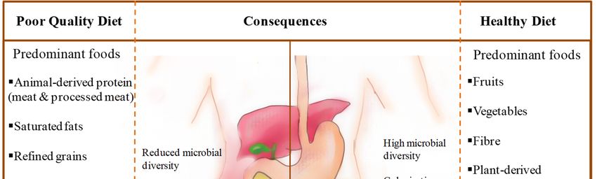
Nutrients 2019, 11, 923                                                                           2 of 45

high-throughput sequencing platform that enables analysis of all the genomes within an ecosystem
sample (shotgun metagenomics), or a description of the taxa within a given community by
sequencing conserved marker genes, such as the 16srRNA gene of bacteria and archaea (marker
gene metagenomics) thus removing the requirement for cultivation of clonal cultures [3]. Indeed,
such is the interest in the gut microbiota, it has been reported that in 2017 alone, approximately 4000
research papers on the topic were published, with more than 12,900 papers published in the
preceding four years [4], and to date the bacterial component has received the most attention. This
vast array of data has contributed to a deeper understanding of the role of the gut microbiome in
health and disease, the factors which shape it, and the potential to harness its therapeutic potential
for optimal health.
     The gut microbiota serves the host by interacting directly or indirectly with host cells, the latter
of which comes about through microbially-produced bioactive molecules, thus the microbiota is
capable of regulating numerous biological pathways involved in immunity and energy homeostasis,
while also protecting the host against pathogens through colonisation resistance. The dysbiotic
microbiota, which has deviated from the ‘healthy’ status in terms of diversity and functionality, has
been implicated in a range of diseases, including inflammatory bowel diseases (IBDs) [5], cancer [6],
neuropsychiatric disorders [7] and cardiometabolic diseases, including obesity, type 2 diabetes and
cardiovascular disease [8]. These diseases tend to inflict those living a Westernised lifestyle. Indeed,
greater economic development has been shown to correlate with significantly lower within-host gut
microbiota diversity, a common feature of the dysbiotic microbiota [9].
     Many researchers have suggested that the gut microbiome can be considered as a virtual organ
but unlike any other organ in the human body, the gut microbiome represents a source of significant
inter-individual variation, rendering the analysis of it ever more complicated. However, this vast
inter-individual variation is now being seized upon as an opportunity to utilize the microbiome for
precision medicine [10,11] and personalised nutrition [12]. Indeed, along with the human genome,
the human microbiome has been implicated as the main source of human variation modulating
dietary responses [12]. This variation presumably goes a long way to explaining the pandemic surge
in metabolic diseases despite the fact that universal advice and education on healthy eating practices
have never been more readily available.
     Therefore, the overall aim of this two-part review is to investigate the potential of the
microbiome for precision microbiomics, particularly in relation to nutrition. In this regard, precision
microbiomics can be described as the use of the gut microbiome as a biomarker to predict the effect
of specific dietary components on host health and the use of these data to design precision diets and
interventions that ensure optimal health.
     In Part I, we describe the development of the gut microbiota throughout life along with the
factors that shape its composition and how it contributes to host health. We look at dietary practices
and specific dietary components that influence the composition and functionality of the gut
microbiota and the consequences for human health with a focus on infection, inflammation and
metabolic disease. In Part II, we focus on the potential use of the microbiome to prevent disease and
promote health. We present research regarding opportunities to modulate the microbiota for
prevention of over- and under- nutrition through probiotics, prebiotics and fibre and we look at the
impact of environment and life-stage on the gut microbiota and health and dietary strategies and
interventions for optimising health. Finally, in Part II, we provide evidence from recent research that
the gut microbiome has potential to serve as a biomarker of human responsiveness to diet and assess
where this current knowledge-base places us in terms of putting precision microbiomics into
everyday practice and discuss the value of present-day commercial microbiome testing.

2. Development of Microbiota from Birth/Young versus Mature versus Aged Microbiota
     Humans encounter their initial gut microbiota in very early life with most studies focusing on
colonisation after birth and the microbial assembly that ensues. Some studies have led to the
hypothesis that colonisation begins in utero by providing evidence of a placental microbiome in
healthy pregnancies, the presence of microorganisms in the amniotic fluid and meconium. These
Nutrients 2019, 11, 923 3 of 45
studies have been presented and critically assessed in a recent review by Perez-Muňoz et al., [13].
The authors concluded that there is insufficient evidence to date to support the ‘in utero colonisation
hypothesis’, since the studies lacked appropriate controls for contamination, the molecular
approaches used were incapable of studying low-biomass microbial populations and evidence of
bacterial viability was not provided. Thus, for the purpose of this review, we have focused on gut
microbiota development from birth onwards.
Although gut microbiota composition varies considerably between infants certain patterns
have been identified. Several factors have been shown to influence microbial colonisation of the gut
at this stage, including gestational age, birth mode, sanitation, antibiotic exposure, feeding regime
and host genetics [14–20]. However, facultative anaerobes have been identified as the initial
colonisers [21]. Depletion of the available oxygen creates the necessary environment for the
establishment of strict anaerobes [16,21]. Anaerobes which colonise the gut in the early days and
weeks of life include Bifidobacterium, Bacteroides, Clostridia and Parabacteroides [16,18,21,22]. Although
the diversity of the microbiota is generally low at this stage, dominated largely by members of the
Actinobacteria phylum in the case of full-term spontaneously vaginally delivered infants, it has been
shown to be greatest in this cohort compared to full-term infants delivered by caesarean section
(dominated by Firmicutes) or pre-term infants (dominated by Proteobacteria) at one week old [16].
Interestingly, by week 24, no significant differences in alpha diversity were recorded between any of
these groups [16]). Diversity increases with age with a gradual increase in the presence of Firmicutes
and Bacteroidetes by the first year [17] (Canadian infants); [20] (Canadian infants); [18] (Swedish
infants) where the introduction of solid food to the diet has been identified as an important step in
the succession of the microbiota [22] (Spanish infants), and [23] (Danish infants). In terms of
metabolic function, genes involved in the de novo biosynthesis of folate have been shown to be
enriched in the infant microbiome across three different populations (Malawian rural communities,
Amerindians from the Amazonas of Venezuela, families from the USA) relative to adults [24]. By
three years of age, obligate anaerobes have been shown to dominate the microbiota in breast-fed
infants [21] which is trending towards an adult-like composition. The establishment of a stable
adult-like microbiota occurs between 2–5 years of age and is dominated by Firmicutes and
Bacteroidetes [25–27].
Few studies have specifically investigated the pre- adolescent and adolescent microbiota of
healthy humans. However, those which have investigated these age groups indicate that the
microbiota has not yet reached the adult-state and is providing essential functions towards the
developmental process of its human host. The gut microbiota of children is more stable than that of
infants with composition largely influenced by dietary habits and geography [28]. The
pre-adolescent microbiota (7–12 years of age) is still in a state of immaturity and based on
observations from a group of children from Houston, Texas, has been shown to be more diverse and
harbor significantly greater abundances of Firmicutes and Actinobacteria than observed in healthy
adults [26]. The pre-adolescent microbiome was also found to be enriched in functions potentially
involved in ongoing development, such as vitamin B12 synthesis and de novo folate synthesis
relative to the adult microbiome. In terms of the adolescent microbiota, Agans et al., [29] identified a
core microbiota of 46 species common to both adults and adolescents (11–18 years) who consumed a
standard Western diet, however, the abundances of the genera Bifidobacterium and Clostridium were
significantly higher in adolescents relative to adults.
The healthy adult gut microbiota is composed primarily of the phyla Firmicutes and
Bacteroidetes, and to a lesser extent the phyla Actinobacteria, Proteobacteria, and Verrucomicrobia
[30,31]. As with any age-group, due to the extensive inter-individual variation, it has been virtually
impossible to define the composition of the ‘healthy’ adult gut microbiota. However, the
‘enterotype’ concept was introduced in 2011 [32]) when faecal metagenomes of individuals from
America, Europe and Japan were found to be dominated by one of three different bacterial
communities, namely Bacteroides (enterotype 1), Prevotella (enterotype 2) or Ruminococcus (enterotype
3). The enterotype ‘concept’ has since been used in other studies when evaluating the gut microbiota
as we will see further on, although further analysis has resulted in the identification of only twoNutrients 2019, 11, 923 4 of 45
enterotypes, one of which is dominated by Prevotella and the other by Bacteroides which have been
linked to long term carbohydrate-, or animal fat and protein-rich diets, respectively [33]. More
recently, it has been suggested that Prevotella and Bacteroides should be interpreted as ‘biomarkers’ of
diet, lifestyle and disease state given that gradients of both Bacteroides and Prevotella have been found
within gut communities as opposed to distinct and consistent community taxa [34].
Reports on the estimated number of species and strains in an individual have varied greatly but
in a study examining the stability of the gut microbiota in 37 US adults over a five year period, Faith
et al., [35] reported an average of 101 ± 27 species and 195 ± 48 strains in the faecal gut microbiota of
each individual with family members sharing strains, which was not observed in unrelated
individuals. However, other studies estimate the number of species to be greater than 1000 [36,37].
More recently, Forster et al., [38] presented the Human Gastrointestinal Bacteria Culture Collection
which consisted of 737 whole-genome-sequenced bacterial isolates, representing 273 different
species, including 105 novel species from 31 families found in the human gastrointestinal
microbiota. The healthy adult microbiota has been shown to be stable over long periods of time [35]
but can be influenced by a number of factors. These include geographical location [24,39] albeit diet
would appear to be an important contributing factor in this regard [40–42], direct antibiotic usage
[43], and indirectly by consumption of antibiotic containing animal derived products, such as beef
and chicken as a result of their use in livestock production [44], non-antibiotic drugs [45], illness,
injury [46,47] and hormonal changes [48]. The healthy gut microbiota is generally characterised by
rich species diversity [49] which has been found to be reduced/altered in individuals with certain
diseases, particularly those typified by a dysregulated immune response (as discussed further on).
Ageing has a significant impact on the gut microbiota with dramatic compositional and
functional changes observed in the elderly microbiota (in general >65 years). Several physiological
and lifestyle changes associated with the ageing process may be contributing factors resulting in
changes in dietary habits and ultimately nutrition, including a decline in dentition and salivary
function, a reduction in digestion and absorption, due to gastrointestinal dysmotility, changes in
appetite as a result of prescribed drugs and psychological state, or changes in living conditions, such
as residential care or hospitalisation [50]. Gastric hypochlorhydria which is associated with ageing
and is prevalent in individuals experiencing or who have experienced Helicobacter pylori infection
can cause malabsorption and bacterial overgrowth in the small intestine [50,51].
In general, the elderly microbiota has been characterised by a decline in microbial diversity, an
increase in the abundance of opportunistic pathogens and a decrease in species associated with short
chain fatty acid (SCFA) production, in particular butyrate [52]. The inter-individual variation
observed in the gut microbiota of adults is even greater in the elderly cohort. Indeed, while
Bacteroidetes was found to be the dominant phylum in an elderly Irish cohort (age 65 years and
older), the proportion of Bacteroidetes within individual composition datasets ranged from 3% to
92%, while the proportion of Firmicutes ranged from 7% to 94% [53]. Mariat et al., [54] reported a
change in the ratio of Firmicutes: Bacteriodetes with age, which was recorded as 0.4 in infants (3
weeks to 10 months old), 10.9 in adults (25–45 years old) and 0.6 in the elderly cohort (70-90 years), of
which the latter two consumed an unrestricted Western-type diet. In a study involving 178 elderly
Irish subjects (65–96 years), Claesson et al., [55] identified distinct microbiota composition groups as
a result of residence location (community versus day-hospital versus rehabilitation versus long-stay
residential care) which also overlapped with diet (low fat/high fibre versus moderate fat/high fibre
versus moderate fat/low fibre versus high fat/low fibre, respectively). The gut microbiota of people
in long-stay care was found to be significantly less diverse than that of healthy community dwellers,
which was found to be more similar to healthy young adults. Furthermore, increased frailty as
observed in the less healthy, long-stay subjects correlated with a loss of community-associated
microbiota. Indeed, a distinct negative correlation between frailty and gut microbiota alpha diversity
has been reported [56]. In the same study, the species Eubacterium dolichum and Eggerthella lenta were
found to be more abundant with frailty, while a Faecalibacterium prausnitzii operational taxonomic
unit (OTU) was less abundant in frailer individuals. Interestingly, F. prausnitzii is an important
butyrate producer [57], while E. lenta is considered a pathogen [58]. More recently, Haran et al., [59]Nutrients 2019, 11, 923 5 of 45
also reported lower abundances of butyrate-producing organisms in the microbiota of American
elderly nursing home residents (age 65 years or older) consuming a low-fibre, typical nursing home
diet with increasing frailty and higher abundances of recognised dysbiotic species. Increasing age
was also associated with a decrease in the abundance of microbiome-encoded genes and pathways
associated with vitamin B, a nitrogenous base and essential amino acid production.
Interestingly, the centenarian microbiota (99–104 years) has been shown to significantly differ
from that of young adults (25–40 years) and even elderly subjects (63–76 years) [60] (Italian subjects),
being characterised by an enrichment in facultative anaerobes, mainly pathobionts, and a
rearrangement in the Firmicutes population with a marked decrease in symbiotic species associated
with anti-inflammatory properties e.g. F. prausnitzii and relatives. Increased inflammatory markers
were also identified in centenarian blood samples, indicative of increased inflammatory status.
Rampelli et al., [61] also reported a rearrangement in Firmicutes in Italian centenarians (99–102
years), which was not observed in 70-year-old subjects. In this study a functional description of the
coding capacity of an ageing human cohort revealed a decrease in saccharolytic potential and an
increased abundance of proteolytic functions in the centenarian microbiome. Remarkably, a study
investigating the semi-supercentarian microbiota (105–109 years old) revealed not only a decrease in
saccharolytic butyrate producers (Faecalibacterium, Coprococcus, Roseburia) and an increase in
potential opportunistic bacteria, as expected, but also an enrichment and/or higher prevalence in
health associated bacterial groups, including Akkermansia, Bifidobacterium and Christensenellaceae.
Whether these species were present at a younger age or related to past lifestyle or genetics is not
known [62].
In certain regions of the world, “healthy” ageing is the norm and the average lifespan of
residents surpasses general averages. Goatian village in Liuyang city of Hunan province in
mid-South China is such an example, which boasts an average lifespan of 92, much higher than the
average 74.83 years for China in general, along with a lack of chronic diseases amongst its people.
An analysis of the gut microbiota in its long-living elderly residents (ages spanning from 50 to >90
years) revealed much greater species diversity than that observed in the control group (healthy
subjects from other areas in China, average age of 50 years) [63]. Similarly, Bian et al., [64] reported
that the overall gut microbiota composition of healthy aged Chinese subjects was similar to that of
much younger people, reporting little differences between individuals from 30 to >100 years which
the authors speculate may be a consequence of the healthy lifestyle and diet of the study subjects.
3. Role of Microbiome in Health, Development and Immune Functioning
The gut microbiome is integral to the health of its host, serving a myriad of functions. It
provides essential nutrients and bioactive metabolites, which can be produced directly by the
microorganisms or indirectly by microbial conversion of host or environmental molecules. It is
involved in energy regulation (as discussed in Section 5). It prevents pathogen colonisation directly
or indirectly through a phenomenon referred to as colonisation resistance. It sustains the integrity of
the mucosal barrier and is an essential component in the orchestration of immune functioning within
the gut. The bidirectional interactions within the brain-gut-microbiome axis, in which the gut
microbes communicate to the central nervous system, have been demonstrated largely by preclinical
and some clinical studies [65]. Alterations in brain-gut-microbiome communication have been
implicated in various disease states, from irritable bowel syndrome to psychiatric and neurologic
disorders, and this is an area of research that has the potential for identification of novel therapeutic
targets and therapies [66]
3.1. Nutrients and Bioactive Metabolites
Fruits, vegetables and cereals are major components of the human diet providing essential
carbohydrates and dietary fibres, although digestion of the latter is beyond the scope of the human
genome [67]. Cantarel et al., [68] identified only 17 enzymes within the human genome to
breakdown carbohydrate nutrients which included starch, lactose and sucrose. Thus, plant cell wall
polysaccharides and resistant starch, which constitute most dietary fibres and cannot be digested orNutrients 2019, 11, 923 6 of 45
absorbed in the small intestine, enter the large intestine and undergo microbial breakdown and
subsequent fermentation [67]. The microbiota also feeds on animal-derived dietary carbohydrates
(glycosaminoglycans and N-linked glycans from cartilage and tissue), host epithelial glycome, and
microbe-derived carbohydrates from resident gut microbes or foodborne microbes [69,70].
Collectively, the carbohydrates consumed by the microbiota have been termed “microbiota
accessible carbohydrates” (MACs) [70].
Carbohydrate active enzymes (CAZymes) breakdown MACs into fermentable
monosaccharides [67]. For example, the gut bacterium Bacteroides thetaiotaomicron was recently
shown to metabolise the most structurally complex plant polysaccharide known,
rhamnogalacturonan-II, using a highly specific enzyme system [71]. Bifidobacterium longum strains
derived from the infant gut were shown to be capable of metabolising human milk oligosaccharides
[72]. In-silico analysis of CAZyme profiles in the guts of 448 individuals from diverse geographies
and age groups revealed 89 CAZyme families which were present across 85% of the gut microbiome
and revealed several geography/age-specific trends in the CAZyme repertoires of individuals [73].
The major end products of microbial fermentation of the resulting monosaccharides are the SCFAs,
including butyrate, propionate, and acetate which reach a combined concentration of 50–150 mM in
the colon at a ratio of 1:1:3, respectively [74]. They are rapidly absorbed by the intestinal epithelial
cells where they are involved in a number of cellular and regulatory processes [75,76] with only 5%
excreted in faeces [77]. Butyrate is mainly produced by Firmicutes, propionate by Bacteroidetes, and
acetate by most gut anaerobes [78]. Butyrate is the main energy source for the epithelial cells [79] and
plays an important role in brain function [80]. It is also known for its anti-cancer [81–83] and
anti-inflammatory properties [77,84] and for its role in the development of the intestinal barrier
[85–87]. Propionate contributes to gluconeogenesis in the liver [86] and, along with butyrate, has
been shown to activate intestinal gluconeogenesis, albeit both use different circuits [88]. Propionate
derived from the gut microbiota has also been shown to reduce cancer cell proliferation in the liver
[89]. The SCFAs are also involved in regulating immune responses, a topic which has been
extensively reviewed by Corrêa et al., [90]. For example, acetate has recently been shown to promote
intestinal antibody IgA responses to the gut microbiota via the G protein coupled receptor GPR43
[91]. Intestinal IgA is specialised in protecting the mucosa [92]. These SCFAs also stimulate secretion
of gut hormones, such as glucagon-like peptide 1 (GLP-1) and plasma peptide YY (PYY) involved in
appetite regulation and satiety from enteroendocrine cells [93,94] proposedly through the SCFA
receptors GPR41 and GPR43 [95,96], thus playing a role in energy regulation in the body.
Unsurprisingly, changes in the production of these compounds as a result of disturbances to the gut
microbiota can result in pathological consequences for the host. As an example, increased acetate
production from an altered gut microbiota in a rodent model was shown to promote metabolic
syndrome [97].
The gut microbiota is also responsible for the biosynthesis of several essential vitamins,
including B vitamins, such as cobalamin, folic acid, biotin, thiamine, riboflavin, nicotinic acid,
pyrodixine and pantothenic acid, as well as vitamin K [98]. Interestingly, Arumugam et al., [32]
observed vitamin biosynthesis pathways across the three identified enterotypes; however,
enterotype 1 was enriched in the biosynthesis of riboflavin, biotin, ascorbate and pantothenic acid,
while enterotype 2 was enriched in the biosynthesis of thiamine and folic acid.
Primary bile acids are produced in the liver from dietary cholesterol and cholesterol derived
from hepatic synthesis, and their main function is to aid absorption of dietary lipids and lipid
soluble nutrients [99]. However, bile acids are also important signalling molecules and are known to
activate a number of nuclear receptors, including farnesoid X receptor (FXR), preganane X receptor,
and vitamin D receptor, as well as the G-protein-coupled receptor TGR5, and cell signalling
pathways in the liver and GIT thus modulating their own biosynthesis, as well as glucose, lipid, and
energy metabolism [100]. In humans, 200-800 mg of bile acids escape enterohepatic circulation every
day, pass into the colon and are metabolised by bacteria to secondary bile acids [99]. In a mouse
model, such secondary bile acids produced through the action of bacterial bile salt hydrolase (BSH)
have been shown to regulate weight gain, lipid metabolism and cholesterol levels via regulation ofNutrients 2019, 11, 923 7 of 45
key genes in the liver or small intestine [101]. The gut microbiota has also been shown to inhibit bile
acid synthesis in the liver by alleviation of FXR inhibition in the ileum [102].
In recent years, there has been a growing appreciation for the ability of the gut microbiota to
produce neurochemicals that can influence the peripheral enteric and central nervous systems [103].
For example, gamma amino butyric acid (GABA) is a major inhibitory neurotransmitter in the brain
[104] and neuropsychiatric disorders, including anxiety and depression have been linked to GABA
system dysfunction [105]. Strains of culturable lactobacilli and bifidobacteria from the human
intestine were shown to produce GABA, namely Lactobacillus brevis, Bifidobacterium dentium,
adolescentis and infantis [106]. Furthermore, GABA was found to serve as a growth factor for a
previously uncultured gut bacterium, Flavonifractor sp. which was shown to ferment GABA [107]. In
the same study, the authors identified several gut bacteria capable of producing GABA which
included Bacteroides, Dorea, Parabacteroides, Alistipes and Ruminococcus species. More recently, a
co-culture experiment revealed that GABA produced by Bacteroides fragilis was essential for the
growth of a gut isolate termed KLE1738 which is believed to be an unreported bacterial genus [108].
This led to the isolation of a variety of GABA-producing bacteria and the Bacteriodes species in
particular were found to produce large quantities of GABA. Furthermore, in the same study relative
abundance levels of faecal Bacteriodes negatively correlated with brain signatures associated with
depression in patients with major depressive disorder. Bacterially-produced GABA is generated by
the enzyme glutamate decarboxylase (GAD) which catalyzes the irreversible α-decarboxylation of
glutamate to GABA and is believed to protect the microorganism against stomach acidity [109].
GAD is also found in higher plants and animals [110]. Interestingly, daily consumption of a
GABA-producing Bif. dentium gut isolate was found to modulate sensory neuron activity in a rat
faecal retention model of visceral hypersensitivity revealing that bacterially-produced GABA can
modulate abdominal pain [111]. GABA-enriched black soybean milk fermented with a
GABA-producing fish gut isolate, L. brevis, generated similar antidepressant activity in rats as the
common antidepressant drug, fluoxetine, but without the side effects normally associated with the
drug, such as appetite loss and reduced body weight [112].
Serotonin (5-hydroxytryptamine, 5-HT) is a brain neurotransmitter and performs regulatory
functions in the gut and other organ systems [113]. It is derived from the amino acid tryptophan and
plays an important role in the regulation of mood [114] such that several antidepressants act on
serotonin transporters in the brain [115]. Yano et al., [113] demonstrated that human- and
mouse-derived gut bacteria promote serotonin biosynthesis in colonic enterochromaffin cells which
supply serotonin to the lumen, mucosa and circulating platelets. Spore forming bacteria, dominated
by clostridial species, were found to elicit this effect. Furthermore, conventional mice were found to
have 2.8 times more plasma serotonin levels than their germ-free counterparts [116]. This
peripherally produced molecule does not pass the blood brain barrier under physiological
conditions [117] but is an important signalling molecule in the gut involved in peristalsis, secretion,
vasodilation, pain perception and nausea, as well as promoting inflammation and being involved in
neuron development and maintenance in the enteric nervous system, while platelet serotonin
derived from the gut influences bone development amongst other functions [118,119]. The
mechanisms involved in gut microbiota-mediated serotonin biosynthesis have not yet been fully
elucidated but are thought to be linked to microbial metabolite-stimulation of the enzyme
tryptophan hydroxylase 1 in enterochromaffin cells which produces a serotonin precursor that is
subsequently metabolised to serotonin [113]. In particular, rectal injection with the microbial
metabolites deoxycholate, p-aminobenzoate, α-tocopherol and tyramine, were shown to increase
colonic and serum concentrations of serotonin in mice [113].
Metabolism of tryptophan in the gut results in tryptophan catabolites which have profound
effects on the host [120]. Direct transformation of tryptophan by intestinal microbes results in the
formation of several molecules, including ligands for the ligand-activated aryl hydrocarbon
receptors (AhRs) [117]. AhRs, which are transcription factors, are expressed by several cells of both
the adaptive and innate immune systems [121]. Upon binding of AhR to its ligand molecule, the
activated transcription factor translocates into the nucleus of the cell where it mediates cell-specificNutrients 2019, 11, 923 8 of 45
transcriptome changes [122]. Thus, AhR signalling plays a key role in immune functioning in health
and disease. Lamas et al., [123] showed that intestinal inflammation in mice harbouring a microbiota
incapable of metabolizing tryptophan was attenuated following treatment with Lactobacillus strains
capable of activating AhRs through the production of tryptophan metabolites. In the same study,
faecal samples from healthy subjects induced significantly greater AhR activation compared to
faecal samples from IBD subjects, the latter of which harbored significantly less tryptophan and
tryptophan metabolites. The tryptophan-metabolising strain Peptostreptococcus russellii provided a
protective effect against colitis in mice, which the authors suggest is linked to its ability to produce
the tryptophan metabolite and AhR ligand, indoleacrylic acid which mitigates inflammatory
responses and promotes intestinal barrier function [124]. The authors also noted a diminishment of
tryptophan-metabolising capacity in stool samples of IBD patients. The metabolite indole has been
shown to enhance epithelial barrier function and attenuate indicators of inflammation [125,126].
3.2. Colonisation Resistance
The intestinal microbiota protects its host against colonisation by exogenous pathogens and
prevents the overgrowth of potentially pathogenic endogenous members, referred to as colonisation
resistance [127]. This phenomenon is elicited through competition for nutrients and colonisation
sites, direct inhibition of pathogens through the production of antimicrobial substances, and
indirectly through modulation of the luminal environment and via host-commensal interactions
involving the epithelial barrier function, modulation of the host cell surface and the host immune
system [128].
Members of the established microbiota are controlled by ‘substrate competition,’ defined as the
superior ability of a species/strain to utilise one or a few substrates over other species and the control
of that population by the limited concentration of these substrates [129]. Furthermore, one
microorganism’s by-product can serve as substrate for another [127]. In this regard, nutrient
resources in the gut are in huge demand and simultaneously limited, making it challenging to
become established or outcompete with resident microbiota. Indeed, nutrient utilisation by the
colonic microbiota of mice was shown to restrain the growth of Clostridium difficile, since it was
incapable of competing with the mouse microbiota for the available carbon sources [130].
Unfavourable environmental conditions created as a result of commensal fermentations can also
inhibit the growth of undesirable microorganisms. The utilisation of human milk oligosaccharides,
particularly the dominant secretor associated oligosaccharide 2’-fucosyllactose, by infant strains of
bifidobacteria led to an increase in their proportions, an increase in lactate concentration and a
subsequent reduction in pH which was shown to decrease the proportions of Escherichia coli and
Clostridium perfringens during in vitro anaerobic fermentations [131]. Furthermore, consumption of
butyrate by epithelial cells as an energy source has been implicated as important in maintaining a
hypoxic environment in the gut lumen [132]. Decreased intestinal butyrate levels due to depletion of
a commensal butyrate producer in a mouse model resulted in increased epithelial oxygenation and
aerobic expansion of Salmonella enterica serovar Typhimurium [133]. Butyrate has also been shown to
down-regulate virulence gene expression in Salmonella [134].
In terms of niche competition, Lee et al., [135] identified specific colonisation factors conserved
within the Bacteroides genus, one of the most prominent genera of the human microbiota. The
specific genetic locus referred to as commensal colonisation factors (ccf) was shown to be
up-regulated in Bac. fragilis during gut colonisation, especially at the colonic surface such that the
strain was able to reside deep within the crypt channels whereas ccf mutants were defective in terms
of crypt association. Such species-specific physical interactions with the host provide an example of
direct competition for niche occupation.
The gut microbiota is a rich reservoir of bacteriocin producers [136,137]. Bacteriocins are
ribosomally synthesised peptides with antimicrobial activity against either a broad range of species
or a narrow range of closely-related species. Their mode of action varies depending on the
bacteriocin class, but they generally exert their antimicrobial activity by forming pores in the target
cell (Classes I and II), by degrading cell wall peptidoglycan (Class III bacteriolysins) or interferingNutrients 2019, 11, 923 9 of 45
with cellular processes (Class III non-lytic bacteriocins) [138]. The genetic machinery for bacteriocin
synthesis is encoded in gene clusters or operons where many of the genes are conserved. Based on
this knowledge, Walsh et al., [139] identified 74 bacteriocin gene clusters within the genomes of the
GIT subset of the Human Microbiome Project’s reference genome database using an in-silico
approach of which the most commonly identified were bacteriolysins, then lantibiotics and
sactibiotics. Thuricin CD is an example of a sactibiotic bacteriocin produced by a human gut isolate,
Bacillus thuringiensis [140]. While it has a narrow spectrum of inhibition, thuricin CD is capable of
killing a wide range of C. difficile isolates, its antimicrobial activity being as potent as the antibiotics
vancomycin and metronidazole, but without the concomitant damage to other members of the
microbiota [141]. Bacteriocin production can also aid niche occupation of the producing strain.
Indeed, bacteriocin production in Enterococcus faecalis harboring the bacteriocin-encoding
conjugative plasmid pPD1 was shown to replace indigenous enterococci and out-competed E. faecalis
strains lacking the plasmid in a mouse model [142]. Supplementing mice with bacteriocin-producing
strains resulted in transient advantageuous changes, such as inhibition of Staphylococcus by
enterocins and Enterococcus by garvicin and promotion of LAB by sakacin, plantaricins and garvicin
[143].
Other antimicrobials generated by the gut microbiota can also aid colonisation resistance. For
example, a single bacterial species, namely Clostridium scindens, was shown to confer colonisation
resistance against C. difficile infection in vivo [144]. In this instance, secondary bile acids generated by
C. scindens from host-derived bile cells were found to inhibit the pathogen.
3.3. Immunity and Mucosal Integrity
Commensal interactions with the host promote immune system maturation and immune
homeostasis through complex microbiota-host networks although much of our understanding of
these mechanisms today have been extrapolated from animal or in vitro studies [145–147]. We have
already seen how several microbial metabolites play essential roles as signalling molecules for the
immune system, such as the SCFAs and tryptophan metabolites. More specifically, a polysaccharide
(PSA) produced by Bac. fragilis was shown to promote cellular and physical maturation of the
developing immune system in mice [148]. Interestingly, this species is an early colonizer of the infant
gut [149] and plays an important role in the development of the infant immune system. A 15 kDa
protein produced by F. prausnitzii, a commensal microbe deficient in Crohn’s disease patients, was
shown to have anti-inflammatory properties, decreasing activation of the NF-κB pathway and also
prevented colitis in an animal model [150]. M-cells are specific phagocytic epithelial cells which
sample particulate antigens [151]. Rios et al., [151] showed that efficient induction of IgA is brought
about by M-cell sampling of commensal microbes. Paneth cells (a specialized intestinal epithelial
lineage) sense enteric bacteria through activation of the MyD88-dependent toll-like receptor which
results in the induction of several antimicrobial factors that are essential for controlling bacterial
translocation across the intestinal barrier [152]. Colonisation by segmented filamentous bacteria was
shown to induce the maturation of T-cell responses in a gnotobiotic mouse model, suggesting these
microbes could play a role in the postnatal maturation of the gut immune system [153]. These
studies provide a snapshot of how commensals modulate host immunity. The importance of the
microbiota for host immunity can also be appreciated from the consequences of its absence in germ
free animals. Indeed, germ free animals exhibit reduced expression of IgA and antimicrobial
peptides and are deficient in Peyers’ patches [128,151,154,155].
Goblet cells are specialised epithelial cells that secrete mucus, composed primarily of
O-glycosylated proteins called mucins, resulting in the formation of a mucus layer whose
composition and density is influenced by the commensal microbiota [128,156,157]. The mucus layer
creates a protective barrier for the epithelial cells making it difficult for pathogens to gain access to
epithelial cell receptors [127]. Germ free mice have been shown to have an extremely thin colonic
mucus layer which can be restored to levels observed in conventional mice following exposure to
bacterial products, including peptidoglycan and lipopolysaccharide [158]. Mice treated with the
antibiotic metronidazole were shown to have a thinning of the mucus layer which correlated with anNutrients 2019, 11, 923 10 of 45
increased attachment of the mouse pathogen Citrobacter rodentium [159]. Certain commensals have
been shown to modulate mucin gene expression and glycosylation patterns [160–162]. This may be
achieved through the activity of SCFAs which have been shown to increase expression of
mucin-associated genes [163]. Furthermore, the SCFA butyrate provides energy for the epithelial
cells and has also been implicated in enhancing the intestinal barrier by up-regulating the tight
junction protein, Claudin-1 [87].
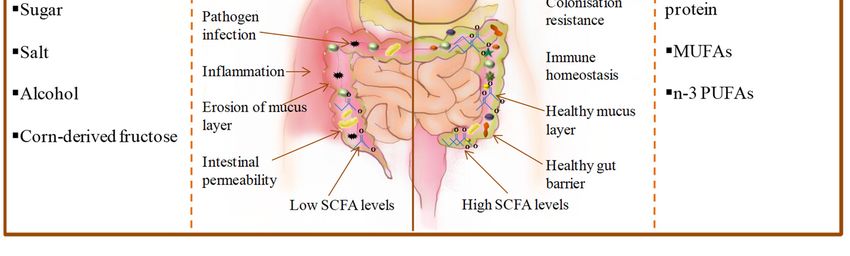
4. Low Diversity of the Microbiota Caused by Poor Quality Diet Linked With Risk of Infections
and Inflammation Predominantly
Dysbiosis is a term used to describe imbalances in gut microbiota communities and is linked
with disease when these imbalances negatively impact microbiota functions required for health or
when they promote disease occurrence [164]. For a number of these diseases, the dysbiotic state is
manifested as a reduction in microbial diversity and often an increase in facultative anaerobes
relative to the ‘healthy’ gut microbiota [164], such as IBD [165–167], cancer [168], liver disease
[169,170] and recurrent C. difficile infection (CDI) [171]. Such diseases tend to primarily inflict those
living a Westernised lifestyle and consuming a Western diet which is characterised by low fruit and
vegetable intake and high consumption of animal-derived protein (meat and processed meat),
saturated fats, refined grains, sugar, salt, alcohol and corn-derived fructose [172–175] (Figure 1).
Whether these changes in microbial diversity are the cause or consequence of these diseases is as yet
unconfirmed, although having reviewed a number of studies, Mosca et al., [176] suggest the
argument for a causal effect is strong in the case of several human conditions, a stance we concur
with in terms of inflammation and infection based on the body of research presented here.
That diet influences the composition of the gut microbiota has been confirmed in several studies
[177–183]. Even short-term dietary changes (four days) have been shown to alter human gut
microbiota composition [178]. In this particular study, David et al., [178] reported that the entirely
animal-based diet (meats, eggs and cheeses) had a greater impact on gut microbiota composition
than the plant-based diet (grains, legumes, fruits and vegetables), resulting in a decrease in plant
polysaccharide-metabolising Firmicutes (Roseburia, Eubacterium rectale, and Ruminococcus bromii) and
an increase in the abundance of bile-tolerant microorganisms presumably owing to the increase in
bile acid secretion as a result of high fat intake [184]. Indeed, the animal-based diet significantly
increased the levels of faecal deoxycholic acid (DCA), a secondary bile acid produced by microbial
dehydroxylation of bile, and has been shown to promote liver cancer in mice [185], and inhibit the
growth of Bacteroidetes and Firmicutes members in rats [186]. Microbial genes required for DCA
production exhibited significantly higher expression on the animal-based diet. Furthermore, the
abundance and activity of the sulfite-reducing bacterium, Bilophila wadsworthia, was also shown to be
increased on the animal-based diet.Nutrients 2019, 11, 923 11 of 45
Figure 1. Comparison of consequences of poor-quality diet versus a healthy diet on the gut and gut
microbiota (MUFAs = monounsaturated fatty acids; PUFAs = polyunsaturated fatty acids).
In mouse models, this particular microorganism has been shown to cause IBD, which is thought
to be due to its production of hydrogen sulfide which inflames intestinal tissue [187]. Overall
microbial gene expression was strongly linked to diet, thus as expected, the animal-based diet
resulted in lower levels of carbohydrate fermentation end-products, the SCFAs. Despite the increase
in dietary fibre, four days of the plant-based diet did not increase the microbiota diversity of
participants, most likely owing to the short time frame. Two days after the animal-based diet ended,
the gut microbiota of participants reverted to their original structure. In a study investigating the
associations between long-term dietary habits and lifestyle, and short-term dietary changes, with gut
microbiota composition, Klimenko et al., [179] reported that alpha diversity was positively linked to
the number of vegetables consumed in long-term dietary patterns.
In certain rural regions of the world, communities continue to live a traditional lifestyle and
thus consume diets resembling those of our early ancestors which are naturally high in fibre. For
example, inhabitants of a rural African village in Burkina Faso still consume a high fibre diet similar
to that of early human settlements at the time of the birth of agriculture. A comparative study
examining the gut microbiota composition of the children of Burkina Faso versus European children
(from Florence, Italy) consuming a Western diet (age of participants, 1−6 years) revealed a
significantly higher richness and biodiversity in the gut microbiota of the Burkina Faso group [180].
The African children also displayed an enrichment of Bacteroidetes and depletion of Firmicutes
relative to their European counterparts. Within the Bacteroidetes, the genera Prevotella and
Xylanibacter were uniquely abundant in the African children being absent in the European children.
These specific bacteria harbor genes for cellulose and xylan hydrolysis. In contrast, the potentially
pathogenic Enterobacteriaceae (Shigella and Escherichia) were significantly over-represented in
European children compared to their African counterparts. Furthermore, SCFAs were significantly
more abundant in the African children. The authors hypothesise that the gut microbiota of the
Burkina Faso children evolved with their polysaccharide-rich diet and protects them from
inflammation and non-infectious colonic diseases. The traditionally-living Hadza people of
Tanzania are one of the last hunter-gatherer communities in the world. A recent study reported no
evidence of cardiovascular disease risk factors in this population [188] and older studies reported
that this group of people had relatively low rates of metabolic diseases, infectious diseases or
nutritional deficiencies compared to other settled groups in the surrounding regions [189–191]. A
comparison of the gut microbiome of the Hadza people with an Italian urban cohort revealed higherNutrients 2019, 11, 923 12 of 45
levels of microbial richness and diversity in the Hadza group [192]. These studies suggest that the
Western microbiota, even in a healthy person, may in fact be dysbiotic in terms of microbial
diversity owing to the low consumption of MACs, and predisposes its host to a range of diseases,
particularly those which are characterised by an inappropriate immune response, a theory which
has been proposed by Sonnenburg and Sonnenburg [70].
The link between inflammation and low microbiota diversity was corroborated in a study
involving 123 non-obese and 169 obese Danish individuals [193]. Within this collective group of 292
subjects, two groups could be differentiated by the number of gut microbial genes and thus bacterial
richness. The ‘low gene count’ (LGC) group represented 23% of the total population studied and
included a significantly higher proportion of obese subjects. The LGC group was characterised by a
more pronounced inflammatory phenotype, marked overall adiposity, insulin resistance, and
dyslipidaemia. Obese individuals within the LGC group were found to gain more weight over time.
Only a few bacterial species were sufficient to distinguish between the LGC group and the ‘high
gene count’ (HGC) group but interestingly anti-inflammatory species, such as F. prausnitzii [194]
were more prevalent in HGC individuals while potentially pro-inflammatory species associated
with IBD, Bacteroides and Ruminococcus gnavus [195–197], were more frequently found in LGC
individuals. In an accompanying intervention study involving 49 obese and overweight subjects of
whom 40% was defined as LGC, a similar phenomenon in terms of clinical parameters and
inflammatory status was observed such that the authors concluded that LGC individuals are at
increased risk of obesity-associated co-morbidities [177]. Members of the LGC group were found to
consume fewer fruits and vegetables and fewer fishery products than the HGC group. An
energy-restricted diet with increased fibre intake for six weeks resulted in an increase in microbial
gene richness in the LGC group which approached but remained significantly different to that of the
HGC group. Both groups showed a loss in body fat mass and an improvement in clinical phenotypes
(lipid and insulin levels and insulin resistance) and a trend towards a decrease in inflammation (as
measured by highly sensitive C-reactive protein) though the effects were more pronounced for the
HGC group. Although this was a short-term intervention study, it suggests that measures of gene
richness and microbial diversity may help predict the efficacy of interventions. Furthermore, it
seems that long term improvements to dietary habits may be required to improve and stabilise gut
microbial diversity which agrees with the observations of Klimenko et al., [179] who reported
considerable correlations between long-term dietary habits and gut community structure.
Loss of microbial diversity is also associated with increased risk of infection, presumably due to
loss of colonisation resistance. For example, human studies have shown that the presence of the gut
pathogen C. difficile is associated with decreased gut microbiota diversity in CDI patients [198–201],
as well as in asymptomatic carriers [199]. Gu et al., [201] also reported a dramatic increase in
endotoxin-producing opportunistic pathogens and lactate-producing phylotypes in CDI patients.
Community richness and diversity were significantly lower in the gut microbiota of
methicillin-resistant Staphylococcus aureus (MRSA)-positive patients compared to individuals
without MRSA [202]. The alpha diversity of the gut microbiota of children suffering from acute
infectious diarrhoea caused by rotavirus was significantly less diverse than those of healthy children
[203]. In this case, probiotic intervention for five days resulted in recovery from diarrhoea. By day 3,
diarrhoea symptoms had ceased, by days 10 and 30 after the intervention, microbiota diversity had
increased to the point that it was no longer significantly different from healthy children. Future
studies are required to determine the exact role of the microbiota in diarrhoea-related processes.
Other viral infections have also been associated with low-diversity dysbiosis, including hepatitis C
[204] and HIV [205].
While the exact mechanisms underlying the link between low microbial diversity, diet and
disease are not fully understood, SCFAs undoubtedly play a role given that low MAC diets are
directly linked to low SCFA levels [70,180,182]. Livanos et al., [206] reported a significant decrease in
the proportion of the SCFA-producing Clostridial Clusters IV/XIVa in intensive care unit patients 72
hrs following hospital admission which was associated with reduced gut microbiota diversity and
community stability over time. Simultaneously, the facultative anaerobe Enterococcus significantlyNutrients 2019, 11, 923 13 of 45
expanded. These changes were associated with receipt of broad-spectrum antibiotics. The depletion
of SCFAs and in particular butyrate producers has been reported in cases of CDI and asymptomatic
carriage of C. difficile, in sufferers of nosocomial diarrhoea and in MRSA-positive patients [199–202].
The potential importance of butyrate in maintaining an oxygen-depleted environment in the lumen
and impeding colonisation by facultative anaerobes has already been mentioned [133]; however, its
specific role, if any, in colonisation resistance against C. difficile infection has not yet been elucidated
and thus the viable bacteria themselves are most likely the responsible agents [200,207]. Indeed, the
loss of a specific butyrate-producing species, C. scindens, a member of the Clostridium XIVa clade
[208], was directly associated with susceptibility to C. difficile infection in a mouse model as a result
of antibiotic treatment [144]. Administration of C. scindens alone or in combination with three other
bacteria to antibiotic-treated mice ameliorated CDI. In this case, secondary bile acids produced by C.
scindens were found to be responsible for the anti-C. difficile effect. However, a recent study reported
the presence of C. scindens and C. difficile in the same stool sample and suggested that the former
does not inhibit the latter [209] but the study does not provide data on bile acid profiles or the 7-α
-dehydroxylating activity of C. scindens, the enzyme responsible for secondary bile acid production.
Furthermore, Sonnenburg et al., [210] showed that in the absence of dietary polysaccharides, a
human gut microbe turned to host mucus glycans as a nutrient source. Mouse models have shown
that a defective mucus barrier enables contact between epithelial cells and bacteria resulting in
spontaneous colitis in mice [211], a feature shared with sufferers of ulcerative colitis [212]. But a
direct connection between dietary fibre and the status of the colonic mucosal barrier was more
recently presented in a mouse study by Desai et al., [213]. In this study, a dietary-fibre deprived gut
microbiota resorted to host-secreted mucus glycoproteins, which resulted in the erosion of the
colonic mucus barrier and enabled the gut pathogen Cit. rodentium greater access to the epithelial
cells, resulting in lethal colitis. In another study, mice fed a Western style diet presented with an
altered gut microbiota composition that resulted in increased permeability and reduced growth rate
of the inner mucus layer compared with mice fed a CHOW diet [214]. However, administration of
Bif. longum or the fibre inulin prevented mucus defects. Inulin prevented penetrability of the inner
colonic mucus layer while Bif. longum restored mucus growth.
The Winning the War on Antibiotic Resistance (WARRIOR) project which is being undertaken
by researchers from the University of Wisconsin aims to investigate the relationship between dietary
fibre intake, the gut microbiota and colonisation by multi-drug resistant microorganisms using 600
randomly selected Wisconsin residents over the age of 18 of which the main results will be published
in a peer-reviewed journal [215]. The results of this study should help us to further delineate the role
of diet and the microbiota in protecting against pathogen invasion.
5. Diet-Derived Microbial Metabolites - Many Are Beneficial but Specific Metabolites Are
Associated with Risk of Metabolic Disease
Metabolic disease refers to any disease in which the normal metabolic processes in the body are
disrupted and examples include obesity, type 2 diabetes and metabolic syndrome, all of which are
risk factors for cardiovascular disease. Obesity and overweight are described as abnormal or
excessive fat accumulation that negatively impacts health [216] and according to the WHO, obesity
has tripled since 1975. Indeed, in 2016, 39% of adults aged 18 years and over were described as
overweight and 13% as obese [216]. A recent study found that body mass index (BMI) had a J-shaped
association with overall mortality among 3.6 million adults in the UK [217]. Obesity results from
ingestion of excess energy which does not get expended and is a strong risk factor for type 2
diabetes, the latter of which is characterised by high blood sugar levels, insulin resistance and
relative lack of insulin [218]. In 2014, the WHO [219] estimated that 422 million adults had diabetes,
of which the majority were inflicted with type 2 diabetes. Diabetic dyslipidaemia which describes
high levels of triglycerides in blood plasma, increased levels of small, dense, low-density lipoprotein
(LDL) cholesterol and decreased levels of high-density lipoprotein (HDL) cholesterol is associated
with insulin resistance and is a risk factor for cardiovascular disease in diabetic individuals
[218,220]. Metabolic syndrome describes the abnormal metabolism of glucose and lipids and isYou can also read