A Combination of Coconut Fiber Suture and Tamarind Seed Gel with Dehydrated Human Amnion Membrane for Wound Surgery in Rats
←
→
Page content transcription
If your browser does not render page correctly, please read the page content below
Hindawi
Advances in Materials Science and Engineering
Volume 2021, Article ID 8122989, 12 pages
https://doi.org/10.1155/2021/8122989

Research Article
A Combination of Coconut Fiber Suture and Tamarind Seed
Gel with Dehydrated Human Amnion Membrane for Wound
Surgery in Rats

          Raghu Babu Pothireddy ,1 Angeline Julius,2 Manu Thomas Mathai,1
          Ganesh Lakshmanan,3 and Beimnet Asfaw Hailemariam 4
          1
            Department of Zoology, Madras Christian College, Affiliated to University of Madras, Chennai 600059, Tamil Nadu, India
          2
            Centre for Materials Engineering and Regenerative Medicine, Bharath Institute of Higher Education and Research,
            Chennai 600126, Tamil Nadu, India
          3
            Department of Anatomy, Asan Memorial Dental College and Hospital, Chennai 603105, Tamil Nadu, India
          4
            Institute of Biotechnology, Addis Ababa University, Addis Ababa, Ethiopia

          Correspondence should be addressed to Raghu Babu Pothireddy; raghubabu_84@yahoo.com and Beimnet Asfaw Hailemariam;
          beimnet.asfaw@aau.edu.et

          Received 8 July 2021; Accepted 10 August 2021; Published 19 August 2021

          Academic Editor: Ravichandran M

          Copyright © 2021 Raghu Babu Pothireddy et al. This is an open access article distributed under the Creative Commons Attribution
          License, which permits unrestricted use, distribution, and reproduction in any medium, provided the original work is
          properly cited.

          Today, there are over 2,000 different biomaterials used for various medical applications, but none of these biomaterials are 100%
          compatible with all human beings. Coconut fiber is widely available but has not been tested as a safe natural alternative for sutures.
          Immature coconut fiber is nonabsorbable and is effective for cuts and open wounds when used in combination with dehydrated human
          amnion membrane (dHAM). Immature coconut fiber, tamarind seed polysaccharide (TSP), and dHAM were prepared to test their
          combinational effect on wound healing in rats. TSP enhanced cell viability, proliferation, and migration in human skin cells and cured
          wounds both individually and in combination with dHAM. An antibiotic-free combination of the human amniotic membrane with
          intact epithelium, tamarind seed polysaccharide, and immature coconut fiber provided faster wound healing. Significantly higher wound
          healing was seen on the 11th day based on an initial 10 mm biopsy punch surgery in Wistar rats compared to control groups. Histological
          studies revealed thickened dermis edges with more neutrophil infiltration. Collagen deposition in the dermis was homogeneous across
          the excised skin tissue in the test group, again attesting to the utility of this procedure. This research signifies the use of TSP gel together
          with the amnion membrane representing a “smart patch” with wound healing potential, which would encourage further research on the
          smart patch made using a combination of plant and animal biological materials.

1. Introduction                                                               various ailments since ancient time [3]. Wound dressings
                                                                              made of pectin and collagen enhance wound healing but are
Wound healing is a natural and complex process of tissue                      highly expensive [4]. Identification of potent and effective
recovery of injured tissues involving growth factors and                      natural compounds for wound healing would benefit in the
cytokines, released at the injured site. Delayed or impaired                  management of wounds in a cost-effective manner.
wound healing may occur due to several reasons like chronic                       The present research relates to the use of plant materials
medical conditions and medications that inhibit the healing                   and biological membrane together as biocompatible bio-
process [1]. Medicinal plants with wound healing properties                   materials for wound healing. Coconut fibers are available
have been used to treat acute and chronic wounds for the                      plenty in India and are used for different purposes. The scope
past three decades [2, 3]. Among the world population, 70 to                  of this research is to come up with this novel use of immature
80% depend on medicinal plants for the management of                          coconut fiber along with other novel combinations of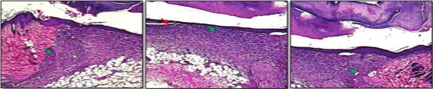
2                                                                            Advances in Materials Science and Engineering

biomaterials for cut and open wound healing studies. Co-         human amniotic membrane (HAM) lacks immunogenicity
conut fiber of green coconuts is immature and tough because       and acts as a substrate for growth, adhesion, and migration
of the presence of lignin [5] and the presence of biode-         [18]. The wound healing ability of HAM accounts for the
gradable hemicellulose and cellulose that contribute to          presence of growth factors such as EGF, KGF, and HGF to
wound healing [6, 7]. The coconut fiber material has never        aid wound healing [19]. The three biomaterials used for the
been thought of as a suture, nor has been used as a cheaper,     study are biowastes, which were used positively for wound
safer, economically viable, and easily available suture ma-      healing, and this research could cause a great impact on the
terial to date. This green, alternative, nonabsorbable suture    identification of novel biomaterials that could work in
(Indian Patent no. 298076) is effective when compared with        combination to provide high healing efficiency.
commercial nonabsorbable sutures such as prolene, silk, and          This study employs plant and animal biomaterials to
nylon.                                                           treat cut and open wounds. Figure 1 illustrates the prepa-
    Xyloglucans of Tamarindus indica L. have been currently      ration of immature coconut fiber, tamarind seed polysac-
explored for its property of wound healing, individually or in   charide, and dehydrated human amnion membrane
combination to heal wounds by enhancing cell viability,          (dHAM) to test wound healing in rats. The use of plant and
proliferation, and migration in human skin keratinocytes         human tissue combinations for wound healing and man-
[8]. Xyloglucans are polysaccharides, which are the main         agement had made this research novel in its attribute that
constituents of the tamarind seed kernel and are rich in         has not been performed or reported before.
xylose and galactoxylose substituents. Due to their me-
chanical properties, they have a wide application in hydrogel    2. Materials and Methods
production, films, and as drug delivery agents for slow drug
delivery [9]. Xyloglucan is abundantly found in plant cell       2.1. Preparation and Evaluation of Physical Parameters of
walls, contains (β1⟶4)-linked d-glucan substituted with               Immature Coconut Fiber
xylose, possesses mucoadhesive properties mainly due to the
mucin-like structure, and belongs to the group of poly-          2.1.1. Preparation of Immature Coconut Fiber Suture.
saccharides, referred to as hemicelluloses [10]. The             The fiber of green coconuts was removed from the shell of
mucoadhesive property of xyloglucan has permitted its use        the nut and was soaked in water for 24 to 48 hours to allow
as an adhesive with antimicrobial property to prevent            the fiber to be separated into strands. The fiber strands are
bacterial adherence and invasion [11]. Xyloglucans when          then soaked into 70% isopropyl alcohol for decolourization
introduced into nanofibrillated cellulose (NFC) through           for 5 hours and dried in a hot air oven between 40 and 50°C
adsorption and presorption to strengthen the NFC revealed        for 1 hour.
highest adsorption, reinforcement, enhancement of cell
growth, and proliferation for wound healing [12].                2.1.2. Determination of Tensile Strength of Coconut Fiber
    Hemicellulose films have proved to be haemostatic,            Using Universal Testing Machine (UTM). The thickness of
absorptive, and bactericidal and have shown effective epi-        each fiber was measured (in diameter) using a dial thickness
thelial wound healing in leukaemia patients with herpes          gauge. The average diameter of the fibers (n � 3) was noted to
zoster infections [13]. A natural hydrogel from honey in         determine the tensile strength. Each fiber was inserted into
combination with polyvinyl pyrrolidone, polyethylene gly-        the universal testing machine and ensured that the ends were
col, and agar solution showed a significant wound healing         gripped symmetrically so that the tension force was dis-
effect compared to the control groups. The hydrogel dem-          tributed uniformly over the cross section. The load cell value
onstrated histopathologically confirmed reduction in wound        was set to zero, and the speed of the moving grip was 10 mm/
size and has been recommended for burn injuries due to a         min. Changes in the test length were noted throughout the
high fluid absorption rate [14]. Furthermore, a porous            test and were continued until the break of the test sample.
hydrogel (size: 32.8–101.6 μm) from a mixture of chitosan        Three samples of thin and thick immature coconut fibers
and xyloglucan with good mechanical properties has en-           were taken in comparison with prolene and silk sutures [20].
hanced the properties of chitosan with the addition of
xyloglucan, without affecting its antimicrobial activity for
wound dressing [15]. Since xyloglucans have shown positive       2.1.3. Skin Holding Effect of Coconut Fiber, Prolene, and Silk
effects on wound healing [16], hydrogels of xyloglucans           Sutures in Rats. Sprague Dawley (SD) rats (14 numbers)
could exhibit wound healing action and also act as a vector      were used for the study, and they were anesthetized with
for slow drug delivery to aid healing. This work focuses on      ketamine and xylazine and acclimatized for 7 days. Animals
extraction, identification of polysaccharide consisting of        were randomly divided into two groups, with 7 animals in
xyloglucan from the kernel of tamarind, preparation of the       each group. Group 1 was tested with thin coconut fiber in
wound gel by crosslinking with epichlorohydrin, and pro-         comparison with the prolene suture. Group 2 was tested with
viding a platform for the intervention of efficacious and cost-    thick coconut fiber in comparison with silk suture.
effective wound healing agent.                                        Two 3.5 cm long parallel full-thickness skin incisions
    The human amniotic membrane (HAM) has been                   were made under aseptic conditions on the back of the
proved to be an excellent source of material for wound           experimental rat. The incisions were closed immediately by 4
therapy [17], since it induces reepithelialization meanwhile     simple sutures (Figure 2). Rats were sacrificed by carbon
processing antiangiogenic and antimicrobial properties. The      dioxide inhalation. The skin wounds were removed from the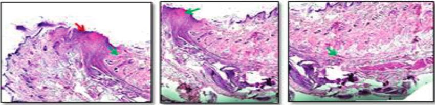
Advances in Materials Science and Engineering                                                                                    3

                                                  Tamarind seed kernel powder           Collection of the placental
                        Preparation of                                                           sample
                     Immature thick and
                      thin coconut fiber
                                                      Extraction of TSP[30]

                                                                                         Preparation of Amnion
                                                                                           from chorian [39]
                   Tensile strength testing       Identification of Xyloglucan

                                               FTIR          NMR              TGA
                                                                                            Gamma sterilization
                    Histological studies
                        in rat model
                    (Immature Coconut         TSP cross-linking with Epichlorohydrin
                     fiber, prolene and                                                       Animal studies
                        silk sutures)                                                        (Biopsy punch)-
                                                                                                 Wound
                                                           MTT Assay                         measurement and
                                                                                               Histological
                                                                                                 studies

                                                         Animal studies
                                                        (Biopsy punch)-
                                                       Wound measurement
                                                        and Histological
                                                            studies

Figure 1: Combination therapy for cut and open wounds. TSP: tamarind seed polysaccharide; FTIR: Fourier-transform infrared spec-
troscopy; NMR: nuclear magnetic resonance; TGA: thermogravimetric analysis; MTT: 3-(4,5-dimethylthiazol-2-yl)-2,5-diphenylte-
trazolium bromide.

                                                                     its spine below the neck region in the dorsal aspect [21, 22].
                                                                     One side of the excision was treated with the prepared TSP
                                                                     gel (approximately 0.25 ml of thawed gel twice daily for 5
                                                                     days) and the other side was not treated (control).
                                                                          Postcreation of the wound, the animals were given a
                                                                     broad-spectrum antibiotic, amoxicillin (0.001 mL/kg b.w,
                                                                     intramuscularly, single dose), and anti-inflammatory/anal-
                                                                     gesic agent, piroxicam (3 mg/kg b.w, intramuscularly daily
Figure 2: SD rat sutured with immature coconut fiber and silk         for 3 days), and monitored for any signs of active infection
suture.                                                              for the first two days. At the end of the study period (after 7
                                                                     days), the animals were euthanized using a gas chamber
                                                                     filled with isofluorane fumes. The wound area with the
body after 24, 48, 72, 96, 120, 144, and 168 h (n � 1/group/         surrounding tissue was excised to its full depth and fixed in
time point). Histopathological analyses were performed               10% neutral buffered formalin and processed for routine
using hematoxylin-eosin, Azur, PAS, and van Gieson stained           histopathology.
slides.

                                                                     2.3. Combined Wound Surgery with the Prepared Biomaterials
2.2. TSP Gel Wound Healing Ability in Wistar Rats. Male              in the Rat Model. The wound was created using a 10 mm
Wistar albino rats (90 days old) weighing around 200 g to            biopsy punch on animals used for the study. One of the
250 g were used for the study. The animals (n � 3) were fed          excised wounds was treated with the prepared biomaterials
with standard laboratory diet in the pellet form, and the rats       (application of approximately 0.25 ml TSP gel on wound
had access to drinking water and libitum. Under intra-               area with dHAM placed on top and sutured with immature
muscular injections of a combination of ketamine (40 mg/kg           coconut fiber). Another excision wound was untreated
body weight (b.w) and xylazine (15 mg/kg b.w), the dorsal            (control) in Wistar rats of group 1 (n � 3). Similarly, one
aspect of the rats was shaved. An excision punch biopsy was          excised wound of group 2 animals (n � 3) was treated with
done passing through both sides of the lifted midline,               TSP gel, applied on the surface of the wound area (ap-
achieving two 8 mm diameter excision wound side by side to           proximately 0.25 ml) with dHAM placed on top and sutured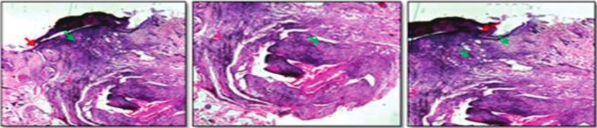
4                                                                                Advances in Materials Science and Engineering

with immature coconut fiber. The other wound incision was            3.3. Histological Investigation. The epidermis of the control
treated with the commercial silicone gel membrane and               animals was thickened at its cut edges. The dermis close to
sutured with the commercial silk suture.                            the excision area showed rich polymorph nuclear infiltra-
    Postcreation of the wound, the animals were given a             tion. A demarcation line was formed, which separated the
broad-spectrum antibiotic, amoxicillin (0.001 mL/kg b.w,            necrotic slough tissue from viable tissue. Mild fibroblast
intramuscularly, single dose), and anti-inflammatory/anal-           proliferation was noted in the dermis region beneath the
gesic agent, piroxicam (3 mg/kgb.w, intramuscularly daily           wound. Neovascularisation in the form of capillary blood
for 3 days), and monitored for any signs of active infection        vessel formation was noted. However, new collagen for-
for the first two days. At the end of the study period (after 11     mation was minimally seen (Figure 8(a)).
days), the animals were euthanized using a gas chamber                  In TSP gel treated animals, the wound edges are ap-
filled with isofluorane fumes. The wound area with the                proximated and the dermis edges are thickened with more
surrounding tissue was excised to its full depth and fixed in        polymorph nuclear infiltration. Fibroblast proliferation is
10% neutral buffered formalin and processed for routine              well noted with collagen deposition noted along the wound
histopathological examinations.                                     area. New blood vessel formation was well marked
                                                                    (Figure 8(b)).
3. Results and Discussion
                                                                    3.4. Combination Therapy Involving Natural Biomaterials for
3.1. Analysis of Immature Coconut Fiber
                                                                    Wound Management. Group I animals (n � 3), 10 mm
3.1.1. Mechanical Testing of Immature Coconut Fiber.                wounds, sutured with immature coconut fiber with prepared
The tensile strength of the immature coconut fiber was tested        dHAM and TSP gel in combination, had better healing after
using the universal testing machine (UTM).Table 1 lists the         11 days. Profound wound reduction was observed in the
parameters for tensile strength estimation.                         treated wound area with a measurement of 2 mm (±0.10),
                                                                    differing much with the nontreated control measuring 6 mm
                                                                    (±0.10) (Figure 9(c)) Difference between the two groups was
3.1.2. Histopathological Examination of Sutures in Wistar           tested using the Student t-test and was found to be statis-
Rats. The skin sections from all the groups revealed spurs of       tically significant (p < 0.001). Following the study, the an-
epithelial cell migration towards the wound edges in the            imals were euthanized, and the wound area was processed
epidermal layer and acute neutrophilic infiltration in the           for histopathological examinations.
dermis and presence of necrotic myofibers of the injured                 Group II animals (n � 3) with 10 mm wound excisions
skeletal muscles in the deepest part of the wounds from day 1       treated with dHAM, TSP gel, and immature coconut fiber
to day 3 with similar severity grades.                              (Figure 10(a)) were compared with 10 mm wound excisions,
    On days 4 to 6, the epithelial cell proliferation resulted in   treated with silicone gel membrane and silk suture as the
a thickened epidermal layer. While in the dermis, neutro-           positive control. Wound measuring 1.5 mm (±0.17) at the
philic infiltrations were largely replaced by macrophages            test site (dHAM + TSP gel + coconut fiber) in comparison
along with the formation and invasion of granulation tissue.        with the positive control (silicone gel membrane + Silk su-
Maximal neovascularization and collagen production were             ture) after 11 days of treatment reveal the wound healing
observed in all three sutured skin samples.                         potency of the biomaterials tested (Figure 10(c)). There is no
    On day 7, the epidermal layer recovered its normal              statistical difference between the groups (p > 0.05)..
thickness (re-epithelialization) and differentiation with self-
keratinisation for the immature coconut fiber treated groups
equal to the silk and prolene treated groups. The dermal layer      3.5. Histopathology Investigation of Group I Samples.
revealed the remodelling phase with the presence of diffused         Control animals exhibited thickened epidermis at its cut
and organized collagen fibers with granulation tissue for-           edges (Figure 11(a)). The dermis close to the excision area
mation (Figures 3–6).                                               showed rich polymorph nuclear infiltration. A demarcation
                                                                    line was formed, which separated the necrotic slough tissue
                                                                    from the viable tissue. Mild fibroblast proliferation was
3.2. TSP Gel Wound Healing Ability in Wistar Rats. TSP gel          noted in the dermis region beneath the wound. Neo-
application on an 8 mm wound on the right side of the               vascularisation in the form of capillary blood vessel for-
animals (n � 3) resulted in gradual wound reduction com-            mation was noted. However, new collagen formation was
pared to the nontreated control. The wound site measure-            minimally seen.
ment of the control and the treated on the 7th day reveal               In test (dHAM + TSP + coconut fiber treated) animals, the
exceptional wound healing property of TSP. The treated site         wound edges were thickened with high epithelialization features
had its size reduced to 3.5 mm (±0.13) on an average, while         (Figure 11(b)). The PMNL infiltration was seen in clusters and
the nontreated control had a wound size of 6.5 mm (±0.22),          evenly dispersed across the wound area. Fibroblast proliferation
which is almost double the size of the TSP treated site,            was high in the dermis region with added neovascularisation
revealing the wound healing ability of TSP. Difference be-           across the dermis and also in the underlying subcutaneous
tween the two groups was tested using the students t-test and       matrix. The dermis edges are thickened with more polymorph
was found to be statistically significant (p < 0.001), revealing     nuclear infiltration. Circular clusters of collagen deposition in
the wound healing ability of TSP (Figure 7).                        the dermis are noted all over the excised skin tissue.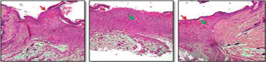
Advances in Materials Science and Engineering                                                                                                5

                                           Table 1: Parameters for tensile strength estimation.
Material                                     Average diameter (mm)           Average length     Rate (Speed) (mm/min)     Tensile strength (N)
Coconut thin fiber (Non absorbable)                    0.126                  18 cm to 25 cm               10                      1.63
Prolene (Non absorbable) monofilament                 0.099                        45cm                    10                      1.96
Coconut thick fiber (Non absorbable)                  0.248                   18 cm to 25 cm               10                      7.40
Silk braided (Non absorbable)                        0.225                       45 cm                    10                      20.12

                       (a)                                  (b)                           (c)                           (d)

                                 (e)                                  (f )                                  (g)

Figure 3: Histopathological indications of immature coconut fiber (multifilament) sutured skin area sections. (a) 24 hrs—hematoxylin and
eosin (H and E) 10x, necrosis of epidermal and dermal cells, mild neutrophilic infiltration. (b) 48 hrs—Giemsa 10x, epithelial cell migration
and moderate neutrophilic infiltration. (c) 72 hrs—H and E 10x, moderate epidermal proliferation, mild granulation tissue invasion. (d)
96 hrs—H and E 10x, epidermal layer thickening, moderate granulation tissue formation. (e) 120 hrs—H and E 10x, moderate epidermal
keratinization, mild collagen proliferation and granulation tissue. (f ) 144 hrs—van Gieson 10x, moderate amount of diffuse collagen
deposition with granulation tissue formation. (g) 168 hrs—PAS 10x, moderate amount of new blood vessels with granulation tissue.

3.6. Histopathology Investigation of Group II Samples. In the            Noncontaminated and small skin wounds are ideally
test (dHAM + TSP gel + coconut fiber) treated animals, the                sealed by topical skin adhesives or glues that are cost-
wound edges are thickened with high epithelialization fea-               effective to prevent further infection. Topical skin ad-
tures. The PMNL infiltration was seen in clusters and evenly              hesives are also proved to be effectively used along with
dispersed across the wound area. Fibroblast proliferation                sutures. Since the degree of healing depends on the af-
was high in the dermis region with added neovascularisation              fected area, therapeutic process, and compatible material
across the dermis and also in the underlying subcutaneous                used for treatment, interventions on combination ther-
matrix. The dermis edges are thickened with more poly-                   apy for wound management would be a better option to
morph nuclear infiltration. Collagen deposition in the                    contain and treat wounds in a multidirectional per-
dermis is homogeneous and was noted all over the excised                 spective [24].
skin tissue (Figure 12(a)).                                                  Large size wounds pose a serious problem and, prefer-
    In the positive control (silicone gel membrane + silk                ably, an autograft is installed at the wound site. Minimal or
suture) treated animals, the edges were in close approxi-                lack of graft tissue for treatment had resulted in the use of
mation with a great reduction in wound space. Healing is                 allograft human amnion/chorion tissue as an alternative to
hastened with a good amount of collagen deposition all over              autografts, which could modulate inflammation and en-
the excision space. PMNL infiltration has started to clear off             hance healing of tissues, thus promoting wound healing.
with signs of thickened epithelium development                           Bioavailability of factors of wound healing and the increased
(Figure 12(b)).                                                          shelf life of the naive and immunomodulatory human
    The complex and coordinated process of wound                         amnion membrane have been major reasons for its clinical
healing involves different factors and steps and requires                 use [25].
additional care to prevent the worsening of the wound                        Our multidirectional research has employed a combi-
and abnormal scar development. Though traditional                        national treatment approach for cut and open wound
therapies for wound care have shown beneficial effects,                    management, combining the use of TSP gel, the human
there remain certain challenges that require novel ther-                 amnion membrane, and the novel immature coconut fiber
apeutic approaches. Wound closure techniques have                        suture.
evolved initially from suture materials comprising of                        Though topical therapy is common in wound man-
absorbable and nonabsorbable properties [23].                            agement, our objective was to provide the best natural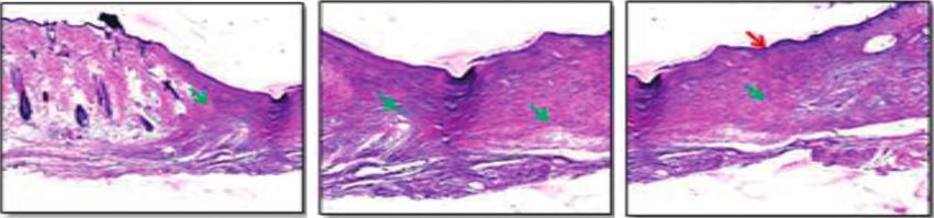
6                                                                                      Advances in Materials Science and Engineering

                 (a)                                (b)                                 (c)                              (d)

                                 (e)                                (f )                                (g)

Figure 4: Histopathological indications on immature coconut fiber (monofilament) sutured skin area sections. (a) 24 hrs—H and E 10x,
necrosis of epidermal and dermal cells with scab formation and severe neutrophilic infiltration. (b) Epithelial cell migration and pro-
liferation with severe neutrophilic infiltration. (c) 72 hrs—H and E 10x, mild epidermal proliferation with granulation tissue invasion. (d)
96 hrs—H and E 10x, epidermal layer thickening, mild granulation tissue formation. (e) 120 hrs—H and E 10x, severe epidermal kera-
tinization, moderate invasion of granulation tissue. (f ) 144 hrs—van Gieson 10x, organized collagen proliferation. (g) 168 hrs—PAS 10x,
moderate amount of neovascularization, granulation tissue formation.

                (a)                               (b)                                 (c)                               (d)

                                       (e)                            (f )                            (g)

Figure 5: Histopathological indications on braided silk sutured skin area sections. (a) 24 hrs—H and E 10x, necrosis of epidermal and
dermal cells with scab formation and mild neutrophilic infiltration. (b) 48 hrs—Giemsa 10x, epithelial cell migration with moderate
neutrophilic infiltration. (c) 72 hrs—H and E 10x, epidermal proliferation, mild granulation tissue invasion. (d) 96 hrs—H and E 10x,
epidermal layer thickening, mild granulation tissue formation. (e) 120 Hrs—H and E 10x, severe epidermal keratinization, moderate
collagen production. (f ) 144 hrs—van Gieson 10x, diffuse moderate collagen production. (g) 168 hrs—PAS 10x, severe collagen production,
moderate neovascularization in the dermis.

alternative to the available treatment options involving                   keloid scars in 85% of the cases treated with silicone gel
synthetic materials in wound treatment. Porous silicone                    sheet [28] though reported with skin irritation, a well-
membranes play a dual role, serving as epidermal barriers                  known side effect. dHAM allografts have been employed
and as a scaffold for delivering therapy to the affected                     to heal wounds without any complications or rejection
area. Collagen-based silicone gel sheet, comprising of a                   even in elderly individuals. The usage of dHAM further
porous silicone sheet coated with collagen, had been                       can rule out inconveniences and pain, mainly due to the
proved to heal different grades of the wound in several                     anti-inflammatory properties of the membrane and its
studies and decreased hypertrophic scarring when ap-                       action as a barrier covering the nociceptors [17].
plied to surgical wounds [26]. Treatment using topical                         Our research uses three different biomaterials for
silicon sheets date back to the early 1980s, where silicone                wound therapy, each having its own medicinal value
sheets were used to treat hypertrophic and keloids scars                   contributing to the wound healing effect. Our novel study
[27]. Studies indicate the improvement of hypertonic and                   investigates and evaluates the use of combination therapy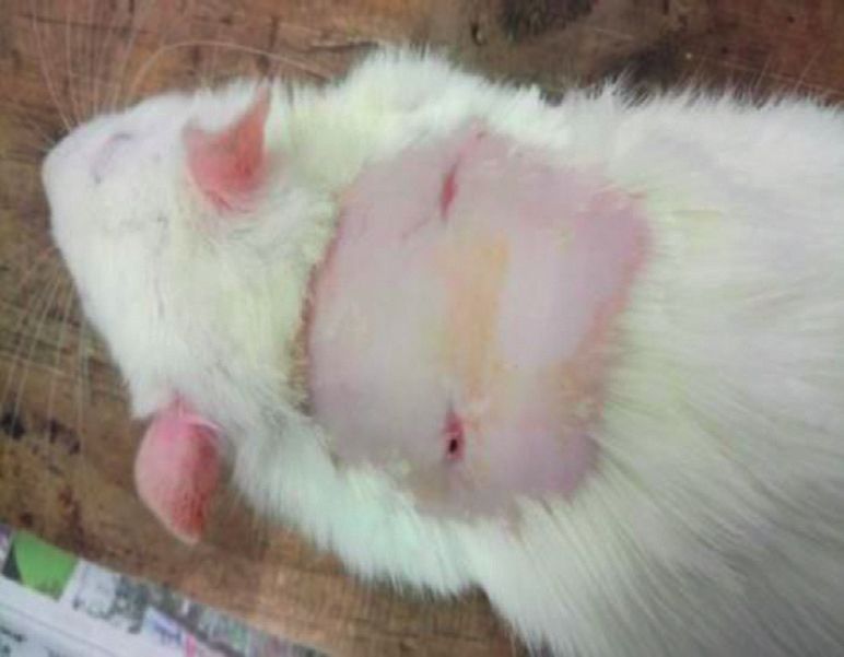
Advances in Materials Science and Engineering                                                                                               7

               (a)                                (b)                                 (c)                                 (d)

                                    (e)                                  (f )                              (g)

Figure 6: Histopathological indications on Prolene suture skin area sections. (a) 24 Hrs—H and E 10x, necrosis of epidermal and dermal
cells, severe neutrophilic infiltration. (b) 48 hrs—Giemsa 10x, epithelial cell migration, severe neutrophilic infiltration. (c) 72 hrs—H and E
10x, moderate amount of epidermal proliferation and thickening. (d) 96 hrs—H and E 10x, epidermal layer thickening, mild granulation
tissue formation. (e) 120 hrs—H and E 10x, severe epidermal keratinization, moderate granulation tissue formation. (f ) 144 hrs—van Gieson
10x, diffuse fibroblast proliferation in the dermis. (g) 160 hrs—PAS 10x, severe collagen production, maximal neovascularization in the
dermis.

                                            (a)                                                (b)

                                            (c)                                                (d)

Figure 7: TSP gel wound healing ability in Wistar rats. (a) TSP gel application site (right side), (b) wound site reduction at TSP gel applied
site (after 7 days), (c) wound site measurement of control (6.5 mm) at 7th day, and (d) wound site measurement of TSP gel applied site
(3.5 mm) at 7th day. Values are expressed as mean ± SE (n � 3), where p ≤ 0.001.

in wound healing and management of cut and open                          recovery of the epidermal layer to its normal thickness on the 7th
wounds.                                                                  day of the study in all three suture treated groups.
    Immature coconut fiber, both thin and thick, had satisfying               Tamarind seed xyloglucan of tamarind seed kernel
skin holding capacity equal to prolene, a monofilament suture,            powder act as a drug vehicle and influence cell viability, cell
and silk, a multifilament suture. Neovascularisation, collagen            migration, and gene expression of human skin keratinocytes
production, and re-epithelialization were observed with the              and fibroblasts [8]. The use of noncarcinogenic TSP in the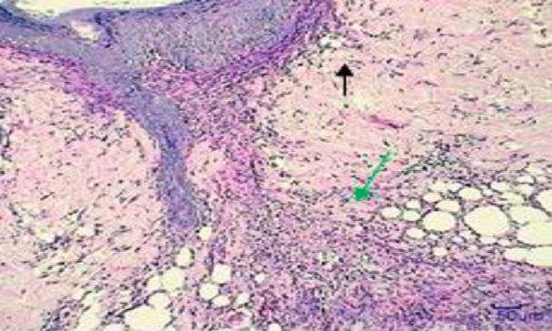
8 Advances in Materials Science and Engineering
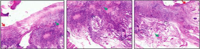
(a)
(b)
Figure 8: (a) Control (untreated) and (b) TSP gel treated: photomicrographs of H &E-stained images showing the wound edges and wound
crater. Epithelialization is marked by a red arrow and collagen formation by green arrows.
(a) (b)
(c) (d)
Figure 9: Group I animals tested with a combination of dHAM, TSP gel, and immature coconut fiber compared with untreated (control).
(a) dHAM surgery on the right side with TSP gel and coconut fiber suture for 10 mm biopsy punch diameter wound. (b) After 11 days,
wound reduction site on the right side is better than the nontreated control. (c) Wound site measurement (6 mm) at the nontreated control
site after 11 days. (d) Wound site measurement (2 mm) at the test site (dHAM + TSP gel + coconut Fiber) after 11 days. Values are expressed
as mean ± SE (n � 3), where p ≤ 0.001.
drug delivery system accounts for its mucoadhesive property its bioadhesive nature has been exploited in the development
and drug holding ability [29]. of polymeric films in the treatment of candida vaginitis using
The gel-like consistency of TSP, when mixed with water, nystatin as the drug [31]. Its high drug holding nature has
add additional advantage to its wound healing property in facilitated its use as carriers to substantiate the sustained
retaining its characteristics during treatment [30]. It also acts release of drugs.
as a carrier in drug delivery as reported by several studies A combination of immature coconut fiber, dHAM, and
[31, 32]. Its slow drug-delivering action ensures proper and TSP gel healed wounds much faster than the nontreated
timely delivery with its elasticity, mimicking a scaffold that control, with wound measurements 2 mm (±0.10) and 6 mm
would benefit in gripping of the treatment site. Additionally, (±0.10), respectively. Similarly, a 10 mm wound treated with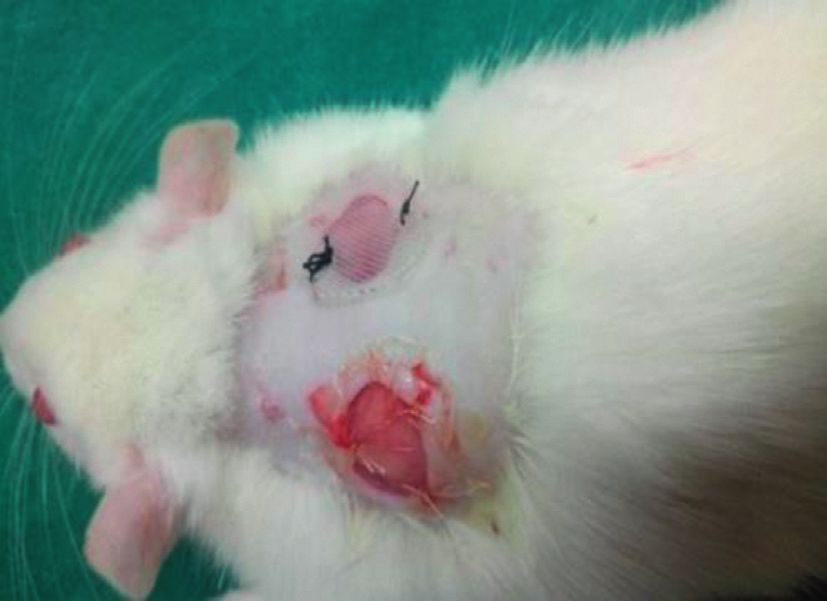
Advances in Materials Science and Engineering                                                                                             9

                                             (a)                                             (b)

                                                                    (c)

Figure 10: Group II animals tested with a combination of dHAM, TSP gel, and immature coconut fiber compared with silk suture and
silicone gel membrane (positive control). (a) dHAM + TSP gel + coconut fiber (Left side) and silicone gel membrane + silk suture (right
side). (b) After 11 days, the test sample on the left side showed healing in comparison with completely healed positive control on the right
side. (c) Wound site measurement (1.5 mm) at the test site (dHAM + TSP gel + coconut fiber) after 11 days. The silicone gel membrane + silk
suture site (positive control) was completely healed after 11 days, and hence no measurement was taken. Values are expressed as mean ± SE
(n � 3), where p ≤ 0.05).

                                                                    (a)

                                                                    (b)

Figure 11: (a). Control (untreated) and (b) test (dHAM + TSP gel + coconut fiber): photomicrographs of H and E-stained images showing
the wound edges and wound crater. Epithelialization is marked by a red arrow and collagen formation by green arrows.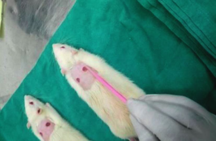
10                                                                                  Advances in Materials Science and Engineering

                                                                  (a)

                                                                  (b)

Figure 12: (a) Test (dHAM + TSP gel + coconut fiber) and (b) positive control (silicone gel membrane with silk suture): photomicrographs
of H and E-stained images showing the wound edges and wound crater. Epithelialization is marked by a red arrow and collagen formation by
green arrows.

the biological preparation had rapid healing, having a                  array with responsive drug delivery with the application of
wound measurement of 1.5 mm (±0.17), compared to the                    hydrogel has been proved beneficial in wound healing [35].
completely healed wound treated with the positive control,              Wound patches with artificial intelligence have wide ap-
i.e., silk suture with silicone gel membrane. The rigidity,             plication in various disciplines, especially it can be used to
thickness, and the direct pasting of the silicone gel sheet             monitor and promote wound healing [36]. On the other
(positive control) during surgery might account for better              hand, advanced, multifunctional, next generation smart
healing compared to the individual application of test                  bandages that could deliver and monitor oxygen in the
materials (TSP gel and thin dHAM). TSP acts as a carrier for            wound site are under research to be made available as a low-
the transport of growth factors and cytokines from the                  cost alternative for quick healing [37].
dHAM, and the hydrating potential acts as a shield for                      Smart hydrogel wound patches can act as a carrier for
preventing skin irritation. Histopathological examinations              drug delivery with a combination of drugs and also as a
revealed high epithelialization and thickened wound edges               wound healing indicator when incorporated with modified
and were similar to the positive control with collagen de-              pH indicator dyes to monitor the tissue healing process by
position all over the excised skin tissue. Our study proves the         the colour transition of the hydrogel patch [38]. This re-
treatment efficiency of the biological preparation comprising             search signifies the use of TSP gel together with the amnion
of dehydrated human amnion membrane, TSP gel, and fiber                  membrane representing a “smart patch” with wound healing
suture in wound healing. Animal study results clearly in-               potential, which would encourage further research on the
dicate the potency of natural biomaterials in wound healing,            smart patch made using a combination of plant and animal
resembling treatment with commercial and synthetic                      biological materials.
biomaterials.
     Conventional tissue adhesive patches serve wound                   4. Conclusion
management and fixation of medical devices. Tissue adhe-
sives, butylcyanoacrylate and octylcyanoacrylate, were not              The natural novel combination of biomaterials (dHAM, TSP
efficient in decreasing the wound closure time when com-                  gel, and immature coconut fiber suture) showed better
pared with the tissue bandages [33]. In contrast to the                 wound healing ability than nontreated controls and closely
conventional patches, multifunctional smart skin adhesive               similar wound healing activity to commercial biomaterials in
patches serve multiple functions of being thin, flexible, and            animal studies. Natural biomaterials were tested individually
incorporate monitoring technology [34]. Smart patches with              and also in combination and compared with commercial
capabilities of preventing wound infections and the pro-                biomaterials used in wound management. The materials are
motion of tissue remodelling are of high value. Recently,               safer and are easily available and can be greatly used by the
smart patches consisting of biomass chitosan microneedle                medical/veterinary community in the near future.
Advances in Materials Science and Engineering 11
The unresolved wound treatment challenges can be [8] W. Nie and A. M. Deters, “Tamarind seed xyloglucans pro-
solved by using different treatment approaches involving mote proliferation and migration of human skin cells through
natural substances as substitutes or adjuvant therapy in internalization via stimulation of proproliferative signal
current wound care procedures. A “smart patch” consisting transduction pathways,” Dermatology Research and Practice,
of natural wound healers is the need of the hour, and the vol. 2013, Article ID 359756, 14 pages, 2013.
combination of plant and animal biological materials would [9] V. Gupta, R. Puri, S. Gupta, S. Jain, and G. K. Rao, “Tamarind
kernel gum: an upcoming natural polysaccharide,” Systematic
improve the search for novel natural materials for wound
Reviews in Pharmacy, vol. 1, no. 1, 2010.
healing.
[10] N. Piqué, M. D. C. Gómez-Guillén, and M. P. Montero,
“Xyloglucan, a plant polymer with barrier protective prop-
5. Limitations erties over the Mucous Membranes: an overview,” Interna-
tional Journal of Molecular Sciences, vol. 19, 2018.
The length of the immature coconut fiber suture could not be [11] K. P. R. Chowdary and Y. Srinivasa Rao, “Mucoadhesive
more than 25 cm, while commercial sutures are available in microspheres for controlled drug delivery,” Biological and
different lengths. Treatment procedures involved the ap- Pharmaceutical Bulletin, vol. 27, no. 11, pp. 1717–1724, 2004.
plication of 0.25 ml TSP gel to the wound and can be tried [12] J. Liu, G. Chinga-Carrasco, F. Cheng et al., “Hemicellulose-
with different volumes for its best use and effectiveness. reinforced nanocellulose hydrogels for wound healing ap-
plication,” Cellulose, vol. 23, no. 5, pp. 3129–3143, 2016.
Data Availability [13] J. Chacon and L. Ferreira, “Hemicellulose dressing for skin
lesions caused by herpes zoster in a patient with leukemia-an
Data used to support the findings of this study are available alternative dressing,” Wounds: A Compendium of Clinical
from the corresponding author upon request. Research and Practice, vol. 21, no. 1, pp. 10–14, 2009.
[14] R. Mohd Zohdi, Z. Abu Bakar Zakaria, N. Yusof, N. Mohamed
Conflicts of Interest Mustapha, and M. N. H. Abdullah, “Gelam (Melaleuca spp.)
honey-based hydrogel as burn wound dressing,” Evidence-
The authors declare that they have no conflicts of interest. Based Complementary and Alternative Medicine, vol. 2012,
Article ID 843025, 7 pages, 2012.
[15] D. M. Martı́nez-Ibarra, D. I. Sánchez-Machado, J. López-
Acknowledgments Cervantes, O. N. Campas-Baypoli, A. Sanches-Silva, and
The authors thank Madras Christian College, affiliated to T. J. Madera-Santana, “Hydrogel wound dressings based on
University of Madras for providing research facilities. De- chitosan and xyloglucan: development and characterization,”
Journal of Applied Polymer Science, vol. 136, no. 12, Article ID
partment of Scientific and Industrial Research (DSIR),
47342, 2019.
Government of India, provided funds for coconut fiber [16] S. Burgalassi, L. Raimondi, R. Pirisino, G. Banchelli,
research: DSIR/tepp/861/2010, Government of India. E. Boldrini, and M. F. Saettone, “Effect of xyfoglucan (tam-
arind seed polysaccharide) on conjunctival cell adhesion to
References laminin and on corneal epithelium wound healing,” European
Journal of Ophthalmology, vol. 10, no. 1, pp. 71–76, 2000.
[1] N. Izzah Ibrahim, S. Wong, I. N. Mohamed et al., “Wound [17] A. B. Lyons, L. K. Chipps, R. L. Moy, and J. L. Herrmann,
healing properties of selected natural products,” International “Dehydrated human amnion/chorion membrane allograft as
Journal of Environmental Research and Public Health, vol. 15,
an aid for wound healing in patients with full-thickness scalp
no. 11, p. 2360, 2018.
defects after Mohs micrographic surgery,” JAAD Case Reports,
[2] T. V. A. Lordani, C. E. De Lara, F. B. P. Ferreira et al.,
vol. 4, no. 7, pp. 688–691, 2018.
“Therapeutic effects of medicinal plants on cutaneous wound
[18] C. Malhotra and A. K. Jain, “Human amniotic membrane
healing in humans: a systematic review,” Mediators of In-
transplantation: different modalities of its use in ophthal-
flammation, vol. 2018, Article ID 7354250, 12 pages, 2018.
mology,” World Journal of Transplantation, vol. 4, no. 2,
[3] C. Agyare, A. J. Akindele, and V. Steenkamp, “Natural
products and/or isolated compounds on wound healing,” p. 111, 2014.
Evidence-Based Complementary and Alternative Medicine, [19] N. Koizumi, T. Inatomi, C. Sotozono, N. J. Fullwood,
vol. 2019, Article ID 4594965, 3 pages, 2019. A. J. Quantock, and S. Kinoshita, “Growth factor mRNA and
[4] J. S. Boateng, K. H. Matthews, H. N. E. Stevens, and protein in preserved human amniotic membrane,” Current
G. M. Eccleston, “Wound healing dressings and drug delivery Eye Research, vol. 20, no. 3, pp. 173–177, 2000.
systems: a review,” Journal of Pharmaceutical Sciences, vol. 97, [20] T. Rihayat, S. Suryani, T. Fauzi et al., “Mechanical properties
no. 8, pp. 2892–2923, 2008. evaluation of single and hybrid composites polyester rein-
[5] Y. Yan, “Developments in fibers for technical nonwovens,” forced bamboo, PALF and coir fiber,” IOP Conference Series:
Advances in Technical Nonwovens, Elsevier, Amsterdam, Materials Science and Engineering, IOP Publishing, vol. 334, ,
Netherlands, pp. 19–96, 2016. Article ID 12081, 2018.
[6] L. Del Valle, A. Dı́az, and J. Puiggalı́, “Hydrogels for bio- [21] S. Lodhi, R. S. Pawar, A. P. Jain, and A. K. Singhai, “Wound
medical applications: cellulose, chitosan, and protein/peptide healing potential of Tephrosia purpurea (Linn.) Pers. in rats,”
derivatives,” Gels, vol. 3, no. 3, p. 27, 2017. Journal of Ethnopharmacology, vol. 108, no. 2, pp. 204–210,
[7] D. Melandri, A. De Angelis, R. Orioli et al., “Use of a new 2006.
hemicellulose dressing (Veloderm) for the treatment of split- [22] S. Murthy, M. K. Gautam, S. Goel, V. Purohit, H. Sharma, and
thickness skin graft donor sites,” Burns, vol. 32, no. 8, R. K. Goel, “Evaluation of in vivo wound healing activity of
pp. 964–972, 2006. Bacopa monniera on different wound model in rats,” BioMed12 Advances in Materials Science and Engineering
Research International, vol. 2013, Article ID 972028, 9 pages,
2013.
[23] L. Al-Mubarak and M. Al-Haddab, “Cutaneous wound clo-
sure materials: an overview and update,” Journal of Cutaneous
and Aesthetic Surgery, vol. 6, no. 4, p. 178, 2013.
[24] G. Han and R. Ceilley, “Chronic wound healing: a review of
current management and treatments,” Advances in Therapy,
vol. 34, no. 3, pp. 599–610, 2017.
[25] S. Kogan, A. Sood, and M. S. Granick, “Amniotic membrane
adjuncts and clinical applications in wound healing: a review
of the literature,” Wounds: A Compendium of Clinical Re-
search and Practice, vol. 30, no. 6, pp. 168–173, 2018.
[26] J. M. Zurada, D. Kriegel, and I. C. Davis, “Topical treatments
for hypertrophic scars,” Journal of the American Academy of
Dermatology, vol. 55, no. 6, pp. 1024–1031, 2006.
[27] I. Westra, H. Pham, and F. B. Niessen, “Topical silicone sheet
application in the treatment of hypertrophic scars and ke-
loids,” Journal of Clinical and Aesthetic Dermatology, vol. 9,
no. 10, p. 28, 2016.
[28] J. E. Fulton, “Silicone gel sheeting for the prevention and
management of evolving hypertrophic and keloid scars,”
Dermatologic Surgery, vol. 21, no. 11, pp. 947–951, 1995.
[29] M. Sano, E. Miyata, S. Tamano, A. Hagiwara, N. Ito, and
T. Shirai, “Lack of carcinogenicity of tamarind seed poly-
saccharide in B6C3F1 mice,” Food and Chemical Toxicology,
vol. 34, no. 5, pp. 463–467, 1996.
[30] R. B. Pothireddy, M. T. Mathai, and A. Julius, “Dual property
of tamarind seed polysaccharide aid wound healing,” Inter-
national Journal of Advanced Science and Technology, vol. 28,
no. 20, pp. 1130–1141, 2019.
[31] P. Bassi and G. Kaur, “Polymeric films as a promising carrier
for bioadhesive drug delivery: development, characterization
and optimization,” Saudi Pharmaceutical Journal, vol. 25,
no. 1, pp. 32–43, 2017.
[32] A. K. Shukla, R. S. Bishnoi, M. Kumar, V. Fenin, and
C. P. Jain, “Applications of tamarind seeds polysaccharide-
based copolymers in controlled drug delivery: an overview,”
Asian Journal of Pharmacy and Pharmacology, vol. 4, no. 1,
pp. 23–30, 2018.
[33] J. C. Dumville, P. Coulthard, H. V Worthington et al., “Tissue
adhesives for closure of surgical incisions,” Cochrane Data-
base of Systematic Reviews, vol. 11, 2014.
[34] I. Hwang, H. N. Kim, M. Seong et al., “Multifunctional smart
skin adhesive patches for advanced health care,” Advanced
Healthcare Materials, vol. 7, no. 15, Article ID 1800275, 2018.
[35] J. Chi, X. Zhang, C. Chen, C. Shao, Y. Zhao, and Y. Wang,
“Antibacterial and angiogenic chitosan microneedle array
patch for promoting wound healing,” Bioactive Materials,
vol. 5, no. 2, pp. 253–259, 2020.
[36] Y. Wang, M. Guo, B. He, and B. Gao, “Intelligent patches for
wound management: in situ sensing and treatment,” Ana-
lytical Chemistry, vol. 93, no. 11, pp. 4687–4696, 2021.
[37] M. Ochoa, R. Rahimi, J. Zhou et al., “Integrated sensing and
delivery of oxygen for next-generation smart wound dress-
ings,” Microsystems & Nanoengineering, vol. 6, no. 1, pp. 1–16,
2020.
[38] L. Liu, X. Li, M. Nagao, A. L. Elias, R. Narain, and H.-J. Chung,
“A pH-Indicating colorimetric tough hydrogel patch towards
applications in a substrate for smart wound dressings,”
Polymers, vol. 9, no. 11, p. 558, 2017.
[39] R. B. Pothireddy, M. T. Mathai, and A. Julius, “Antibiotic free
dehydrated human Amnionmembrane from C-section deliveries
to accelerate wound healing,” International Journal of Advanced
Science and Technology, vol. 28, no. 20, pp. 212–217, 2019.You can also read