A Novel Technique for Intraoral Ultrasound-Guided Aspiration of Peritonsillar Abscess - MDPI
←
→
Page content transcription
If your browser does not render page correctly, please read the page content below
diagnostics
Article
A Novel Technique for Intraoral Ultrasound-Guided
Aspiration of Peritonsillar Abscess
Tobias Todsen 1,2, *     ID
                              , Mads Georg Stage 1 and Christoffer Holst Hahn 1
 1    Department of Otorhinolaryngology, Head and Neck Surgery & Audiology,
      Rigshospitalet University Hospital, 2100 Copenhagen, Denmark; Mads.Georg.Stage.01@regionh.dk (M.G.S.);
      Christoffer.Holst.Hahn.01@regionh.dk (C.H.H.)
 2    Department of Otorhinolaryngology and Maxillofacial Surgery, Zealand University Hospital,
      4600 Køge, Denmark
 *    Correspondence: tobiastodsen@gmail.com; Tel.: +45-5184-7468

 Received: 23 June 2018; Accepted: 31 July 2018; Published: 2 August 2018

 Abstract: Peritonsillar abscess (PTA) is a common complication to acute tonsillitis. The treatment is
 drainage of the abscess, but many needle aspirations are unsuccessful due to a low diagnostic accuracy
 based on oral examination only. In this article, we describe how intraoral ultrasound can be added
 to improve the diagnostic work-up of PTA and present a novel technique for ultrasound-guided
 aspiration of PTA, using a small pencil-shaped transducer. We present our first clinical experiences
 with this technique and describe how it could be integrated in a clinical setting to guide safe and
 successful needle aspirations of PTA.

 Keywords: point-of-care ultrasound; intraoral ultrasound; peritonsillar abscess; ultrasound-guided aspiration

1. Introduction
     Peritonsillar abscess (PTA) is a common deep infection in relation to the palatine tonsil with
an incidence of 30 cases per 100,000 people per year in the United States [1]. Early treatment,
in form of drainage of the pus, is important to avoid spreading into the surrounding tissue and
fatal complications [2]. The formation of an abscess is often preceded by an acute tonsillitis that
progresses to a peritonsillar cellulitis and further to a PTA. The patients typically complain about
unilateral sore throat, fever, ipsilateral ear pain and decreased oral intake [3]. The oral examination may
find muffled voice, trismus and unilateral erythematous and bulging palate with the corresponding
tonsil displaced to the midline or beyond. Most patients with PTA can be treated in outpatient clinic
by an otolaryngologist or an emergency physician with needle aspiration (or incision) using local
anesthetic [1,4]. The landmark technique is traditional used to determine the point with the maximum
bulging and fluctuance—usually in the superior pole of the tonsil—where the needle is inserted for
“blind” aspiration [3,5]. If the aspiration is unsuccessful, further attempts will be conducted typical
in the middle and lower poles of the tonsil [6]. However, the diagnostic accuracy of PTA, based on
physical examination only, is low (sensitivity of 78% and specificity of 50%) [7], and may lead to many
unnecessary attempts at drainage of peritonsillar cellulitis with no therapeutic effect [8]. Computed
tomography (CT) with contrast has a high sensitivity for PTA, but is expensive and exposes young
patients to ionizing radiation. Instead, intraoral ultrasound can provide ionized-free, low-cost and
real-time imaging of PTA, though it may be difficult to use in patients with severe trismus and active
oral tongue musculature [9,10]. Most studies only use intraoral ultrasound as a static diagnostic
image modality, and afterwards, perform a “blind” needle aspiration of the PTA, as a two-step
maneuver [7,9,11–17]. A few case reports describe the use of an endocavity transducer, designed
for transvaginal examination for real-time image guidance of the PTA needle aspiration [18–20].

Diagnostics 2018, 8, 50; doi:10.3390/diagnostics8030050                      www.mdpi.com/journal/diagnostics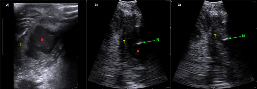
Diagnostics 2018, 8, x FOR PEER REVIEW 2 of 7
Diagnostics 2018, 8, 50 2 of 7
transvaginal examination for real‐time image guidance of the PTA needle aspiration [18–20].
However,
However,the the
sizesize
of the endocavity
of the endocavitytransducer
transducer makes
makes ititdifficult
difficulttotohandle
handle in in
thethe
oraloral cavity
cavity without
without
triggering the the
triggering gaggagreflex of of
reflex thethe
patients.
patients.Instead,
Instead, we
we have developeda anovel
have developed novel technique
technique using
using a smaller
a smaller
pencil‐shaped
pencil-shaped transducer
transducer (originally
(originallydeveloped
developed for neurosurgical
neurosurgicalimagingimaging through
through a burr
a burr holehole in the
in the
skull)skull)
for intraoral ultrasound‐guided
for intraoral ultrasound-guidedaspiration
aspiration of PTA
PTA(see(seeFigure
Figure 1).1).
We We
willwill in this
in this article
article describe
describe
our our intraoral
intraoral ultrasound approach
ultrasound approach and andpresent somesome
present illustrative images from
illustrative imagesclinical
frompractice. Further,
clinical practice.
we will present a case report and discuss the potential impact of care for patients with
Further, we will present a case report and discuss the potential impact of care for patients with PTA. PTA.
Figure 1. Different types of transducers that can be used for intraoral ultrasound (Left). A Burr‐Hole,
Figure 1. Different types of transducers that can be used for intraoral ultrasound (Left). A Burr-Hole,
a Hockey Stick
a Hockey andand
Stick a transvaginal/rectal
a transvaginal/rectaltransducers
transducers were fromBK
were from BKUltrasound
Ultrasound (Analogic,
(Analogic, Peabody,
Peabody,
MA, MA,
USA). A needle
USA). guide
A needle attached
guide attachedtotothe
the Burr‐Hole transducer(Middle).
Burr-Hole transducer (Middle). AnAn intraoral
intraoral ultrasound
ultrasound
examination conducted with a Burr‐Hole transducer with a cover (Right).
examination conducted with a Burr-Hole transducer with a cover (Right).
2. Materials andand
2. Materials Methods
Methods
Topical
Topical anesthetic
anesthetic (lidocaine,
(lidocaine, 1010 mg/dose)
mg/dose) shouldbe
should besprayed
sprayed to to the
the posterior
posteriorpharynx
pharynxininorder
order to
to decrease
decrease the gagthereflex
gag reflex
beforebefore intraoral
intraoral ultrasoundis
ultrasound is conducted.
conducted.AApencil-shaped
pencil‐shaped Burr-Hole 8863 8863
Burr‐Hole
transducer
transducer (BK(BK Ultrasound,
Ultrasound, Peabody,MA,
Peabody, MA,USA)USA) isis suitable
suitableforforintraoral
intraoral ultrasound
ultrasound with the small
with the small
curved head placed on the edematous palatoglossal arch and swiped from the cranial to caudal
curved head placed on the edematous palatoglossal arch and swiped from the cranial to caudal end of
end of the tonsil. An abscess cavity can be seen as a hypoechoic area in relation to the tonsil
the tonsil. An abscess cavity can be seen as a hypoechoic area in relation to the tonsil (see Figure 2). If
(see Figure 2). If there are any doubts about the presence of an abscess, Power Doppler should be used
there(Figure
are any doubts about the presence of an abscess, Power Doppler should be used (Figure 3) or
3) or ultrasound of the opposite tonsil should be performed as a reference. Further, a linear
ultrasound
Hockeyof thetransducer
Stick opposite tonsil
can also should
be usedbeforperformed as a reference.
improved intraoral imagingFurther, a linear Hockey
to help differentiate severeStick
transducer can also from
tonsillitis/cellulitis be used for Figure
a PTA (see improved intraoral
4). Local anesthesiaimaging
(e.g., 2% to help with
lidocaine differentiate
5 microgram severe
tonsillitis/cellulitis from a PTA (see Figure 4). Local anesthesia (e.g., 2% lidocaine
epinephrine) should be infiltrated in the mucous membrane if a PTA is confirmed. A needle guide with 5 microgram
epinephrine)
is attached should
to thebe infiltrated
Burr-Hole in the mucous
transducer with anmembrane if a PTA isneedle
on-screen ultrasound confirmed. A needle
guideline guide is
to ensure
that small and deep PTAs are precise and safely drained. When the needle
attached to the Burr‐Hole transducer with an on‐screen ultrasound needle guideline to ensure that tip is visualized into
smallthe
andabscess
deep cavity,
PTAs are the precise
assistingandnurse can drained.
safely aspirate the pusthe
When intoneedle
a syringe
tip until the hypoechoic
is visualized into thearea
abscess
disappear on the ultrasound image (see Figure 5). A larger PTA cavity can
cavity, the assisting nurse can aspirate the pus into a syringe until the hypoechoic area disappearalso be aspirated with
on the
a free-hand ultrasound-guided technique with use of a syringe holder for aspirating without the help
ultrasound image (see Figure 5). A larger PTA cavity can also be aspirated with a free‐hand ultrasound‐
from an assistant. A pean or knife might be used to open the abscess cavity for further drainage after
guided technique with use of a syringe holder for aspirating without the help from an assistant. A pean
aspiration. The patient—who is not airway compromised—can now be discharged with oral antibiotics
or knife might be used to open the abscess cavity for further drainage after aspiration. The patient—
and painkillers for follow-up in the outpatient clinic.
who is not airway compromised—can now be discharged with oral antibiotics and painkillers for
follow‐up in the outpatient clinic.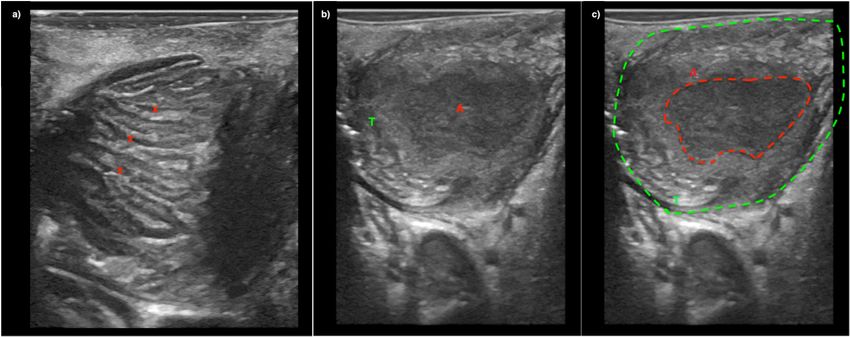
Diagnostics 2018, 8, 50 3 of 7
Diagnostics 2018, 8, x FOR PEER REVIEW 3 of 7
Diagnostics 2018, 8, x FOR PEER REVIEW 3 of 7
Figure 2. A2.static ultrasound
A static ultrasound
image with
imagewith
a Burr‐Hole
withaaBurr‐Hole
N11C5sBK
Burr-Hole N11C5s
BK Ultrasound transducer of a left‐side
Figure
Figure 2. A static ultrasound image N11C5s BKUltrasound
Ultrasoundtransducer of a of
transducer left-side
a left‐side
peritonsillar abscess
peritonsillar seen
abscess asas
seen the
themeasured
measured hypoechoic area.
hypoechoic area.
peritonsillar abscess seen as the measured hypoechoic area.
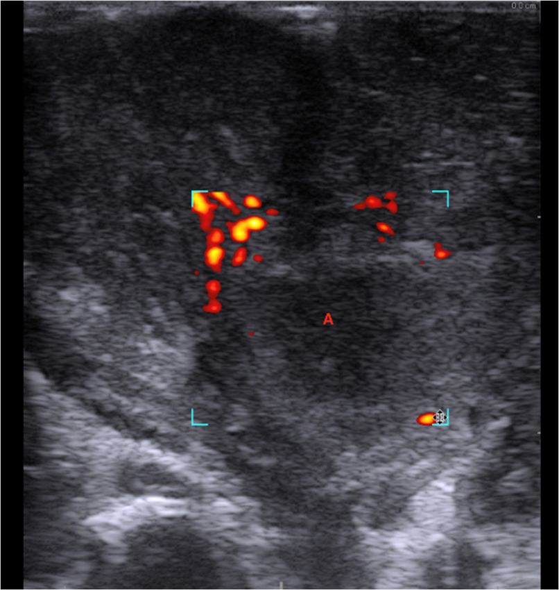
Figure 3. Power Doppler demonstrated no vascular activity in the ill‐defined hypoechoic area (A),
Figure 3. Power Doppler demonstrated no vascular activity in the ill‐defined hypoechoic area (A),
supporting
Figure 3.the diagnose
Power Dopplerofdemonstrated
a peritonsillar abscess. activity
no vascular Ultrasound
in the image with
ill-defined a Hockeyarea
hypoechoic Stick
(A),8809
supporting the the
supporting diagnose
diagnoseofofa aperitonsillar
peritonsillar abscess.
abscess. Ultrasoundimage
Ultrasound imagewith with
a a Hockey
Hockey Stick
Stick 8809 8809
transducer and a Flex Focus 800 BK Ultrasound machine (Analogic, Peabody, MA, USA).
transducer andand
transducer a Flex Focus
a Flex 800
Focus 800BK
BKUltrasound machine(Analogic,
Ultrasound machine (Analogic, Peabody,
Peabody, MA,MA, USA).
USA).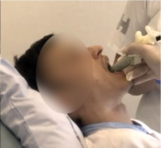
Diagnostics 2018, 8, 50 4 of 7
Diagnostics 2018, 8, x FOR PEER REVIEW 4 of 7
Diagnostics 2018, 8, x FOR PEER REVIEW 4 of 7
Figure 4. Image (a) A patient with severe tonsillitis presenting with peritonsillar swelling. Tonsillar
Figure 4. Image (a) A patient with severe tonsillitis presenting with peritonsillar swelling. Tonsillar
Figure 4. Image
hypertrophy (a) Aonpatient
is seen with severe
ultrasound tonsillitis
with inflamed presenting
tonsillar with
crypts (x) peritonsillar
but without swelling. Tonsillar
abscess. Image (b)
hypertrophy is seen on ultrasound with inflamed tonsillar crypts (x) but without abscess. Image (b) and
hypertrophy
and is seenimage
(c) Ultrasound on ultrasound withpatient
from another inflamed tonsillar
showing thecrypts (x)tonsil
palatine but without
(T) withabscess. Image
an abscess (b)
cavity
(c) Ultrasound image from another patient showing the palatine tonsil (T) with an abscess cavity (A).
and All
(A). (c) Ultrasound
imaged were image fromwith
captured another patientStick
a Hockey showing the palatine
transducer tonsil
and a GE (T) S7
Logiq with an abscessSystem
Ultrasound cavity
All imaged were captured with a Hockey Stick transducer and a GE Logiq S7 Ultrasound System
(A). All imaged were
(GE_Healthcare captured
Chicago, withIL,
Chicago, a Hockey
USA). Stick transducer and a GE Logiq S7 Ultrasound System
(GE_Healthcare Chicago, Chicago, IL, USA).
(GE_Healthcare Chicago, Chicago, IL, USA).
Figure 5. (Image (A)) A well‐defined hypoechoic peritonsillar abscess (A) seen with a linear Hockey
Figure
Stick 5. (Image(BK
transducer (A))Ultrasound)
A well‐defined hypoechoic
in relation to theperitonsillar abscess
Palatine Tonsil (A) seen(B))
(T). (Image with a linear Hockey
Ultrasound image
Figure 5. (Image (A)) A well-defined hypoechoic peritonsillar abscess (A) seen with a linear Hockey
Stick transducer
from (BK Ultrasound)
the same patient in relation
using a convex array to the Palatine
Burr‐Hole transducer T). (Image
Tonsil (with needle(B)) Ultrasound
guide. The tip image
of the
Stick transducer (BK Ultrasound) in relation to the Palatine Tonsil (T). (Image (B)) Ultrasound image
from the
needle sameas
is seen patient using a reflection
a hypoechoic convex array Burr‐Hole
(N) in transducer
the abscess with
cavity (A). needle
(Image guide.
(C)) The tip image
Ultrasound of the
from the same patient using a convex array Burr-Hole transducer with needle guide. The tip of the
needle
after is seen asaspiration
successful a hypoechoic
fromreflection (N)cavity
the abscess in theemptied
abscess of
cavity
pus. (A). (Image (C)) Ultrasound image
needle is seen as a hypoechoic reflection (N) in the abscess cavity (A). (Image (C)) Ultrasound image
after successful aspiration from the abscess cavity emptied of pus.
after successful aspiration from the abscess cavity emptied of pus.
3. Results
3. Results
A young man in his early 30s was referred to the Department of Otorhinolaryngology, Head and
3. Results
NeckASurgery
young man in his earlyRigshospitalet
& Audiology, 30s was referred to suspicion
with the Department
of a PTAof Otorhinolaryngology, Head and
by the emergency department.
A young man in his early 30s was referred to the Department of Otorhinolaryngology, Head and
Neck Surgery & Audiology, Rigshospitalet with suspicion of a PTA by the emergency
The initial clinical exam confirmed the suspicion of left‐side PTA, and three blind needle aspiration department.
Neck Surgery & Audiology, Rigshospitalet with suspicion of a PTA by the emergency department.
The initialwere
attempts clinical exam confirmed
performed using the the suspicion
traditional of left‐side
landmark PTA, and
technique three aspiration
without blind needle aspiration
of pus. A CT
The initial clinical exam confirmed the suspicion of left-side PTA, and three blind needle aspiration
attempts were
examination performed
with contrastusing the traditional
was ordered landmark
and a deep abscesstechnique without
in relation to theaspiration of tonsil
left palatine pus. AwasCT
attempts were performed using the traditional landmark technique without aspiration of pus.
examination with contrast was ordered and a deep abscess in relation to the left
found (see Figure 6). The patient was therefore planned for an acute tonsillectomy in general palatine tonsil was
A CT examination with contrast was ordered and a deep abscess in relation to the left palatine tonsil
found (see(Quincy
anesthesia Figure 6). The patienttowas
tonsillectomy) therefore
ensure planned
drainage of the for an acute
abscess cavity.tonsillectomy
However, due in to
general
other
was found (see Figure 6). The patient was therefore planned for an acute tonsillectomy in general
anesthesia (Quincy
emergency surgical tonsillectomy)
procedures, the to operation
ensure drainage of the abscess
was postponed. cavity.
Instead, However, due toneedle
ultrasound‐guided other
anesthesia (Quincy tonsillectomy) to ensure drainage of the abscess cavity. However, due to other
emergency as
aspiration, surgical procedures,
described the operation
in the method wassuccessfully
section, was postponed.performed
Instead, ultrasound‐guided
with local anesthetic needle
(see
emergency surgical procedures, the operation was postponed. Instead, ultrasound-guided needle
aspiration,
Video as described in the
S1, Supplementary). method section,
Afterwards, was successfully
the patient was discharged performed
with oralwith local anesthetic
antibiotics (see
and follow‐
aspiration, as described in the method section, was successfully performed with local anesthetic
Video S1, Supplementary).
up in the outpatient clinic. Afterwards, the patient was discharged with oral antibiotics and follow‐
(see Video S1, Supplementary). Afterwards, the patient was discharged with oral antibiotics and
up in the outpatient clinic.
follow-up in the outpatient clinic.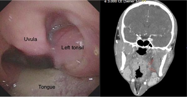
Diagnostics 2018, 8, 50                                                                                             5 of 7
Diagnostics 2018, 8, x FOR PEER REVIEW                                                                              5 of 7

      Figure 6. The oral examination with left‐side peritonsillar swelling (Left). A deep left‐side
      Figure 6. The oral examination with left-side peritonsillar swelling (Left). A deep left-side peritonsillar
      peritonsillar abscess, indicated by red arrows in the computed tomography (CT) image (Right).
      abscess, indicated by red arrows in the computed tomography (CT) image (Right).

4. Discussion
4. Discussion
      In this article, we described a new technique for point‐of‐care intraoral ultrasound of the palatine
      In this article, we described a new technique for point-of-care intraoral ultrasound of the palatine
tonsils. We presented its use in a case with a small deep PTA, where aspiration with traditional
tonsils. We presented its use in a case with a small deep PTA, where aspiration with traditional
landmark technique was unsuccessful, but ultrasound‐guided needle aspiration succeeded instead.
landmark technique was unsuccessful, but ultrasound-guided needle aspiration succeeded instead.
We used a new small pencil‐shaped transducer that allowed for visualization of the tonsil,
We used a new small pencil-shaped transducer that allowed for visualization of the tonsil, palatoglossal
palatoglossal arch and decreased patient discomfort. Compared to other studies, our technique
arch and decreased patient discomfort. Compared to other studies, our technique allows real-time
allows real‐time needle guidance to ensure safe and complete drainage of the abscess cavity, which
needle guidance to ensure safe and complete drainage of the abscess cavity, which is also suitable for
is also suitable for patients with trismus. We believe this technique can be used to decrease the
patients with trismus. We believe this technique can be used to decrease the number of unsuccessful
number of unsuccessful needle aspirations compared to the landmark technique, while the real‐time
needle aspirations compared to the landmark technique, while the real-time ultrasound guidance can
ultrasound guidance can ensure precise needle incision and avoid damage to vascular structures. We
ensure precise needle incision and avoid damage to vascular structures. We only recommend the
only recommend the intraoral ultrasound technique performed on adults [21], while transcutaneous
intraoral ultrasound technique performed on adults [21], while transcutaneous cervical ultrasound is
cervical ultrasound is better tolerated for children and should be preferred for these cases instead
better tolerated for children and should be preferred for these cases instead [22]. Another limitation
[22]. Another limitation of our technique is the use of a special neurosurgical transducer, which is not
of our technique is the use of a special neurosurgical transducer, which is not traditionally used in
traditionally used in Emergency Medicine or Otolaryngology. A linear Hockey Stick transducer is
Emergency Medicine or Otolaryngology. A linear Hockey Stick transducer is more commonly available
more commonly available and will actually provide better image resolution of the tonsils due to more
and will actually provide better image resolution of the tonsils due to more transducer crystals
transducer crystals and higher frequency, compared to the Burr‐Hole transducer (see Figure 5) [23].
and higher frequency, compared to the Burr-Hole transducer (see Figure 5) [23]. Most ultrasound
Most ultrasound manufacturers have Hockey Stick transducers available, and we have good
manufacturers have Hockey Stick transducers available, and we have good experiences with equipment
experiences with equipment from both GE (GE_Healthcare Chicago, Chicago, IL, USA) and BK
from both GE (GE_Healthcare Chicago, Chicago, IL, USA) and BK Ultrasound (Analogic, Peabody,
Ultrasound (Analogic, Peabody, MA, USA) in our departments. The GE transducer provides the best
MA, USA) in our departments. The GE transducer provides the best image quality of the tonsils
image quality of the tonsils (see Figure 3), while the Hockey Stick transducer from BK has a flexible
(see Figure 3), while the Hockey Stick transducer from BK has a flexible tip, making it very suitable
tip, making it very suitable for intraoral use. However, due to the size of the Hockey Stick transducer,
for intraoral use. However, due to the size of the Hockey Stick transducer, we could use it as a static
we could use it as a static imaging modality, while the needle incision afterwards is performed
imaging modality, while the needle incision afterwards is performed “blind” [17]. The intraoral
“blind” [17]. The intraoral ultrasound examinations illustrated in this article were all obtained by
ultrasound examinations illustrated in this article were all obtained by Tobias Todsen, who is
Tobias Todsen, who is an experienced ultrasound resident in Otolaryngology, and certified in head
an experienced ultrasound resident in Otolaryngology, and certified in head and neck ultrasound.
and neck ultrasound. However, point‐of‐care ultrasound is a very user‐dependent image modality,
However, point-of-care ultrasound is a very user-dependent image modality, requiring both technical
requiring both technical and image interpretation skills by the physician [24,25]. It is therefore
and image interpretation skills by the physician [24,25]. It is therefore unknown if our initial results can
unknown if our initial results can be generalized to other settings with physicians’ without intraoral
be generalized to other settings with physicians’ without intraoral ultrasound experience, and further
ultrasound experience, and further studies are needed to assess the learning curves [26,27].
studies are needed to assess the learning curves [26,27].
      This article describes a new method for ultrasound‐guided aspiration of PTA, which may
      This article describes a new method for ultrasound-guided aspiration of PTA, which may decrease
decrease patient discomfort and the number of unsuccessful needle aspiration attempts. However,
patient discomfort and the number of unsuccessful needle aspiration attempts. However, we only
we only described our initial clinical experiences with this technique and future randomized
described our initial clinical experiences with this technique and future randomized controlled trials
controlled trials are needed to explore the patient outcome and cost‐effectiveness.
are needed to explore the patient outcome and cost-effectiveness.
Supplementary Materials: The following are available online at www.mdpi.com/xxx/s1

Author Contributions: Conceptualization, T.T., M.G.S. and C.H.H.; Methodology, T.T.; Data Curation, T.T.;
Writing‐Original Draft Preparation, T.T.; Writing‐Review & Editing, T.T., M.G.S. and C.H.H.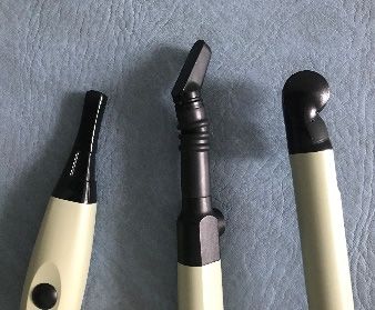
Diagnostics 2018, 8, 50                                                                                           6 of 7

Supplementary Materials: The following are available online at http://www.mdpi.com/2075-4418/8/3/50/s1.
Author Contributions: Conceptualization, T.T., M.G.S. and C.H.H.; Methodology, T.T.; Data Curation, T.T.;
Writing-Original Draft Preparation, T.T.; Writing-Review & Editing, T.T., M.G.S. and C.H.H.
Funding: This proof-of-concept study received no external funding.
Acknowledgments: The Burr-Hole ultrasound transducer was borrowed from BK Ultrasound (Analogic, Peabody,
MA, USA) for the study period.
Conflicts of Interest: The authors declare no conflicts of interest.

References
1.    Herzon, F.S.; Harris, P. Peritonsillar abscess: Incidence, current management practices, and a proposal for
      treatment guidelines. Laryngoscope 1995, 105, 1–17. [CrossRef] [PubMed]
2.    Page, C.; Biet, A.; Zaatar, R.; Strunski, V. Parapharyngeal abscess: Diagnosis and treatment. Eur. Arch.
      Oto-Rhino-Laryngol. 2008, 265, 681–686. [CrossRef] [PubMed]
3.    Passy, V. Pathogenesis of peritonsillar abscess. Laryngoscope 1994, 104, 185–190. [CrossRef] [PubMed]
4.    Powell, J.; Wilson, J.A. An evidence-based review of peritonsillar abscess. Clin. Otolaryngol. 2012, 37, 136–145.
      [CrossRef] [PubMed]
5.    Shaul, C.; Koslowsky, B.; Rodriguez, M.; Schwarz, Y.; Muahnna, N.; Peleg, U.; Sichel, J.Y. Is needle aspiration
      for peritonsillar abscess still as good as we think? A long-term follow-up. Ann. Otol. Rhinol. Laryngol. 2015,
      124, 299–304. [CrossRef] [PubMed]
6.    Roberts, J.R.; Hedges, J.R. Clinical Procedures in Emergency Medicine; Elsevier Health Sciences: Amsterdam,
      The Netherlands, 2009.
7.    Scott, P.M.J.; Loftus, W.K.; Kew, J.; Ahuja, A.; Yue, V.; Van Hasselt, C.A. Diagnosis of peritonsillar infections:
      A prospective study of ultrasound, computerized tomography and clinical diagnosis. J. Laryngol. Otol. 1999,
      113, 229–232. [CrossRef] [PubMed]
8.    Froehlich, M.H.; Huang, Z.; Reilly, B.K. Utilization of ultrasound for diagnostic evaluation and management
      of peritonsillar abscesses. Curr. Opin. Otolaryngol. Head Neck Surg. 2017, 25, 163–168. [CrossRef] [PubMed]
9.    Nogan, S.; Jandali, D.; Cipolla, M.; DeSilva, B. The use of ultrasound imaging in evaluation of peritonsillar
      infections. Laryngoscope 2015, 125, 2604–2607. [CrossRef] [PubMed]
10.   Rehrer, M.; Mantuani, D.; Nagdev, A. Identification of peritonsillar abscess by transcutaneous cervical
      ultrasound. Am. J. Emerg. Med. 2013, 31, 267.e1–267.e3. [CrossRef] [PubMed]
11.   Filho, B.C.A.; Sakae, F.A.; Sennes, L.U.; Imamura, R.; de Menezes, M.R. Intraoral and transcutaneous cervical
      ultrasound in the differential diagnosis of peritonsillar cellulitis and abscesses. Braz. J. Otorhinolaryngol.
      2006, 72, 377–381. [CrossRef]
12.   Fordham, M.T.; Rock, A.N.; Bandarkar, A.; Preciado, D.; Levy, M.; Cohen, J.; Safdar, N.; Reilly, B.K.
      Transcervical ultrasonography in the diagnosis of pediatric peritonsillar abscess. Laryngoscope 2015, 125,
      2799–2804. [CrossRef] [PubMed]
13.   Salihoglu, M.; Eroglu, M.; Yildirim, A.O.; Cakmak, A.; Hardal, U.; Kara, K. Transoral ultrasonography in the
      diagnosis and treatment of peritonsillar abscess. Clin. Imaging 2013, 37, 465–467. [CrossRef] [PubMed]
14.   Kew, J.; Ahuja, A.; Loftus, W.K.; Scott, P.M.; Metreweli, C. Peritonsillar abscess appearance on intra-oral
      ultrasonography. Clin. Radiol. 1998, 53, 143–146. [CrossRef]
15.   Strong, E.B.; Woodward, P.J.; Johnson, L.P. Intraoral ultrasound evaluation of peritonsillar abscess.
      Laryngoscope 1998, 105, 779–782. [CrossRef] [PubMed]
16.   Ahmed, K.; Jones, A.S.; Shah, K.; Smethurst, A. The role of ultrasound in the management of peritonsillar
      abscess. J. Laryngol. Otol. 1994, 108, 610–612. [CrossRef] [PubMed]
17.   Costantino, T.G.; Satz, W.A.; Dehnkamp, W.; Goett, H. Randomized Trial Comparing Intraoral Ultrasound to
      Landmark-based Needle Aspiration in Patients with Suspected Peritonsillar Abscess. Acad. Emerg. Med.
      2012, 19, 626–631. [CrossRef] [PubMed]
18.   Lyon, M.; Blaivas, M. Intraoral ultrasound in the diagnosis and treatment of suspected peritonsillar abscess
      in the emergency department. Acad. Emerg. Med. 2005, 12, 85–88. [CrossRef] [PubMed]
19.   Blaivas, M.; Theodoro, D.; Duggal, S. Ultrasound-guided drainage of peritonsillar abscess by the emergency
      physician. Am. J. Emerg. Med. 2003, 21, 155–158. [CrossRef] [PubMed]
Diagnostics 2018, 8, 50 7 of 7
20. Haeggström, D.A.; Gustafsson, D.O.; Engquist, D.S.; Engström, D.C.-F. Intraoral Ultrasonography in the
Diagnosis of Peritonsillar Abscess. Otolaryngol. Head Neck Surg. 1993, 108, 243–247. [CrossRef] [PubMed]
21. Buckley, A.R.; Moss, E.H.; Blokmanis, A. Diagnosis of peritonsillar abscess: Value of intraoral sonography.
Am. J. Roentgenol. 1994, 162, 961–964. [CrossRef] [PubMed]
22. Huang, Z.; Vintzileos, W.; Gordish-Dressman, H.; Bandarkar, A.; Reilly, B.K. Pediatric peritonsillar abscess:
Outcomes and cost savings from using transcervical ultrasound. Laryngoscope 2017, 127, 1924–1929.
[CrossRef] [PubMed]
23. Prokofieva, A.; Modayil, V.; Chiricolo, G.; Ash, A.; Raio, C. Ultrasound-guided drainage of peritonsillar
abscess: Shoot with your hockey-stick. Int. Emerg. Med. 2016, 11, 883–884. [CrossRef] [PubMed]
24. Todsen, T. Surgeon-performed ultrasonography. Dan. Med. J. 2017, 64. PMID:29115210.
25. Tolsgaard, M.G.; Todsen, T.; Sorensen, J.L.; Ringsted, C.; Lorentzen, T.; Ottesen, B.; Tabor, A. International
multispecialty consensus on how to evaluate ultrasound competence: A delphi consensus survey. PLoS ONE
2013, 8, e57687. [CrossRef] [PubMed]
26. Todsen, T.; Melchiors, J.; Charabi, B.; Henriksen, B.; Ringsted, C.; Konge, L.; von Buchwald, C.
Competency-based assessment in surgeon-performed head and neck ultrasonography: A validity study.
Laryngoscope 2018, 128, 1346–1352. [CrossRef] [PubMed]
27. Todsen, T.; Tolsgaard, M.G.; Olsen, B.H.; Henriksen, B.M.; Hillingsø, J.G.; Konge, L.; Jensen, M.L.; Ringsted, C.
Reliable and valid assessment of point-of-care ultrasonography. Ann. Surg. 2015, 261, 309–315. [CrossRef]
[PubMed]
© 2018 by the authors. Licensee MDPI, Basel, Switzerland. This article is an open access
article distributed under the terms and conditions of the Creative Commons Attribution
(CC BY) license (http://creativecommons.org/licenses/by/4.0/).You can also read