Beyond Pulse Oximetry to Pulmonary Gas Exchange Measurement in COVID-19 - MediPines
←
→
Page content transcription
If your browser does not render page correctly, please read the page content below
Beyond Pulse Oximetry to Pulmonary Gas
      Exchange Measurement in COVID-19
Standalone Pulse Oximetry, Friend or Foe?                          the tissue level, it must be released from the
There is no question that stand-alone pulse                        hemoglobin molecule into the plasma so it can
oximetry has become a standard in modern                           diffuse into the cells. To understand how well
healthcare. It is as ubiquitous as the stethoscope,                oxygen is binding to hemoglobin, oxygen saturation
                                                                   (SO2) is measured and to understand how much
and as heavily relied upon as the
                                                                   oxygen is available for cells to use, partial pressure
electrocardiogram. That makes it all the more
                                                                   of gas (PO2) is measured.
concerning that suddenly, in the midst of the
largest respiratory crisis the world has ever faced,               Oxygen saturation (the ratio of oxygen bound to
healthcare providers are wondering if this long-                   hemoglobin to total hemoglobin) is measured using
held lynchpin of healthcare monitoring is leading                  a pulse oximeter that attempts to measure the
them astray. Simply put, the peripheral saturation                 blood oxygen saturation ratio non-invasively.
readings from stand-alone pulse oximetry (i.e.,                    When this saturation (SaO2) is measured using a
SpO2% values) are not correlating with the clinical                pulse oximeter, it is denoted as SpO2. A
realities of the patients in front of them.                        commercially available “medical grade” pulse
Sometimes they see patients with good saturations                  oximeter reading should equate SpO2 (measured) =
collapse suddenly, while others with poor                          SaO2 (blood based actual) with reasonable
saturations can hold a casual conversation with                    acceptability within +/-3%, as standardized by
ease. Healthcare providers want answers to                         regulatory agencies such as the FDA. Notice
explain these confusing signals. This has resulted in              saturation (SpO2) is a ratio expressed in % terms,
medical staff developing complex theories about                    not a specific oxygen level in the blood.
what COVID-19 might be doing to blood and its                      The saturation ratio (referred to as “O2 Sat”) is NOT
ability to carry oxygen. However, many of these                    a measure of oxygen concentration. To
theories have been dismissed. Is there something                   approximate oxygen gas concentration in the lung
more fundamental going on here? Is there                           (air) and in liquid form (circulating blood), one
something healthcare workers have failed to                        must measure partial pressure of gas which is
recognize, even before COVID-19?                                   directly proportional to oxygen concentration, and
Hypoxemia is Defined by PO2 (PaO2) and Not by                      is expressed in mmHg (or Torr). The partial
Saturation (SpO2)                                                  pressure of oxygen gas in the arterial blood is
                                                                   expressed as PaO2. Healthy lungs should provide
It is well-established that the majority of oxygen
                                                                   arterial blood with approximately 90-100 mmHg of
carried in the blood is bound to hemoglobin, a
                                                                   O2. Moderate hypoxemia is defined as a PaO2 of
protein in red blood cells that is an efficient carrier
of oxygen. In order for cells to use that oxygen at                less than 60 mmHg. In other words, hypoxemia is
                                                                   NOT based on “O2 Sat” but based on “PO2 values.”

1                                              MediPines Scientific Series
COVID-19, the SpO2 value alone can be critically
misleading.
SpO2 90% Rule is Insufficient
Today, many healthcare workers are wedded to a
90% SpO2 threshold. If a patient is above 90%
SpO2, common practice is to not escalate care for
that patient. If they are below 90%, the clinical
staff often takes action by escalating care, such as
by offering supplemental oxygen or other
treatments. This rule of thumb presumes a
standard Oxygen Dissociation Curve relationship
with normal arterial CO2 tension values of 40
mmHg, which is often not the case with COVID
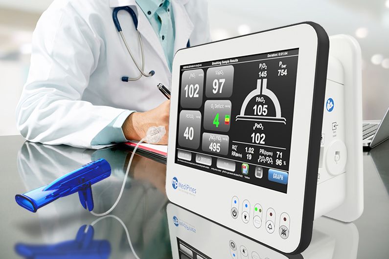
Figure 1 Oxygen-Hemoglobin Dissociation Curve Modified patients.
from John B. West’s, Respiratory Physiology: The Essentials1 When PaO2 drops as a consequence of pneumonia,
which commonly occurs in COVID-19, increased
The Oxygen Dissociation Curve Shifts Due to
ventilation is stimulated. There are two primary
Abnormal Carbon Dioxide Levels in Covid-19 Cases
ways patients increase ventilation: either by
The oxygen saturation (SaO2) and partial pressure increasing their respiratory rate and/or by
of arterial oxygen (PaO2) have a sigmoidal increasing the depth or volume of air with each
relationship known as the Oxygen Dissociation breath.
Curve (Figure 1)1. An interesting feature of the
The latter can be very challenging for clinicians to
curve is that it flattens as saturations rise above
detect. Regardless of the mechanism, such
90% and steeply falls off below 90%.
elevations in ventilation can rapidly reduce CO2.
Understanding this curve is particularly useful
This reduction in CO2 shifts the oxygen-hemoglobin
because one could use the saturation SaO2 to
dissociation curve to the left (Figure 2). The end
estimate the PaO2 under standard physiologic
result is that for a given saturation, there is a lower
conditions. Given that obtaining a PaO2
PaO2, which is the oxygen available for the body to
measurement requires arterial blood gas sampling,
use. This lower PaO2 is the result of an increased
which is invasive, time consuming and logistically
affinity of hemoglobin for oxygen, which has
challenging, the industry invented a non-invasive
benefits for oxygen uptake in the lung, but can
method of estimating functional oxygen saturation
subsequently reduce oxygen unloading at the
of arterial hemoglobin using a pulse oximeter
tissue level. As previously mentioned, hospital
(SpO2) to infer oxygen level (PaO2) from the Oxygen
providers often set a SpO2 threshold of 90% as
Dissociation Curve.
significant, since they assume that at 90%
Once an SpO2 reading is obtained, clinicians often saturation, the patient’s PaO2 is near 60 mmHg,
infer a certain PaO2 from the saturation reading. which is the associated threshold for moderate
This convenient, non-invasive way of estimating hypoxemia. This is typically known as the “60-90
arterial oxygenation to approximate moderate rule.” However, this can be a flawed assumption
hypoxemia (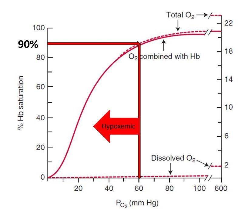
100

                                                     95                     Actual
                                                                                                         Right shift

                                                     90                                          Expected normal

                                 Hb Saturation (%)
                                                     85        Left shift

                                                     80

                                                     75
                                                                                                 High CO2
                                                     70
                                                                                                 Normal CO2 (40)
                                                     65
                                                                                                 Low CO2
                                                     60
                                                          20            40            60            80             100
                                                                                 PaO2 (mmHg)

         Figure 2: The impact of left and right shifts on the oxygen hemoglobin-dissociation curve due to CO2.1
 standard carbon dioxide level of 40 mmHg; this is                                     patient. The “sudden” deterioration of the patient
often not the case in COVID patients where CO2                                         can be confusing or surprising to the care team
values may have deviated substantially from                                            when the warning signs of respiratory compromise
normal values.                                                                         are missed.
COVID-19 Patient Case
In a case where a patient has 91% saturation, it is
assumed that the patient has a PaO2 in the mid-to-
upper 60s. However, in an actual COVID patient
case (Figure 3), the PaO2 is 57mmHg, not in the
mid-60s as you would expect! What happened
here? Failure to correct for the left-shifting effect
of the oxygen dissociation curve due to a changing
CO2 level in the blood increases error in inferring
PaO2 based on saturation. The standard
assumption creates a potential blind spot regarding
                                                                                       Figure 3: MediPines Respiratory Monitoring System
the patient’s actual oxygenation.                                                      AGM100® results screen from an actual COVID-19
Therefore, the hypoxemic patient’s developing                                          patient.
respiratory impairment (i.e., high A-a gradient) may
                                                                                       The Importance of Measuring A-a Gradient (or
go undetected and they may experience delayed
                                                                                       Oxygen Deficit) for COVID-19
intervention as the patient’s pulse oximeter
reading of 91% may not normally warrant concern.                                       Dr. Martin J. Tobin, in his recent clinical study on
When oxygen consumption increases, due to the                                          the respiratory management of coronavirus stated,
condition of the patient or from exertion, oxygen                                           The dominant respiratory feature of severe
availability can become severely compromised.                                               coronavirus disease 2019 (COVID-19) is arterial
This may lead to rapid destabilization of the                                               hypoxemia…Hypoxemia accompanied by a

3                                                                  MediPines Scientific Series
normal alveolar-to-arterial oxygen gradient and               the advent of new medical technology, the degree
    increase in PaCO2 signifies hypoventilation.                  of respiratory impairment can now be rapidly
    Hypoventilation is uncommon with Covid-19.                    detected with a patient’s exhaled gas samples.
    Instead, hypoxemia is usually accompanied by
                                                                  Practical Gas Exchange Measurements for
    an increased alveolar-to-arterial oxygen                      Respiratory Impairment: MediPines AGM100®
    gradient, signifying either ventilation-perfusion
    mismatch or intra-pulmonary shunting.2                        The shape of the curve and the disproportionate
                                                                  impact that even a small shift can have on
Currently, A-a Gradient is obtained from the                      estimating PaO2 have historically made the
Arterial Blood Gas sampling method (ABG) which is
                                                                  calculation of the PaO2 from the saturation
an alternative method to the pulse oximeter, and is               problematic. However, the advent of portable gas
the most reliable procedure to determine oxygen                   analyzers, fast and refined algorithm processors,
and CO2 levels in the blood. However, this process                and the technological advancement of steady state
is invasive, logistically complicated, and time                   gas measurements have finally provided a highly
consuming, requiring an experienced clinician to
                                                                  accurate4 and cost-effective way to detect
perform an arterial blood extraction and then
                                                                  respiratory impairment in a portable device
transport the sample to a centralized lab for
                                                                  package. The MediPines AGM100® (Figure 4) is a
testing. Furthermore, the alveolar oxygen value is                respiratory device designed to provide a
calculated using standardized assumptions from
                                                                  comprehensive set of respiratory parameters and
arterial blood values from ABG. This process can
                                                                  indicators via sampling from a patient’s breath in a
take hours, which is inefficient for hospital
                                                                  non-invasive test. It is the only commercially
workflow.                                                         available, portable pulmonary gas exchange
Is There a Better Way?                                            monitor that provides Oxygen Deficit, a method
So how does one obtain the alveolar-to-arterial
oxygen gradient other than using arterial blood gas
measurements and performing the computation
using standard assumptions in a busy hospital
setting? Fortunately, there is a newly validated,
non-invasive approach to obtaining a comparable
measurement known as “Oxygen Deficit.”
Oxygen Deficit is similar to traditional A-a gradient
(from arterial blood gas sampling using a
standardized equation) but done non-invasively
from a breath-sampling method. Oxygen Deficit
highly correlates to A-a gradient,3 and is a measure              Figure 4: MediPines AGM100® Respiratory Monitoring
                                                                  System – A Portable Respiratory Monitor
of pulmonary gas exchange inefficiency. The
higher the Oxygen Deficit (i.e., similar to high A-a
                                                                  pioneered by Dr. John B. West, an eminent
gradient), the higher the degree of gas exchange
                                                                  respiratory physiologist.
inefficiency. For healthy adults at sea level, the
Oxygen Deficit is typically in the single digits of               The MediPines AGM100® Respiratory Monitoring
mmHg. Simply put, Oxygen Deficit measures the                     System focuses on detecting and measuring
degree of efficiency (or inefficiency) of gas                     pulmonary gas exchange. It is the only US FDA
exchange between the lungs and the blood. With                    cleared, Health Canada COVID-19 emergency

4                                             MediPines Scientific Series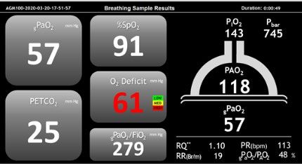
authorized gas exchange monitoring system                         seems relatively normal with a slightly low
currently on the market that provides accurate                    saturation (SpO2 91-93%) that is above the
correction for the curve shift for non-invasive PaO2              desaturation threshold (~90%), which is certainly
calculation (Figure 4). MediPines AGM100®                         not enough to warrant intervention. Again, a pulse
directly measures the lung’s oxygen and carbon                    oximeter alone only measures oxygen saturation
dioxide gas levels from taking steady state                       and misses critical CO2 and other developing
breathing gas samples, and then calculating the                   respiratory impairment. Therefore, a pulse
arterial oxygen level (PaO2) adjusted for the Bohr                oximeter by itself is an insufficient tool to detect
effect. It integrates these direct and calculated                 impending respiratory decline (i.e., gas exchange
measurements on a single device. The respiratory                  inefficiency).
monitor performs with comparable accuracy and                     Underneath the surface “COVID pneumonia” can
precision within acceptable bias against the current              be readily ascertained by detecting gas exchange
gold standard, arterial blood gas values, in normal               impairment. When dealing with gas exchange
to hypoxic physiology scenarios.4                                 impairment (i.e., inefficiency), it is necessary to ask
Accurate Detection Imperative                                     three fundamental questions that have a direct
                                                                  impact on the proper assessment, treatment
The primary function of the lung is to conduct gas
                                                                  choices and patient outcome:
exchange efficiently, but COVID-19 infection
disrupts this efficiency. In hospitalized COVID-19                      1. Is there a respiratory impairment? If so,
cases, the dominant symptoms are arterial                                  what is the degree of that impairment? The
hypoxemia combined with abnormal alveolar to                               degree of impairment (i.e., gas exchange
arterial oxygen gradient (Oxygen Deficit), typically                       inefficiency) can be answered by
accompanied with low CO2 (Figure 4).                                       referencing the Oxygen Deficit (O2 Deficit).
                                                                        2. Is blood oxygenation sufficient or does
An example of an actual hospitalized COVID patient
                                                                           developing hypoxemia exist? This is best
case monitored over multiple days is provided in
                                                                           measured using blood oxygen level
Figure 5. On the surface, the patient on room air
                                                                           (gPaO2TM).

    Figure 5: Actual COVID-19 patient case during hospitalization days.
    Note: The unit of measure for SpO2 is % and the units of measure for PO2, PCO2, O2 deficit values are in
    mmHg (partial pressure); however, they are displayed on one graph for convenience.
5                                             MediPines Scientific Series
3. Is ventilatory effort adequate? Is it high Definitions
(i.e., hypoventilation) or is it too low (i.e., SaO2 – Oxygen saturation of hemoglobin obtained from
hyperventilation)? It is best to refer to arterial blood gas (ABG) method.
carbon dioxide level (PETCO2). SpO2 – Oxygen saturation of hemoglobin obtained non-
In the patient case shown in Figure 5, by using invasively from a pulse oximeter. A pulse oximeter utilizes a
validated light-based method that relates light absorption to
these three core measurements (gPa02TM, Oxygen empirical oxygen saturation using the co-oximetry method.
Deficit, and PETCO2) the medical team was able to
PaO2 – Partial pressure of arterial oxygen; the oxygen level in
readily recognize the need for treatment. They
the blood, measured in mmHg, obtained from arterial blood
were able to monitor the patient’s response to gas (ABG) method.
therapy and successfully reverse the decline,
gPaO2TM – Partial pressure of arterial oxygen obtained non-
ultimately saving the patient’s life. Had the invasively from calculations and breathing gas sampling
medical team only relied on saturation provided by methods. Measured in mmHg. Exclusively provided by FDA-
a pulse oximeter, they would have missed the cleared MediPines AGM100®.
declining respiratory status of the patient and PETCO2 – End-tidal carbon dioxide, commonly denoted as
would not have been able to intervene in time. etCO2, a measure of ventilation; the partial pressure of
carbon dioxide at the end of an exhaled breath, measured in
To make a difference in time-sensitive respiratory
mmHg.
patient care, the ideal monitor should provide
Oxygen Deficit (O2 Deficit) – A-a gradient (AaDO2) equivalent
objective respiratory measurements that
measured non-invasively. The difference between the
immediately answer these three fundamental alveolar (lung) and arterial (blood) levels of oxygen that
questions. represents the degree of respiratory gas exchange
inefficiency, a measurement of respiratory impairment,
Summary
measured in mmHg.
In a busy clinical setting, healthcare providers strive References
for balance between convenience and accuracy,
1. West, John Burnard. Respiratory physiology: the
and are often wedded to a traditional framework
essentials. Lippincott Williams & Wilkins, 2012.
such as the 60-90 rule. Although this rule is quite 2. Tobin, Martin J. "Basing respiratory management of
convenient and can be a good rule of thumb COVID-19 on physiological principles." (2020): 1319-
approach in normal cases, the very tools that 1320.
provide some advantages can be critically 3. West, John B., et al. "A new, noninvasive method of
measuring impaired pulmonary gas exchange in lung
misleading when dealing with the impending
disease: an outpatient study." Chest 154.2 (2018): 363-
decline of patients with respiratory diseases such 369.
as COVID-19. The assessment of respiratory gas 4. Howe, Connor A., et al. "Validation of a Non-invasive
impairment is now imperative to support sound Assessment of Pulmonary Gas Exchange During Exercise
clinical assessment in patients presenting with in Hypoxia." Chest (2020).
COVID-19. © 2020 MediPines Corporation. All rights reserved.
MediPines logo and AGM100 are trademarks of MediPines Corporation.
MPJuly13, 2020
MediPines.com
6
info@medipines.com (949) 398-4670
MediPines Scientific SeriesYou can also read