A Novel Threshold based Method for Vessel Intensity Detection and Extraction from Retinal Images
←
→
Page content transcription
If your browser does not render page correctly, please read the page content below
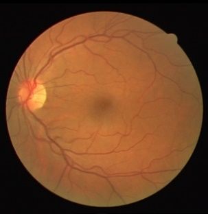
(IJACSA) International Journal of Advanced Computer Science and Applications,
 Vol. 12, No. 6, 2021

A Novel Threshold based Method for Vessel Intensity
 Detection and Extraction from Retinal Images
 Farha Fatina Wahid1, Sugandhi K2
 Department of Information Technology, Kannur University, Kannur, Kerala, India

 Raju G3 Biswaranjan Acharya5
 Department of Computer Science and Engineering School of Computer Engineering
 Faculty of Engineering, Christ (Deemed to be University) KIIT Deemed to be University
 Bengaluru, Karnataka Bhubaneswar, Odisha, India

 Debabrata Swain4 Manas Ranjan Pradhan6
 Department of Computer Science and Engineering School of Information Technology
School of Technology, Pandit Deendayal Energy University Skyline University College
 Gandhinagar, Gujarat, India Sharjah, UAE

 Abstract—Retinal vessel segmentation is an active research vessel attributes such as its tortuosity, vessel width, diameter,
area in medical image processing. Several research outcomes on bifurcation, etc. These abnormalities are identified by the
retinal vessel segmentation have emerged in recent years. Each ophthalmologist using a detailed analysis of vessel color, shape,
method has its own pros and cons, either in the vessel detection and contrast [2], [4].
stage or in its extraction. Based on a detailed empirical
investigation, a novel retinal vessel extraction architecture is When Intra Ocular Pressure (IOP) is higher than normal, it
proposed, which makes use of a couple of existing algorithms. In leads to ocular hypertension. As a consequence of this situation,
the proposed algorithm, vessel detection is carried out using a the retinal artery narrows considerably and may cause the
cumulative distribution function-based thresholding scheme. The bursting of blood vessels [5]. When a blockage occurs to the
resultant vessel intensities are extracted based on the hysteresis arteries carrying oxygen to the nerve cells, it leads to retinal
thresholding scheme. Experiments are carried out with retinal artery occlusion, which may cause vision loss [6]. Changes in
images from DRIVE and STARE databases. The results in terms the structure of blood vessels may lead to bleeding or leakage of
of Sensitivity, Specificity, and Accuracy are compared with five fluid and vision distortion. This situation, diabetic retinopathy,
standard methods. The proposed method outperforms all arises in the retinal vessels due to diabetes [7]. The obstruction
methods in terms of Sensitivity and Accuracy for the DRIVE of blood flow occurs when blockages are present in arteries or
data set, whereas for STARE, the performance is comparable veins of the retina and lead to the narrowing of blood vessels or
with the best method.
 blood clots. This is normally known as eye stroke in
 Keywords—Retinal images; blood vessel detection; and
 ophthalmology [8].
segmentation; segmentation; hysteresis thresholding; cumulative In the early days, ophthalmologists faced difficulties in
distribution function introduction diagnosing these ophthalmic diseases as manual extraction may
 contain a human error. Also, the entire process of vessel
 I. INTRODUCTION
 extraction is time-consuming. Hence, blood vessel segmentation
 Retinal images are the fundamental diagnostic element to using medical image processing techniques has become
identify various ophthalmic diseases for medical experts. It can significant for an ophthalmologist for accurate diagnostics.
be captured using a specialized camera, fundus camera, with
low power light capable of simultaneously illuminating and Several blood-vessel segmentation algorithms for retinal
capturing the interior part of the eye [1]. The image captured by images have been developed in recent years [2], [9]–[12]. The
a fundus camera includes the retina along with the optic disc, segmentation algorithms can be broadly categorized into
macula, fovea, posterior pole, etc. [2]. algorithms which comes under pattern recognition techniques
 [10], [12], vessel tracking approaches [13], [14], model-based
 A critical component of the retina is the blood vessels which approaches [15], [16], hardware implementation [17], [18] and
are broadly categorized to retinal arteries and veins. The retinal hybrid approaches [19], [20]. Further sub-classification is
artery carries oxygen and nutrients. It is through the optic nerve possible for pattern recognition (PR) techniques and model-
that the retinal artery enters the eye and then splits to superior based methods. The model-based methods can be categorized
and inferior branches [3]. The abnormalities in the structure of into region-based models and edge-based models [2]. On the
blood vessels lead to several ophthalmic diseases. In general, other hand, PR techniques are divided into supervised and
the abnormalities are due to the variations that occur in blood unsupervised algorithms based on the classification

 546 | P a g e
 www.ijacsa.thesai.org
(IJACSA) International Journal of Advanced Computer Science and Applications,
 Vol. 12, No. 6, 2021

methodology [2]. In supervised learning, prior model has to be Heart and Health Study in England (CHASE_DB1), and High-
trained using a set of reference images whereas in unsupervised Resolution Fundus (HRF) databases and claimed that the
learning, there is no need of prior training of samples. In the last proposed method outperforms other existing methods based on
decade, researchers proposed several deep learning models, sensitivity, F1-score, G-mean, and Matthews correlation
specifically convolutional Neural Network models, for the coefficient [12].
segmentation of blood vessels. Compared to classical machine
learning approaches, these methods are resource demanding. A simple retinal vessel segmentation algorithm was
 developed by Dash et al. (2017) [23]. The extracted green
 A comprehensive review of the topic reveals the limitations channel from the retinal image is initially enhanced using the
of existing models in terms of recognition accuracy and CLAHE algorithm in this algorithm. The enhanced image is
algorithm complexity. This motivated the authors to explore the then segmented using mean-c thresholding. Morphological
area. In this paper, a novel retinal vessel segmentation cleaning is carried out on the segmented image to obtain the
framework based on unsupervised learning is proposed to final segmented blood vessels. The authors carried out their
accurately extract blood vessels from fundus images. Vessel experiments on DRIVE as well as CHASE_DB1 databases and
extraction is achieved by combining different segmentation claimed 0.9555 and 0.9540 accuracy, respectively.
steps from well-established unsupervised algorithms. The vessel
detection step is constructed from an unsupervised coarse-to Jiang et al. (2017) proposed a retinal vessel segmentation
fine vessel segmentation method developed by Câmara Neto et algorithm that mainly comprised of capillary detection and
al. using an adaptive threshold [20]. The threshold is based on venules detection [9]. In this algorithm, the input retinal image
the cumulative distribution function of vessel intensity values is converted to gray scale by extracting its green channel. A
[10]. The vessel extraction is carried out using the hysteresis morphological top-hat operation is performed on the extracted
thresholding technique [11]. The proposed vessel segmentation image to obtain characteristic features for the classification of
framework is compared with a set of state-of-the-art methods vessels and non-vessels, followed by empirical intensity
and is found to have the edge over them. thresholding. This leads to venules detection. For capillary
 detection, centerline candidate pixels are highlighted using the
 The paper is articulated as follows. Section 2 gives a first-order derivative of the Gaussian filter by rotating it at
comprehensive review of blood vessel segmentation algorithms different angles with a variation of 180. In their work, instead of
developed in recent years. Section 3 describes the proposed rotating the filter, the image itself is rotated in order to avoid the
methodology followed by experimental results discussed in loss of image information. After applying the filter, a
section 4. Section 5 concludes the paper. connectivity check is performed to remove low connective
 components. Finally, the extracted venules and capillaries are
 II. LITERATURE REVIEW combined and denoised using morphological erosion. The
 In this section, descriptions of prominent retinal vessel authors obtained an accuracy of 95.88% for single database
segmentation algorithms using conventional and Convolutional tests and 95.27% for cross-database tests on experiments carried
Neural Network (CNN) based approaches are discussed, out with images from DRIVE and STARE databases.
highlighting their methodologies and merits. A coarse-to-fine algorithm for vessel segmentation was
 Coye developed a novel vessel segmentation algorithm for developed by Câmara Neto et al. (2017) [10]. In this method, a
retinal images in 2015 [21]. RGB retinal images are converted vessel segmentation algorithm is proposed in which coarse
into LAB color space and enhanced using the CLAHE vessels are detected using intensity thresholding, and vessel
algorithm in the proposed algorithm. Background exclusion is refinement is carried out using the principal curvature method.
carried out using an average filter followed by thresholding In the coarse vessel segmentation stage, the preprocessed retinal
using the IsoData algorithm on the improved image. Further, image is divided into two categories- tissue intensities and
isolated pixels are removed using morphological operations. vessel intensities using an intensity threshold which is obtained
 by maximizing the distance between the extreme intensity
 A retinal vessel segmentation algorithm from color fundus values of an image and its cumulative distribution function
images using an ensemble classifier is developed by Zhu et al.in (CDF). Further, an adaptive threshold is used for the final
2016 [22]. They have used a 36-dimensional feature vector for segmentation of vessels which are refined using curvature
each pixel. These feature vectors are then given as input to the analysis and vessel reconstruction. Balanced accuracies of
Classifier and Regression Tree (CART) classifier. Ultimately, 0.7819 and 0.8702 are reported by the authors for images from
an AdaBoost classifier is constructed by iterative training for DRIVE and STARE databases.
vessel extraction. The authors have experimented with their
algorithm on the Digital Retinal Images for Vessel Extraction In 2018, Oliveira et al. proposed a retinal vessel
(DRIVE) database, and average values of accuracy, sensitivity, segmentation algorithm by combining multi-scale analysis
and specificity of 0.9535, 0.8319, and 0.9607, respectively, are provided by stationary wavelets and multi-scale fully
reported [22]. convolutional neural network [24]. This method used rotation
 operation for data augmentation as well as prediction. The
 Orlando et al., in 2017, developed a segmentation algorithm authors carried out their experiments on DRIVE, STARE, and
for retinal blood vessels via a discriminatively trained fully CHASE_DB1 databases and reported accuracies of 0.9576,
connected conditional random field model. A structured output 0.9694, and 0.9653, respectively.
support vector machine is used to automatically learn the
parameters. The authors carried out their experiments on images Dash and Bhoi (2018) [25] reported a study on
from DRIVE, Structured Analysis of Retina (STARE), Child thresholding, founded on the variational minimax optimization

 547 | P a g e
 www.ijacsa.thesai.org
(IJACSA) International Journal of Advanced Computer Science and Applications,
 Vol. 12, No. 6, 2021

algorithm, to segment retinal blood vessels. In this algorithm, Once features are extracted from convolutional layers, feature
gamma correction is applied on the extracted green channel of maps were added and connected with a fully connected layer.
the fundus image, which is then enhanced using the CLAHE The proposed network consisted of three fully connected layers.
algorithm. On this enhanced image, thresholding is carried out Softmax function was used for classification and the loss
and followed by morphological cleaning to obtain the final function used was cross-entropy. The network was
segmented blood vessels. The authors claimed that the implemented using Anaconda and TensorFlow. The training
experiments carried out on DRIVE and CHASE_DB1 databases process took about 18h to complete. The proposed methods
give an average accuracy of 0.957 and 0.952, respectively. were evaluated on DRIVE and STARE databases with
 sensitivity, specificity, accuracy, and AUC values of 0.843,
 In 2018, Wang et al. proposed a vessel segmentation
 0.980, 0.951, 0.979 on DRIVE and 0.823, 0.978, 0.956, and
algorithm that used Hessian-based linear feature filtering at
 0.974 on STARE databases, respectively.
preprocessing stage followed by fuzzy entropic thresholding.
The authors reported an F1 score of 66.15% for their proposed In [32], a novel multi-label classification scheme for retinal
method [26]. vessel segmentation by a local de-regression model (LODESS)
 designed for multi-labeling was proposed. CNN classifier was
 A vessel segmentation algorithm that used Gray-level Hit or
 designed for multi-label classification and learning of multi-
Miss Transform (GHMT) with multi-scale, iteratively rotated
 label neighborhood relations. The proposed CNN model
multi-structuring elements to extract blood vessels from retinal
 consisted of an encoder with max-pooling layers and a decoder
images was proposed by Pal et al. in (2019) [11]. The vessels
 with upsampling and convolutional layers. A focal loss function
extracted using GHMT are further post-processed using
 was used to solve the imbalanced classification problem. The
hysteresis thresholding. The authors claimed that their method
 algorithm was built on the Keras library with NVIDIA Titan X
gives an average accuracy of 94.31% for images from the
 Pascal GPU. The network was trained for 20 epochs, and a
DRIVE database.
 standard stochastic gradient descent optimizer was used for
 Preity and Jayanthi introduced Multi-threshold along with training. Experiments were conducted on DRIVE and STARE
morphological operations for vessel segmentation [27]. databases. Accuracy, sensitivity, specificity and F1 scores of
Preprocessing was carried out using AHE, CLAHE, and 0.952, 0.776, 0.979 and 0.813 on DRIVE and 0.970, 0.812,
average filter, followed by segmentation using a variant of the 0.990 and 0.855 on STARE databases were reported.
Otsu method followed by post-processing using morphological
 In [33], retinal vessel segmentation using a cross-connected
operations. Average accuracy of 95.3% was obtained using the
 convolutional neural network (CcNet) was proposed. Features
DRIVE database.
 from different layers were fused using a cross-connected
 An adaptive segmentation algorithm was used to extract structure. There were primary and secondary paths in the
retinal blood vessels by Kabir (2020) [28]. Anisotropic network, with each convolutional layer in the primary path
diffusion was applied on grayscale fundus images, followed by connected with all layers in the secondary path. Each cross-
top-hat transformation to enhance the image. Local-property- connection from the primary to the secondary path was
based intensity transformation was introduced on sub-images, processed by a convolution, ReLU, and max-pooling module
and finally, vessels were segmented using k-means clustering (CRM) in order to reduce the network parameters. The outputs
from all sub-images. 95.29% and 95.47% accuracies were of the primary path concatenated with each output of the
obtained on DRIVE and STARE databases, respectively. secondary path. ReLU is the activation function used for the last
 layer. Green channel images were given as input to the network,
 Jadoon et al. (2020) proposed a vessel extraction algorithm and a pre-training step was used to accelerate the convergence
in which the CLAHE algorithm is applied on the green channel of the network. The parameters of these pre-trained modes were
of fundus images, followed by the top-hat operation [29]. employed as the initial values of the final trained model. CcNet
Prominent vessels are obtained using Frangi as well as high was trained using the ADAM optimization method. GeForce
boost filters. The entire vessels were obtained using Isodata GTX 1070 was used to accelerate the computation using the
thresholding. 95.32% and 94.98% accuracies on DRIVE and Caffe toolkit. Based on the experiments carried out on DRIVE
STARE databases are reported. and STARE databases, sensitivities of 0.7625 and 0.7709,
 Alhussein et al. (2020) used hessian along with intensity specificities of 0.9809 and 0.9848, accuracies of 0.953 and
transformation information for blood vessel segmentation from 0.963, and AUC values of 0.968 and 0.970 were obtained,
fundus images. On CLAHE enhanced image, Wiener and respectively.
morphological filter were applied to denoise it [30]. Thick and Retinal vessel segmentation by making use of deep
thin vessel enhanced images were obtained separately by using supervision with historically nested edge detector (HED) and
Eigenvalues of the hessian matrix at two different scales. Otsu smoothness regularization from CRFs was proposed in [34].
and Isodata thresholding were applied to extract thick and thin The network, known as Deeply Supervised and Smoothly
vessels from the respective enhanced images. Accuracies of Regularized Network (DSSRN), was an end-to-end network for
95.59% and 95.01% on DRIVE and CHASE_DB1 databases a pixel to pixel segmentation that combined FCN and CRF
were respectively reported. strengths. Five staged HED network was used, and the
 In [31], a multi-scale convolutional neural network for segmented results from each layer and the last fuse layer were
retinal vessel segmentation was proposed. The network connected to mean-field CRF. The learning rate was initially set
consisted of two consecutive convolution structures. After each to 10-8, momentum and weight decay parameters were set to 0.9
convolution layer, ReLU was used as the activation function. and 0.0002, respectively. The model was implemented on

 548 | P a g e
 www.ijacsa.thesai.org
(IJACSA) International Journal of Advanced Computer Science and Applications,
 Vol. 12, No. 6, 2021

NVIDIA GTX Titan X GPU using the Caffe framework. 12% of total intensities in an image and the CDF becomes
Training model on a single GPU took 12h for completion, smoother when vessel pixels are reached [10].
whereas testing of one retinal image required only 0.3s.
Accuracy and sensitivity values of 0.954, 0.763 on DRIVE, ( ) = ∑ =0 (3)
0.960, 0.742 on STARE and 0.959, 0.782 on CHASE_DB1 where ∈ [0 –(L-1)] and
databases were obtained respectively.
 . ℎ
 Based on the review, it is clear that even though various = (4)

algorithms exist, the majority are computationally expensive.
Moreover, the results obtained can be further enhanced. So, where = 0,1,2, … , ( − 1).
there is a requirement for a better method with maximum true From the CDF, the threshold is computed as follows.
positive values and minimal false negative values. This
motivated the development of a new unsupervised framework | ( )+ ∆ ∗ (1− )−1|
for retinal vessel segmentation. The following section describes = � � (5)
 2
 �1+ ∆
the proposal.
 III. PROPOSED METHODOLOGY where
 A novel blood vessel segmentation framework that ∆ = ( − 1) − (0) (6)
combines a coarse-to-fine vessel extraction algorithm with
hysteresis thresholding is proposed in this section. The vessel intensity image is then extracted based on the
 threshold, , as follows:
 Retinal vessel extraction, in general, is carried out in
multiple steps. For thresholding-based methods, the last steps ( > ) (7)
include thresholding for classification of pixels and post-
 From this vessel intensity image, vessels are segmented
processing for fine-tuning/denoising. We have carried out an
 using hysteresis thresholding. Unlike most of the thresholding
extensive experimental study of chosen retinal vessel
 schemes, which uses a single global threshold or local
segmentation algorithms [10], [11], [21], [23]. Based on the
 threshold, hysteresis thresholding uses two threshold values, say
study, the proposed model is built.
 1 and 2 . All the pixels in whose intensity values are greater
 The separation of vessel intensities from the tissue than or equal to 1 have been considered as vessel pixels, and
intensities as proposed in [10] is found to give a relatively good all the pixels whose intensity values are less than 2 ,
result. Also, the thresholding technique used by Pal et al. in considered as non-vessel pixels. Also, for each pixel whose
their work [11] is simple, efficient, and convincing. The use of intensity values lies in between 1 and 2 , it is considered as a
hysteresis thresholding ensures that final vessel segmentation is vessel pixel if at least one of the pixel in its 8-neighborhood has
not based on a single threshold. Based on these observations, a an intensity value greater than or equal to 1 [11]. The working
novel retinal blood vessel segmentation framework is proposed of hysteresis thresholding for vessel segmentation is
by obtaining vessel intensities as described in [10], followed by mathematically expressed as follows.
the application of hysteresis thresholding [11].
 1, ( , ) ≥ 1
 The working of the proposed algorithm is as follows. Let, ⎧
 0, ( , ) ≤ 2
denote the green channel of the input retinal image. In order to ( , ) (8)
 ⎨1, ( 2 < ( , ) < 1)
make the vessels in brighter than their background non-
 ⎩ ∃ |∅ ( , ) ≥ 1
vessels, is inverted.
 where ∅( , ) denotes the 8-neighborhood of pixel with
 = ( − 1) − (1) coordinate positions (x,y). Fig. 1 depicts the working principle
 of the proposed methodology. The algorithm for the proposed
where L is the number of possible intensity values. framework is as follows.
 is then smoothed using a Gaussian filter to remove noise
 Proposed Algorithm
and further enhanced using morphological top-hat operation.
 • Read input fundus image.
 = − ( ∘ ) (2) • Extract the green channel and invert it.
 where denotes the Gaussian smoothed image, the symbol • Smooth the inverted image using a Gaussian filter.
∘ denotes morphological opening operation, and , its • Enhance the smoothed image using morphological
structuring element, respectively. top-hat operation.
 • Apply contrast enhancement.
 The enhanced image, , further undergoes contrast • Detect retinal blood vessels from the contrast-
enhancement such that the top and bottom 1% of all the pixel enhanced image
values in are saturated [10]. From this contrast-enhanced
 • Threshold-based on CDF of pixel intensities
image, , vessels are extracted using a threshold defined based
on the cumulative distribution function (CDF) of pixel are used.
intensities in . The idea behind the selection of CDF for • Final retinal vessels are extracted by applying
threshold computation is that vessels constitute approximately Hysteresis thresholding.

 549 | P a g e
 www.ijacsa.thesai.org
(IJACSA) International Journal of Advanced Computer Science and Applications,
 Vol. 12, No. 6, 2021

 B. Values for Various Parameters
 The first parameter to be fixed is the sigma value for the
 Gaussian filter, used to remove noise from the inverted green
 channel image. The sigma value is fixed to 0.45, and a filter of
 size 3x3 is used as suggested in [10]. As far as the enhancement
 process is considered, the structuring element selected for the
 top hat filter is a disk with a radius of 6 [10]. For hysteresis
 thresholding, different combinations of threshold values are
 tried, and an optimum threshold value is fixed as 1 = 0.7 and
 2 = 0.2 respectively. If the value of 1 is reduced, it may lead
 to an increase in false-positive values. On the other hand, if the
 value of 2 is increased, it may lead to decrease in true positive
 values.
 Fig. 1. Working Principle of the Proposed Methodology.
 C. Performance Evaluation Measures
 IV. EXPERIMENTAL SETUP “Sensitivity,” “specificity,” and “accuracy” are three
 In this section, details of the data sets used, values of various performance evaluation measures considered for the study.
parameters, and matrices for performance evaluation are These are powerful measures generally used to evaluate
described. segmentation results. Based on the manually segmented vessels
 and the one obtained by applying a segmentation scheme, the
A. Data Sets
 numbers of pixels classified as true positive (TP), true negative
 1) Digital Retinal Image for Vessel Extraction (DRIVE): (TN), false positive (FP), and false-negative (FN) are computed.
DRIVE is a well-known publically available fundus image From these values, the following formulas are derived [37].
database used for comparative studies on retinal blood vessel
segmentation algorithms [35]. The images in the database are
obtained from a Diabetic Retinopathy (DR) screening program = (9)
 +
in the Netherlands with a population that comprised 400
 = (10)
subjects within the age group 25-90 years. From the +
photographs of these subjects, 40 images are randomly +
 = (11)
selected, among which 7 showed signs of mild early diabetic + + +
retinopathy, and the remaining had no signs of DR. Canon V. RESULTS AND DISCUSSION
CR5 3CCD camera with a 45o field of view (FOV) is used for
 Twenty images each from the DRIVE and STARE
capturing the images. Each image in the database has a size of
 databases are selected for the experiments. . The output of blood
584x568. The database is divided into two sets – training and vessel segmentation is analyzed quantitatively using the
testing set. Both the sets contain 20 images each, along with a performance evaluation measures – sensitivity, specificity, and
corresponding mask that describes the FOV. Vasculature’s accuracy.
single manual segmentation is available for the training set,
whereas the testing set contains two manual segmentations – In order to assess the merit of the proposed work, it is
 compared with the following algorithms: (i) Coye (2015):
one is used as gold standard/ ground truth and the other for
 enhancement using CLAHE algorithm followed by
comparison of an algorithm’s performance with that of a segmentation using IsoData thresholding technique [21];
human observer. (ii) Jiang et al. (2017). : segmentation is carried out in two steps
 2) Structured Analysis of Retina (STARE): The STARE – venules detection and capillary detection [9];
database is based on the STARE project initiated by Michael (iii) segmentation technique which uses conditional random
Goldbaum, M.D. in 1975, at the University of California, San field model for discriminative feature extraction developed by
Diego, funded by the U.S. National Institutes of Health [36]. Orlando et al. [12]; (iv) segmentation using mean-c thresholding
The project is contributed by more than 30 people from the [24] and (v) segmentation via variational minimax optimization
field of medicine, science, and engineering. The dataset thresholding [25].
consists of 400 raw images of size 700x605. The images are Among the algorithms considered for implementation, each
captured using the TopCon TRV-50 fundus camera at 35o has its enhancement technique which precedes segmentation.
FOV. For each image in the database, a list of diagnoses and These algorithms are applied on the green channel of color
diagnosis codes are also provided. Expert annotations of (RGB) retinal image and use the CLAHE algorithm for
features visible in the database are also presented in a tabular enhancement. The above algorithms are unsupervised methods
format. For the blood vessel segmentation task, 20 images except the one proposed by Orlando et al. [12], which is a
with manually segmented vasculature from two experts are supervised method.
provided. The database also contains artery/vein labeling of 10 Table 1 gives the list of methods used for the
images by two experts and 80 images with ground truth for experimentations. The average, best and worst performance of
optic nerve head detection. the segmentation algorithms given in Table 1, based on

 550 | P a g e
 www.ijacsa.thesai.org
(IJACSA) International Journal of Advanced Computer Science and Applications,
 Vol. 12, No. 6, 2021

sensitivity, specificity, and accuracy on images from the Analysis of best and worst performance highlights the fact
DRIVE database, is given in Table 2. that variation of the performance of the proposed method with
 respect to images is low, which is a preferred quality.
 From Table 2, it is clear that based on both sensitivity and
accuracy, the proposed method outperforms the other methods From Table 3, it is clear that Method 1, which is a
for images from the DRIVE database. Based on specificity, supervised method, shows good performance in identifying
though the proposed framework performs well, the supervised blood vessels with accuracy (high sensitivity and accuracy).
method based on the conditional random field [12] gives the The proposed method is better in terms of specificity, and
highest value. Table 2 reveals that when the best performance overall performance is very close to Method 1. The
of each algorithm is considered, the proposed method performance of the other four methods is relatively poor.
dominates the existing methods based on accuracy. For Analysis of best and worst performance highlights the fact that
specificity, it performs closer to the method giving the best variation of the performance of the proposed method with
result. Also, it is evident that the worst performance of the respect to images is low, which is a preferred quality.
proposed algorithm is more comparable to its average
performance in terms of performance evaluation measures. The Table 4 gives a comparison of the proposed method with
worst performance evaluation highlights the fact that for images other state-of-the-art retinal vessel segmentation algorithms in
where other methods fail to give a satisfactory result, the terms of sensitivity, specificity, and accuracy on images from
proposed method gives better results, which is a significant the DRIVE database. From Table 4, it is evident that the
achievement. proposed method produces better results compared to other
 state-of-the-art methods.
 In Table 3, the average, best and worst performances of the
segmentation algorithms for images from the STARE database Fig. 2 and Fig. 3 show the segmented vessels obtained using
are given. each algorithm for sample images from DRIVE and STARE
 databases, respectively.
 Methods that perform satisfactorily with the DRIVE dataset
are found to give a poor performance with the STARE dataset. The proposed framework fuses the merits of different works
This is basically due to the difference in the quality of images. and is tested based on images from standard test databases. A
 possible challenge that may affect the algorithm is the quality of
 From Table 3, it is clear that Method 1, which is a real-time input fundus images. We can correlate image statistics
supervised method, shows good performance in identifying with input parameters by mapping image statistics with the
blood vessels with accuracy (high sensitivity and accuracy). The parameters in such cases. It requires extensive experimentation
proposed method is better in terms of specificity, and overall and theoretical modeling and is not considered in the present
performance is very close to Method 1. The performance of the study.
other four methods is relatively poor.
 TABLE I. LIST OF SEGMENTATION ALGORITHMS USED FOR EXPERIMENTATIONS

Sl. No. Method Name Method Abbreviation
1 Conditional Random Field Model Segmentation [12] M1
2 Unsupervised method using variational minimax optimization thresholding algorithm [25] M2
3 Segmentation using mean-c thresholding [23] M3
4 Fast, accurate retinal vessel segmentation [9] M4
5 Taylor Coye algorithm [21] M5
6 Proposed Framework M6

 TABLE II. AVERAGE, BEST AND WORST PERFORMANCE OF SEGMENTATION ALGORITHMS ON IMAGES FROM DRIVE DATABASE

 Sensitivity Specificity Accuracy
Sl. No. Method
 Aver Best Worst Aver Best Worst Aver Best Worst
1 M1 0.7341 0.8245 0.6301 0.9822 0.9899 0.9758 0.9579 0.9648 0.9517
2 M2 0.6968 0.8539 0.3984 0.9752 0.9867 0.9615 0.9472 0.9607 0.9038
3 M3 0.7609 0.8933 0.3368 0.9608 0.9831 0.9115 0.9513 0.9715 0.9067
4 M4 0.7167 0.8419 0.5188 0.9499 0.9617 0.9406 0.9366 0.9471 0.9289
5 M5 0.6975 0.9484 0.1695 0.9641 0.9823 0.9487 0.906 0.9704 0.6834
6 M6 0.7992 0.8850 0.6567 0.9734 0.9850 0.9648 0.9595 0.9794 0.9527

 551 | P a g e
 www.ijacsa.thesai.org
(IJACSA) International Journal of Advanced Computer Science and Applications,
 Vol. 12, No. 6, 2021

 TABLE III. AVERAGE, BEST AND WORST PERFORMANCE OF SEGMENTATION ALGORITHMS ON IMAGES FROM STARE DATABASE

 Sensitivity Specificity Accuracy
Sl. No. Method
 Aver Best Worst Aver Best Worst Aver Best Worst
1 M1 0.8809 0.9541 0.7653 0.9462 0.977 0.8981 0.9427 0.9699 0.9018
2 M2 0.7409 0.9679 0.3343 0.9395 0.9761 0.896 0.9219 0.9657 0.8843
3 M3 0.6622 0.9041 0.2255 0.9220 0.9763 0.8546 0.9083 0.9726 0.8382
4 M4 0.7203 0.9125 0.3564 0.9348 0.9755 0.8785 0.9225 0.95 0.8761
5 M5 0.3821 0.7869 0.1446 0.9568 0.9805 0.9183 0.8079 0.9422 0.6713
6 M6 0.8002 0.9195 0.5983 0.9539 0.9875 0.8929 0.9392 0.9626 0.8912

 TABLE IV. COMPARISON WITH EXISITING METHODS BASED ON PERFORMANCE ON IMAGES FROM DRIVE DATABASE

Sl. No. Author, Year Sensitivity Specificity Accuracy
1. Coye, 2015 0.698 0.964 0.906
2. Orlando et. al., 2017 0.734 0.982 0.958
3. J. Dash and N. Bhoi,2017 0.761 0.961 0.951
4. Jiang et. al., 2017 0.717 0.950 0.937
5. J. Dash and N. Bhoi, 2018 0.697 0.975 0.947
6. Fan et. al, 2019 0.736 0.981 0.960
7. Alhussein et. al., 2020 0.785 0.972 0.956
8. Kabir, 2020 0.784 0.982 0.953
9. Preity and Jayanthi, 2020 0.695 0.985 0.953
10. Jadoon et al., 2020 0.598 0.988 0.953
11. Proposed Method 0.799 0.973 0.960

 (a) (b) (c)

 (d) (e) (f)

 (g) (h)
 Fig. 2. Segmentation Results Obtained for Image 01_test from DRIVE database. (a) Original Fundus Image (b) Manual Segmented Vessels (c) M1[12] (d) M2[25]
 (e) M3[23] (f) M4 [9](g) M5 [21] (h) M6 (Proposed Method).

 552 | P a g e
 www.ijacsa.thesai.org
(IJACSA) International Journal of Advanced Computer Science and Applications,
 Vol. 12, No. 6, 2021

 (a) (b) (c) (d)

 (e) (f) (g) (h)
Fig. 3. Segmentation Results Obtained for Image im_0235 from STARE Database. (a) Original Fundus Image (b) Manual Segmented Vessels (c) M1[12] (d) M2[25]
 (e) M3[23] (f) M4 [9] (g)M5 [21] (h) M6 (Proposed Methodology).

 [6] “Retinal Artery Occlusion - The American Society of Retina Specialists
 VI. CONCLUSION - The American Society of Retina Specialists,” 2019. [Online].
 Available: https://www.asrs.org/patients/retinal-diseases/32/retinal-
 In this paper, a novel retinal vessel segmentation algorithm artery-occlusion. [Accessed: 16-Apr-2019].
by performing vessel detection using an adaptive thresholding [7] “Diabetic retinopathy - Symptoms and causes - Mayo Clinic,” 2019.
technique followed by hysteresis thresholding for final vessel [Online]. Available: https://www.mayoclinic.org/diseases-
segmentation is proposed. Vessel detection is carried out with conditions/diabetic-retinopathy/symptoms-causes/syc-20371611.
an adaptive threshold that depends on the cumulative [Accessed: 16-Apr-2019].
distribution function of image intensity values. This method is [8] “Eye stroke: Symptoms, risks, and treatment,” 2019. [Online].
 Available: https://www.medicalnewstoday.com/articles/317877.php.
capable of detecting almost all vessel intensities within a retinal [Accessed: 16-Apr-2019].
image. The detected vessel intensities are then extracted using [9] Z. Jiang, J. Yepez, S. An, and S. Ko, “Fast, accurate and robust retinal
hysteresis thresholding. The combination of the detection and vessel segmentation system,” Biocybern. Biomed. Eng., 2017.
extraction stage using the two approaches led to the [10] L. Câmara Neto, G. L. B. Ramalho, J. F. S. Rocha Neto, R. M. S. Veras,
development of an innovative idea for the extraction of retinal and F. N. S. Medeiros, “An unsupervised coarse-to-fine algorithm for
blood vessels. blood vessel segmentation in fundus images,” Expert Syst. Appl., 2017.
 [11] S. Pal, S. Chatterjee, D. Dey, and S. Munshi, “Morphological operations
 Experiments carried out with the DRIVE and the state with iterative rotation of structuring elements for segmentation of retinal
databases establish the merit of the proposed algorithm. The vessel structures,” Multidim Syst Sign Process, vol. 30, no. 1, pp. 373–
algorithm extends help to the ophthalmologist in an easy 389, 2019.
diagnosis of retinal diseases as the extracted vessels can identify [12] J. I. Orlando, E. Prokofyeva, and M. B. Blaschko, “A Discriminatively
changes in normal vessel structures and indicate the presence of Trained Fully Connected Conditional Random Field Model for Blood
abnormalities, if any. This opens scope for further research for a Vessel Segmentation in Fundus Images,” IEEE Trans. Biomed. Eng.,
 2017.
better segmentation algorithm. The present study can be further
 [13] C. G. Owen et al., “Measuring retinal vessel tortuosity in 10-year-old
extended by incorporating the influence of image quality on the children: Validation of the computer-assisted image analysis of the
algorithm and making it suitable for all types of images. retina (caiar) program,” Investig. Ophthalmol. Vis. Sci., 2009.
 REFERENCES [14] C. M. Wilson et al., “Computerized analysis of retinal vessel width and
 tortuosity in premature infants,” Investig. Ophthalmol. Vis. Sci., 2008.
[1] “Color Fundus Photography | Department of Ophthalmology.” [Online].
 Available: https://ophthalmology.med.ubc.ca/patient-care/ophthalmic- [15] W. S. Oliveira, J. V. Teixeira, T. I. Ren, G. D. C. Cavalcanti, and J.
 photography/color-fundus-photography/. [Accessed: 26-Nov-2018]. Sijbers, “Unsupervised retinal vessel segmentation using combined
 filters,” PLoS One, 2016.
[2] K. B. Khan et al., “A review of retinal blood vessels extraction
 techniques: challenges, taxonomy, and future trends,” Pattern Anal. [16] A. E. Rad, M. S. Mohd Rahim, H. Kolivand, and I. Bin Mat Amin,
 Appl., pp. 1–36, 2018. “Morphological region-based initial contour algorithm for level set
 methods in image segmentation,” Multimed. Tools Appl., 2017.
[3] “About Retinal Blood Vessels | Retina Doctor Melbourne,” 2019.
 [Online]. Available: https://www.retinadoctor.com.au/about-the-eye/the- [17] F. Argüello, D. L. Vilariño, D. B. Heras, and A. Nieto, “GPU-based
 retinal-blood-vessels/. [Accessed: 16-Apr-2019]. segmentation of retinal blood vessels,” J. Real-Time Image Process.,
 2014.
[4] M. M. Fraz et al., “Blood vessel segmentation methodologies in retinal
 images - A survey,” Comput. Methods Programs Biomed., 2012. [18] D. Koukounis, C. Ttofis, A. Papadopoulos, and T. Theocharides, “A
 high performance hardware architecture for portable, low-power retinal
[5] “Ocular Hypertension: Causes, Symptoms, Tests, and Treatment,” 2019.
 vessel segmentation,” Integr. VLSI J., 2014.
 [Online]. Available: https://www.webmd.com/eye-health/occular-
 hypertension. [Accessed: 16-Apr-2019].

 553 | P a g e
 www.ijacsa.thesai.org
(IJACSA) International Journal of Advanced Computer Science and Applications,
 Vol. 12, No. 6, 2021

[19] K. Jiang et al., “Isotropic Undecimated Wavelet Transform Fuzzy [29] Z. Jadoon, S. Ahmad, M. A. Khan Jadoon, A. Imtiaz, N. Muhammad,
 Algorithm for Retinal Blood Vessel Segmentation,” J. Med. Imaging and Z. Mahmood, “Retinal Blood Vessels Segmentation using
 Heal. Informatics, 2015. ISODATA and High Boost Filter,” in 2020 3rd International Conference
[20] P. Dai et al., “A new approach to segment both main and peripheral on Computing, Mathematics and Engineering Technologies: Idea to
 retinal vessels based on gray-voting and Gaussian mixture model,” PLoS Innovation for Building the Knowledge Economy, iCoMET 2020, 2020.
 One, 2015. [30] M. Alhussein, K. Aurangzeb, and S. I. Haider, “An Unsupervised
[21] W. Zeng and C. Wang, “View-invariant gait recognition via Retinal Vessel Segmentation Using Hessian and Intensity Based
 deterministic learning,” Neurocomputing, vol. 175, no. PartA, pp. 324– Approach,” IEEE Access, vol. 8, pp. 165056–165070, Sep. 2020.
 335, 2016. [31] M. Li, Q. Yin, and M. Lu, “Retinal Blood Vessel Segmentation Based
[22] C. Zhu, B. Zou, Y. Xiang, and J. Cui, “An Ensemble Retinal Vessel on Multi-Scale Deep Learning,” in 2018 Federated Conference on
 Segmentation Based on Supervised Learning in Fundus Images,” Computer Science and Information Systems (FedCSIS), 2018, pp. 117–
 Chinese J. Electron., vol. 25, no. 3, pp. 503–511, 2016. 123.
[23] J. Dash and N. Bhoi, “A thresholding based technique to extract retinal [32] Q. He, B. Zou, C. Zhu, X. Liu, H. Fu, and L. Wang, “Multi-Label
 blood vessels from fundus images,” Futur. Comput. Informatics J., vol. Classification Scheme Based on Local Regression for Retinal Vessel
 2, no. 2, pp. 103–109, 2017. Segmentation,” in 2018 25th IEEE International Conference on Image
 Processing (ICIP), 2018, pp. 2765–2769.
[24] A. Oliveira, S. Pereira, and C. A. Silva, “Retinal vessel segmentation
 based on Fully Convolutional Neural Networks,” Expert Syst. Appl., [33] S. Feng, Z. Zhuo, D. Pan, and Q. Tian, “CcNet: A cross-connected
 vol. 112, 2018. convolutional network for segmenting retinal vessels using multi-scale
 features,” Neurocomputing, 2019.
[25] J. Dash and N. Bhoi, “An Unsupervised Approach for Extraction of
 Blood Vessels from Fundus Images,” J. Digit. Imaging, vol. 31, no. 6, [34] Y. Lin, H. Zhang, and G. Hu, “Automatic Retinal Vessel Segmentation
 pp. 857–868, 2018. via Deeply Supervised and Smoothly Regularized Network,” IEEE
 Access, 2019.
[26] H. Wang, Y. Jiang, X. Jiang, J. Wu, and X. Yang, “Automatic vessel
 segmentation on fundus images using vessel filtering and fuzzy [35] J. Staal, M. D. Abràmoff, M. Niemeijer, M. A. Viergever, and B. Van
 entropy,” Soft Comput., 2018. Ginneken, “Ridge-based vessel segmentation in color images of the
 retina,” IEEE Trans. Med. Imaging, 2004.
[27] Preity and N. Jayanthi, “A Segmentation Technique of Retinal Blood
 Vessels using Multi-Threshold and Morphological Operations,” in 2020 [36] A. Hoover, “STructured Analysis of the Retina.” [Online]. Available:
 International Conference on Computational Performance Evaluation, http://cecas.clemson.edu/~ahoover/stare/.
 ComPE 2020, 2020, pp. 447–452. [37] “Sensitivity and Specificity.” [Online]. Available:
[28] M. A. Kabir, “Retinal Blood Vessel Extraction Based on Adaptive http://www.med.emory.edu/EMAC/curriculum/diagnosis/sensand.htm.
 Segmentation Algorithm,” in 2020 IEEE Region 10 Symposium, [Accessed: 27-Nov-2018].
 TENSYMP 2020, 2020, pp. 1576–1579.

 554 | P a g e
 www.ijacsa.thesai.org
You can also read