A Quantitative Comparison of Multispectral Refraction Topography and Autorefractometer in Young Adults
←
→
Page content transcription
If your browser does not render page correctly, please read the page content below
ORIGINAL RESEARCH
                                                                                                                                      published: 13 September 2021
                                                                                                                                     doi: 10.3389/fmed.2021.715640

                                              A Quantitative Comparison of
                                              Multispectral Refraction Topography
                                              and Autorefractometer in Young
                                              Adults
                         Edited by:           Yunru Liao 1,2† , Zhenlan Yang 1† , Zijing Li 1 , Rui Zeng 1 , Jing Wang 1 , Yichi Zhang 1*‡ and
                       Jorge L. Alio,         Yuqing Lan 1*‡
   Miguel Hernández University, Spain
                                              1
                                               Department of Ophthalmology, Sun Yat-sen Memorial Hospital, Sun Yat-sen University, Guangzhou, China, 2 Department of
                       Reviewed by:
                                              Glaucoma, Zhongshan Ophthalmic Center, Sun Yat-sen University, Guangzhou, China
             Yu Xiang George Kong,
      The Royal Victorian Eye and Ear
                   Hospital, Australia
                                              Purpose: Purpose of this study is to evaluate the measuring consistency of central
                                Yu Su,
           Renmin Hospital of Wuhan           refraction between multispectral refraction topography (MRT) and autorefractometry.
                     University, China
                                              Methods: This was a descriptive cross-sectional study including subjects in Sun Yat-sen
                 *Correspondence:
                        Yichi Zhang
                                              Memorial Hospital from September 1, 2020, to December 31, 2020, ages 20 to 35 years
       zhangych46@mail.sysu.edu.cn            with a best corrected visual acuity of 20/20 or better. All patients underwent cycloplegia,
                         Yuqing Lan
                                              and the refractive status was estimated with autorefractometer, experienced optometrist
             lanyq@mail.sysu.edu.cn
                                              and MRT. We analyzed the central refraction of the autorefractometer and MRT. The
     † These authors have contributed
                                              repeatability and reproducibility of values measured using both devices were evaluated
    equally to this work and share first
                            authorship        using intraclass correlation coefficients (ICCs).
     ‡ These   authors have contributed       Results: A total of 145 subjects ages 20 to 35 (290 eyes) were enrolled. The mean
                    equally to this work
                                              central refraction of the autorefractometer was −4.69 ± 2.64 diopters (D) (range −9.50 to
                    Specialty section:        +4.75 D), while the mean central refraction of MRT was −4.49 ± 2.61 diopters (D) (range
          This article was submitted to       −8.79 to +5.02 D). Pearson correlation analysis revealed a high correlation between the
                        Ophthalmology,
                                              two devices. The intraclass correlation coefficient (ICC) also showed high agreement.
                a section of the journal
                   Frontiers in Medicine      The intrarater and interrater ICC values of central refraction were more than 0.90 in both
             Received: 27 May 2021            devices and conditions. At the same time, the mean central refraction of experienced
         Accepted: 20 August 2021             optometrist was −4.74 ± 2.66 diopters (D) (range −9.50 to +4.75D). The intra-class
      Published: 13 September 2021
                                              correlation coefficient of central refraction measured by MRT and subjective refraction
                              Citation:
 Liao Y, Yang Z, Li Z, Zeng R, Wang J,
                                              was 0.939.
          Zhang Y and Lan Y (2021) A
                                              Conclusions: Results revealed that autorefractometry, experienced optometrist and
           Quantitative Comparison of
  Multispectral Refraction Topography         MRT show high agreement in measuring central refraction. MRT could provide a potential
      and Autorefractometer in Young          objective method to assess peripheral refraction.
         Adults. Front. Med. 8:715640.
     doi: 10.3389/fmed.2021.715640            Keywords: consistency, multispectral refraction topography, autorefractometer, refraction, multispectral imaging

Frontiers in Medicine | www.frontiersin.org                                        1                                      September 2021 | Volume 8 | Article 715640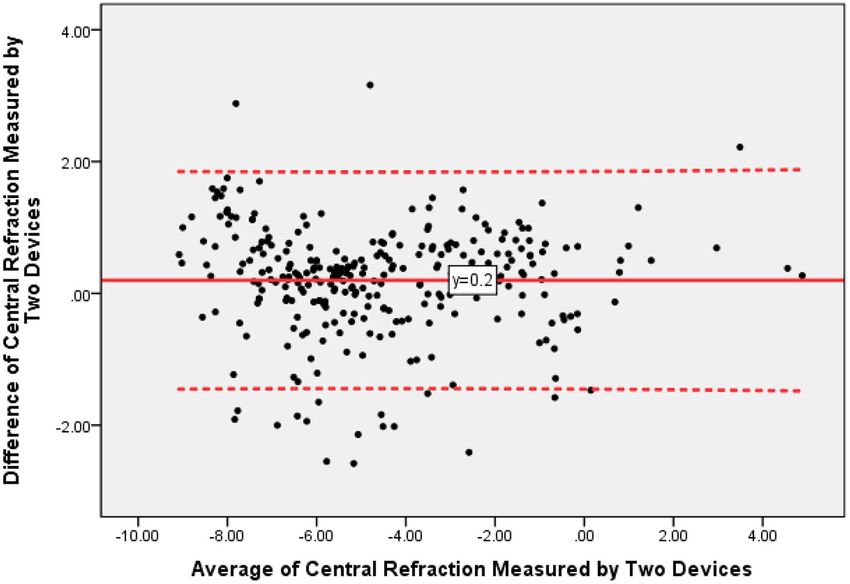
Liao et al.                                                                                                        Comparison of MRT and Autorefractometer

INTRODUCTION                                                                TABLE 1 | Baseline characteristics of the participants.

Myopia is by far the most common refractive error and a                     Characteristics                                                       Value

dominant reason of visual impairment globally (1), with a
                                                                            Subjects (eye)                                                      145 (290)
prevalent rate of 10–30% of adults in many countries and 80–
                                                                            Sex                       Female                                       105
90% of young people in some parts of East and South-East
                                                                                                      Male                                         40
Asia (2, 3). Myopia of −6.00 diopters (D) or more severe
                                                                            Age                       Mean                                    26.23(SD2.62)
is called high myopia and is often causes visual impairment
                                                                                                      Range                                       20–35
due to complications such as posterior uveioma, choroidal new
                                                                            Emmetropia                                                              3
vascularization, retinal detachment and so on (4). Reducing the
                                                                            Ametropia                 Myopia      Low(−0.25∼−3.00D)                61
high myopia incidence rate and improving the quality of life are
                                                                                                                  Medium(−3.25∼−6.00D)             121
the goal of prevention and treatment of myopia.
                                                                                                                  High(< −6.00D)                   93
   Animal experiments have provided details about myopia:
                                                                                                      Hyperopia   Low(+0.25∼+3.00D)                10
hyperopic defocus increases axial elongation, while myopic
                                                                                                                  Medium(+3.25∼+5.00D)              2
defocus decreases axial elongation (4–11). Retinal peripheral
                                                                                                                  High(> +5.00D)                    0
visual signals, which are basically the sum of regions, can contral
central refractive development independent of central visual                SD, standard deviation.
experience. The effectiveness of optical defocus in changing axial
elongation depends on retinal defocus degree (12, 13). However,
there are generally four methods to evaluate eccentric refractive
                                                                            Memorial Hospital from September 1, 2020 to December 31,
errors (14): subjective eccentric refraction (15), wavefront
                                                                            2020 were reviewed. The inclusion criteria were as follows: (1)
measurements with an aHS sensor (16), streak retinoscopy (17),
                                                                            subjects ages 20 to 35 years, (2) subjects with a best corrected
and photo refraction with a power refractor (14). However, these
                                                                            visual acuity of 20/20 or better, (3) subjects with MRT results,
methods can only detect a small area of the retina and cannot
                                                                            (4) subjects with the refraction results of autorefractometer and
accurately detect the peripheral defocus of each region of the
                                                                            experienced optometrists. Exclusion criteria were as follows: (1)
retina. Further, the process has high requirements for patient
                                                                            intraocular pressure higher than 21 mmHg, (2) a history of ocular
cooperation, and it is cumbersome, time-consuming, and difficult
                                                                            diseases or previous ocular surgery that may influence refraction
to adapt to clinical practice (18, 19).
                                                                            or axial length, such as corneal and lens diseases, and (3) a history
   MRT is a new instrument using multispectral imaging
                                                                            of corneal contact lens, such as orthokeratology.
technology (MSI). MSI is an emerging technology based on
                                                                               The refractive errors of all eyes were measured by both
imaging and spectroscopy. It is the result of remote sensing
                                                                            an autorefractometer (AR-360A, NIDEK Co., Ltd, Japan), an
technology as a kind of analysis tool and can obtain information
                                                                            experienced optometrist, and MRT (version 1.0.5T05C; Thondar,
on the measured target simultaneously from the spectral and
                                                                            Inc.) Thirty minutes before examination, a cycloplegic agent
spatial dimensions. MRT can detect the refraction of each part of
                                                                            (one drop of 0.5% tropicamide along with 0.5%) phenylephrine
the retina within a range of 30◦ at the posterior pole of the retina,
                                                                            hydrochloride (Sinqi Pharmaceutical Co. Ltd., Shenyang, China)
especially the refraction of the fovea of the macula.
                                                                            was applied 3 times (with 5 min between each application). The
   The usage of different technologies in the MRT and
                                                                            mean of three consecutive autorefraction readings was collected
other devices above talking about may result in differences
                                                                            as the refractive error value measured by the autorefractometer.
in measurements. Because treatment centers use different
                                                                            MRT measurements use MSI technology for central refraction
topographic devices, differences in such measurements might
                                                                            measurements. To compare the two devices, the central
lead to differences between diagnostic or treatment centers.
                                                                            refraction values from the two devices were analyzed. All
   Therefore, we evaluated agreement between MRT and
                                                                            examinations were performed by the same experienced doctor.
autorefractometer to see whether they could be interchangeable
                                                                               Statistical analysis was performed using SPSS (version
used or not. The data offered by each device should be maintained
                                                                            23). Intra-class correlation coefficient (ICC) and repeated
consistent at different measurements so that results can be used
                                                                            measurement analysis of variance (ANOVA) were used to
in research. Hence, we evaluated the repeatability of the devices’
                                                                            evaluate the repeatability of the equipment. Pearson’s correlation
measurements to decide their effectiveness and availability.
                                                                            coefficient, paired t-test, and Bland-Altman plots were used to
                                                                            compare the two devices. A value of P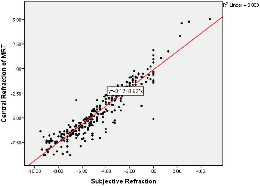
Liao et al. Comparison of MRT and Autorefractometer
FIGURE 1 | The Bland-Altman plots of central refraction measured by multispectral refraction topography (MRT) and autorefractometer.
measured by an autorefractometer was −4.69 ± 2.64 diopters measuring central refraction error in healthy eyes, particularly
(D) (range −9.50 to +4.75 D), while the mean central refraction for mild myopia. Furthermore, the values showed a high
measured by MRT was −4.49 ± 2.61 diopters (D) (range correlation between the two devices. Comparing with
−8.79 to +5.02 D). Pearson correlation analysis revealed a high autorefractometer or experienced optometrist, measured
correlation between MRT and autorefractometer results (R = values by MRT showed a statistically significant shift toward
0.950, P < 0.001). Figure 1 showed the difference values of hyperopia. This difference is about 0.20 D (comparing with
central refraction between MRT and autorefractometer vs. the autorefractometer) to 0.26D (comparing with subjective
average of these two results. The mean difference value was 0.20 refraction). It suggests that the accommodation reflex may
D while the 95% confidence interval was −1.43 to +1.83 D. still have played a role in these participants. We consider the
The correlations of central refraction of the two devices were MRT test a more powerful tool to measure the full hyperopic
shown in Figure 2 (R = 0.947, P < 0.001). refractive error.
Tables 2, 3 showed the description and intraclass correlation To date, current research suggests that the surrounding
coefficient (ICC) for central refraction of autorefractometer and area of the eye also plays an important role in controlling
multispectral refraction topography. The intrarater and interrater the growth of the eye and the development of refractive
ICC values were 0.947 and 0.973, respectively. errors. Peripheral hyperopic defocus of the retina is one of
Then we analyzed the result of MRT measurement and the causes of myopia. If the defocus degree of the retina,
subjective refraction. The mean subjective refraction was −4.74 especially the peripheral defocus, can be measured effectively
± 2.66D, its range was −9.50 to +4.75D. The mean of the and accurately, it will be helpful in preventing myopia. The
difference between the central refraction obtained by the MRT reason for using objective methods to test peripheral defocus
measurement and the experienced optometrist was 0.26 ± is to try to find the “gold standard” compared with how
0.87D, and its 95% confidence interval was −1.44 to +1.95 conventional subjective refraction is used. An ideal screening
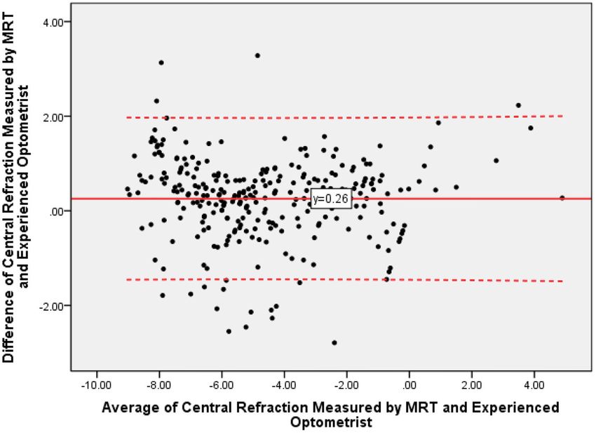
(Figure 3). The intra-class correlation coefficient of central test should be perfect in specificity, sensitivity, and positive
refraction measured by MRT and subjective refraction was 0.939 predictive value, but we still can’t find any screening method
(Figure 4, P < 0.001). that achieves this level of accuracy. The current methods used
to measure peripheral refraction are more difficult to evaluate
DISCUSSION due to poor retinal image quality, optical aberration and low
retinal resolution, which may result in insufficient retinal image
The study demonstrates that the central refraction obtained sampling (14).
by autorefractometer devices, experienced optometrists, MRT is a new instrument using MSI that can accurately
and MRT shows high repeatability and reproducibility. Our measure the refraction of each part of the retina, and in a sense, it
results indicate that MRT is a valid and safe method for can replace the role of autorefractometer measurement. However,
Frontiers in Medicine | www.frontiersin.org 3 September 2021 | Volume 8 | Article 715640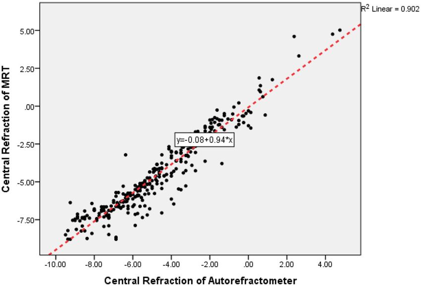
Liao et al.                                                                                                                         Comparison of MRT and Autorefractometer

  FIGURE 2 | The correlations of central refraction between multispectral refraction topography (MRT) and autorefractometer.

TABLE 2 | Measured Values and Paired T-test Results by Autorefractometer and Multispectral Refraction Topography (MRT).

                                                         Mean                       Standard                      Range                                   p-value(2-tailed)
                                                                                    deviation                                                           (paired samples test)

Central refraction of MRT                                −4.49D                        2.61                       −8.79D to +5.02D                               0.000
Central refraction of autorefractometer                  −4.69D                        2.64                       −9.50D to +4.75D

TABLE 3 | Intraclass Correlation Coefficient.

                                      Intraclass                              95% confidence interval                                          F test with true value 0
                                                   b
                                    Correlation                     Lower bound                      Upper bound                      Value                           Sig

Single measures                           0.947a                         0.931                            0.959                       38.740                         0.000
Average measures                          0.973                          0.964                            0.979                       38.740                         0.000

Two-way random effects model where both individual effects and measure effects are random. a The estimator is the same whether the interaction effect is present or not. b Type An
intraclass correlation coefficients using an absolute agreement definition.

because the device is a new technology, its accuracy must be                                confirmed that compared with traditional refractometers, MRT
compared with the traditional gold standard. Autorefractometers                             can accurately measure central refraction, and the results
have been used for several decades. They are used in optometric                             are closely related to those of autorefractometers. There was
practice all around the world, primarily as a starting point for                            no significant difference between the two devices (Pearson
ophthalmologists or optometrists to assess subjective refraction                            correlation coefficient test, P < 0.001). To our knowledge,
(20). Autorefractometers are currently the gold standard for                                this is the first report of the consistency between MRT and
testing refration of the central retina. In young adults, most of                           the autorefractometer.
the time, we would use cycloplegic refraction to detect refractive                             Nevertheless, our experiment has limitations. The subjects
errors, which is also the gold standard now. Hence, if the                                  of this experiment were Asian individuals ages 20–35 who
central refraction measured by MRT and autorefractometers                                   were treated at Sun Yat-sen Memorial Hospital, and no other
is consistent in cycloplegic cases, we can assume that the                                  ethnic groups were involved. Therefore, further experiments are
MRT accurately reflects the level of refraction of each part                                needed to prove whether this instrument is suitable for other
of the retina. MRT is a rapid, accurate, and noninvasive                                    populations. The results included in our study were mostly
refractometer, and it has excellent specificity and sensitivity.                            myopic patients and a few hyperopic patients; there were no
The data of our experiment, under cycloplegic conditions,                                   patients with high hyperopia, because the greatest proportion of

Frontiers in Medicine | www.frontiersin.org                                             4                                          September 2021 | Volume 8 | Article 715640
Liao et al. Comparison of MRT and Autorefractometer FIGURE 3 | The Bland-Altman plots of central refraction obtained by MRT and experienced optometrist. FIGURE 4 | The correlations of central refraction obtained by MRT and experienced optometrist. Frontiers in Medicine | www.frontiersin.org 5 September 2021 | Volume 8 | Article 715640
Liao et al.                                                                                                                    Comparison of MRT and Autorefractometer

myopic patients in China. In the future, we will collect the results                   informed consent for participation was not required for
of hyperopic patients, especially those with high hyperopia, to                        this study in accordance with the national legislation and the
clarify the accuracy of the instrument.                                                institutional requirements.
   In conclusion, results revealed that autorefractometry and
MRT show high agreement in measuring central refraction. MRT                           AUTHOR CONTRIBUTIONS
can accurately reflect the refraction of the retina. It can therefore
be used as a potential objective method to measure peripheral                          YLi, ZY, and YZ contributed to conception and design of the
defocus of the retina.                                                                 study. ZY, RZ, and JW organized the database. YLi and ZL
                                                                                       performed the statistical analysis. YLi wrote the first draft of
DATA AVAILABILITY STATEMENT                                                            the manuscript. RZ, ZY, ZL, and YZ wrote sections of the
                                                                                       manuscript. All authors contributed to manuscript revision, read,
The raw data supporting the conclusions of this article will be                        and approved the submitted version.
made available by the authors, without undue reservation.
                                                                                       FUNDING
ETHICS STATEMENT
                                                                                       This study was funded by the National Natural Science
The studies involving human participants were reviewed                                 Foundation of China (Grant No: 81700833) and the Sun Yat-sen
and approved by the Ethics Committee of Sun Yat-sen                                    Clinical Research Cultivation Program of Sun Yat-sen Memorial
Memorial Hospital at Sun Yat-sen University. Written                                   Hospital, Sun Yat-sen University (No. SYS-C-201705).

REFERENCES                                                                             15. Wang YZ, Thibos LN, Lopez N, Salmon T, Bradley A. Subjective refraction
                                                                                           of the peripheral field using contrast detection acuity. J Am Optom Assoc.
 1. Dolgin E. The myopia boom. Nature. (2015) 519:276–8. doi: 10.1038/519276a              (1996) 67:584–9.
 2. Baird PN, Saw SM, Lanca C, Guggenheim JA, Smith EL III, Zhou X,                    16. Liang J, Grimm B, Goelz S, Bille JF. Objective measurement of wave
    et al. Myopia. Nat Rev Dis Primers. (2020) 6:99. doi: 10.1038/s41572-020-0             aberrations of the human eye with the use of a Hartmann-Shack wave-
    0231-4                                                                                 front sensor. J Opt Soc Am A Opt Image Sci Vis. (1994) 11:1949–
 3. Ding BY, Shih YF, Lin LLK, Hsiao CK, Wang IJ. Myopia among schoolchildren              57. doi: 10.1364/JOSAA.11.001949
    in East Asia and Singapore. Surv Ophthalmol. (2017) 62:677–97.                     17. Jackson DW, Paysse EA, Wilhelmus KR, Hussein MA, Rosby
 4. Grossniklaus HE, Green WR. Pathologic findings in pathologic myopia.                   G, Coats DK. The effect of off-the-visual-axis retinoscopy on
    Retina. (1992) 12:127–33. doi: 10.1097/00006982-199212020-00009                        objective refractive measurement. Am J Ophthalmol. (2004)
 5. Diether S, Schaeffel F. Local changes in eye growth induced by imposed local           137:1101–4. doi: 10.1016/j.ajo.2004.02.012
    refractive error despite active accommodation. Vision Res. (1997) 37:659–          18. Rosen R, Lundstrom L, Unsbo P. Influence of optical defocus on peripheral
    68. doi: 10.1016/S0042-6989(96)00224-6                                                 vision. Invest Ophthalmol Vis Sci. (2011) 52:318–23. doi: 10.1167/iovs.10-
 6. Irving EL, Sivak JG, Callender MG. Refractive plasticity of the developing             5623
    chick eye: a summary and update. Ophthalmic Physiol Opt. (2015) 35:600–            19. Atchison DA, Mathur A, Varnas SR. Visual performance
    6. doi: 10.1111/opo.12253                                                              with     lenses    correcting    peripheral    refractive   errors.    Optom
 7. Smith EL III, Kee CS, Ramamirtham R, Qiao-Grider Y, Hung                               Vis     Sci.    (2013)    90:1304–11.    doi:    10.1097/OPX.000000000000
    LF. Peripheral vision can influence eye growth and refractive                          0033
    development in infant monkeys. Invest Ophthalmol Vis Sci. (2005)                   20. Stoor K, Karvonen E, Liinamaa J, Saarela V. Evaluating
    46:3965–72. doi: 10.1167/iovs.05-0445                                                  refraction and visual acuity with the Nidek autorefractometer
 8. Benavente-Perez A, Nour A, Troilo D. Axial eye growth and refractive error             AR-360A         in     a     randomized       population-based      screening
    development can be modified by exposing the peripheral retina to relative              study. Acta Ophthalmol. (2018) 96:384–9. doi: 10.1111/aos.
    myopic or hyperopic defocus. Invest Ophthalmol Vis Sci. (2014) 55:6765–                13636
    73. doi: 10.1167/iovs.14-14524
 9. McFadden SA, Howlett MH, Mertz JR. Retinoic acid signals the direction             Conflict of Interest: The authors declare that the research was conducted in the
    of ocular elongation in the guinea pig eye. Vision Res. (2004) 44:643–             absence of any commercial or financial relationships that could be construed as a
    53. doi: 10.1016/j.visres.2003.11.002                                              potential conflict of interest.
10. Mutti DO, Gwiazda J, Norton TT, Smith EL III, Schaeffel F. To CH:
    Myopia—yesterday, today, and tomorrow. Optom Vis Sci. (2013) 90:1161–              Publisher’s Note: All claims expressed in this article are solely those of the authors
    4. doi: 10.1097/OPX.0000000000000117                                               and do not necessarily represent those of their affiliated organizations, or those of
11. Smith EL III, Hung LF, Huang J, Arumugam B. Effects of local myopic defocus
                                                                                       the publisher, the editors and the reviewers. Any product that may be evaluated in
    on refractive development in monkeys. Optom Vis Sci. (2013) 90:1176–
                                                                                       this article, or claim that may be made by its manufacturer, is not guaranteed or
    86. doi: 10.1097/OPX.0000000000000038
12. Koumbo Mekountchou IO, Conrad F, Sankaridurg P, Ehrmann K. Peripheral              endorsed by the publisher.
    eye length measurement techniques: a review. Clin Exp Optom. (2020)
    103:138–47. doi: 10.1111/cxo.12892                                                 Copyright © 2021 Liao, Yang, Li, Zeng, Wang, Zhang and Lan. This is an open-access
13. Atchison DA, Pritchard N, Schmid KL, Scott DH, Jones CE, Pope JM. Shape            article distributed under the terms of the Creative Commons Attribution License (CC
    of the retinal surface in emmetropia and myopia. Invest Ophthalmol Vis Sci.        BY). The use, distribution or reproduction in other forums is permitted, provided
    (2005) 46:2698–707.                                                                the original author(s) and the copyright owner(s) are credited and that the original
14. Lundstrom L, Gustafsson J, Svensson I, Unsbo P. Assessment of                      publication in this journal is cited, in accordance with accepted academic practice.
    objective and subjective eccentric refraction. Optom Vis Sci. (2005)               No use, distribution or reproduction is permitted which does not comply with these
    82:298–306. doi: 10.1097/01.OPX.0000159366.61943.62                                terms.

Frontiers in Medicine | www.frontiersin.org                                        6                                          September 2021 | Volume 8 | Article 715640
You can also read