British Intestinal Failure Alliance (BIFA) Guidelines Detection and Treatment of Catheter-Related Thrombosis in Patients Receiving Home Parenteral ...
←
→
Page content transcription
If your browser does not render page correctly, please read the page content below
Date of Preparation September 2021

British Intestinal Failure Alliance (BIFA) Guidelines

Detection and Treatment of Catheter-Related Thrombosis in Patients Receiving
Home Parenteral Support*
Authors**: Simon Gabe, James Willsmore, Siddhartha Oke, Jeremy Nightingale and the BIFA
Committee
**Competing interests: None declared
                                                    September 2021

Aims of the guideline
1. To help the recognition of acute catheter/central vein thrombosis and appreciate that it is a
   medical emergency.
2. To outline the short and long-term prevention and treatment/of catheter/central vein
   thrombosis.
3. To prevent chronic central vein stenosis/occlusion from occurring, and when it does to show
   methods of treating it.

*Disclaimer: BAPEN Position Statements/Guidelines have been prepared as guidance only to assist qualified healthcare
professionals in the decision-making processes surrounding nutritional care. Users of these materials may only do so on the
condition that they exercise their own professional knowledge and skills when applying such guidance to specific
circumstances. Anyone without the appropriate qualifications must seek the advice of a qualified healthcare professional before
taking, or refraining from, any action on the basis of the policies or guidance. BAPEN does not (i) owe a duty of care to users of
the policies or guidance who are not qualified healthcare professionals; and (ii) cannot accept liability to anyone using these
policies or guidance.

Background
Catheter-related thrombosis can include central venous thrombosis (CVT) in the vein in which the
catheter is located, as well as thrombus attached to the end of the catheter (attached to a fibrin
sheath). Central venous stenosis or occlusion can occur as a result.

Catheter-related thrombosis (CRT) is a serious complication of home parenteral support (HPS) (1).
Recurrent CRT can lead to a loss of venous access, which is essential for delivery of HPS to patients
with intestinal failure (IF). Providing and maintaining central venous access is vital in this group of
patients. There is a wide range of reported incidence, ranging from 0.01-0.40 per 1000 catheter
days in adults (2–11)(12–14). Patent central veins are crucial for patients on HPS and the loss of 2 or
more central veins is an indication for consideration of intestinal transplantation (15).
There is little evidence based guidance and the treatment algorithms below are largely based on
expert opinion and clinical experience. In this guidance evidence is drawn from the literature and if
not available from expert opinion.

                  BAPEN Office, Seven Elms, Dark Lane, Astwood Bank, Redditch, Worcestershire, B96 6HB, ENGLAND
                  Tel: +44 (0)1527 457 850         Email: bapen@bapen.org.uk                   www.bapen.org.uk
Date of Preparation September 2021

Catheter Related Thrombosis

Risk factors
The presence of a central venous catheter is the most important risk factor for the development of
CRT. Several mechanisms play a role in the development of CRT including compromised blood flow,
vessel wall injury by the catheter or parenteral nutrition, increasing number of lumens (16),
catheter/vessel ratio (17), use of power injectable devices (18), site of catheter insertion (19), CVC
tip position (16,20), catheter related bloodstream infection (21,22), parenteral nutrition osmolality
(23), prothrombotic tendencies and hypercoagulable states.

A provoked thrombosis can be caused by sepsis and venous trauma.

 1.   If there is a history of recurrent thrombosis, a family history of thrombosis or no clear factor
      causing the thrombosis (unprovoked), then tests should be performed to look for an
      underlying coagulation disorder (Table 1).
 2.   Catheter tip position is crucial for long term catheters. The tip position for long term catheters
      (usually made of polyurethane or silicone) should be in the lower SVC or at the entry to the
      right atrium. CVT rates increase exponentially as the tip becomes located proximally (20).
 3.   It is important that for all patients on HPS that the current status of their central veins are
      clearly documented. This is often overlooked.
 4.   Ultrasound is recommended for all PICC insertions.
 5.   External diameter of the catheter should not exceed ⅓ of the internal diameter of the vein (17).

Symptoms and signs
The majority of CRT are thought to be asymptomatic, perhaps even as high as two thirds (12).
Symptomatic CRT can present with a wide range of symptoms and signs, including CVC dysfunction,
formation of visible collateral vessels, head or upper extremity pain and swelling (when the CVC
accesses a vein leading to the SVC) or pain and swelling in the back, pelvis or legs (when the CVC
accesses a vein leading to the IVC).

Symptoms and signs occur when the thrombosis is acute. However, in patients where the
thrombosis is more indolent or longstanding then there can be no symptoms or signs, although on
close inspection there are often enlarged superficial veins in the territory that the thrombosis
affects.

Often the flow rate through the catheter is not compromised but sometimes there can be occlusion
alarms occurring from the pump, or patients may notice some stiffness and/or discomfort when
flushing the catheter. For patients where the catheter is used on a regular basis to withdraw blood
or an antimicrobial lock a persistent withdrawal occlusion (PWO) may also be present.

               BAPEN Office, Seven Elms, Dark Lane, Astwood Bank, Redditch, Worcestershire, B96 6HB, ENGLAND
               Tel: +44 (0)1527 457 850         Email: bapen@bapen.org.uk                   www.bapen.org.uk
Date of Preparation September 2021

Diagnosis

 6.   A diagnosis of CVT can be made by CT venogram, MR venogram or contrast venogram.
      Ultrasound with doppler can be used but this is only reliable to detect subclavian, jugular or
      femoral thromboses. It is not reliable when the thrombosis is more central.

Management
Where investigations reveal the presence of a new thrombus.
 7. Do not remove CVC immediately; take bloods to test for a coagulation disorder (Table 1) and
     start thrombolysis if within 14 days of onset of symptoms. If >14 days since onset of
     symptoms then consider anticoagulation only.
 8. There should be an early discussion of the patient with the vascular radiological team.
     Thrombolysis should be offered in accordance with the available interventional radiological
     expertise. This is initiated by interventional radiology and the patient should then be
     monitored in an HDU bed.
 9. Subsequently venography is repeated every 24 hours (see flowchart). If the thrombus
     disperses without any central venous distortion, then subsequently the patient should be
     anticoagulated prior to discharge.
 10. If there is a residual central venous stenosis then balloon venoplasty can be considered (with
     or without stenting) and subsequently antiplatelet therapy should be given for 3-6 months,
     with anticoagulation after this.
 11. If thrombolysis does not result in any improvement in the thrombosis with a persistent
     occlusion, then the CVC should be replaced via a different route and subsequently
     anticoagulation should be offered. Occasionally, if very difficult venous access, interventional
     radiology can access the same site and place a new catheter beyond the thrombus.
 12. In some centres central venous recannalisation (venoplasty with or without placement of a
     venous stent) can be considered when there is a chronic stenosis/occlusion. This will
     depend on the venous anatomy as well as the available expertise.
 13. Long-term anticoagulation must be considered, with any decision to cease determined on a
     case-by-case basis involving discussions with an expert in intestinal failure/parenteral
     support.
 14. When 2 of the 4 major supra-diaphragmatic veins have been lost there should be
     consideration for a small bowel transplant.

Central venous thrombosis with a coexistent catheter related infection
A CVT can occur together with a CVC infection in 10% cases (24,25).
 15. As sepsis can be occult, some units recommend blood cultures in all patients with a
      suspected central vein thrombosis as for suspected CRBSI (26,27). If the patient presents
      with a fever together with the thrombosis then antibiotics should be started (according to a
      CVC infection protocol) together with thrombolysis, otherwise a bacteraemia and
      septicaemia may develop.

              BAPEN Office, Seven Elms, Dark Lane, Astwood Bank, Redditch, Worcestershire, B96 6HB, ENGLAND
              Tel: +44 (0)1527 457 850         Email: bapen@bapen.org.uk                   www.bapen.org.uk
Date of Preparation September 2021
Bilateral thrombosis
16. If the patient has a new thrombosis that extends bilaterally then thrombolysis may need to
be applied to both sides (either at the same time or sequentially). This should be discussed
with the radiologist.
Table 1: Checking for a coagulation disorder
Provoked thrombosis
No specific investigations required
(sepsis, venous trauma)
Measure
- Autoantibodies (ANA, anticardiolipin & B2GPI antibodies)
- Lupus anticoagulant
- Factor V leiden
- Antithrombin III
Unprovoked Initial tests - Test for paroxysmal nocturnal haemaglobinuria
thrombosis, family - Protein gene 20210A mutation
history or recurrent - If mesenteric or portal thrombosis in the past, check
thrombosis JAK2 mutation (if positive for lifelong anticoagulation)
Consider underlying malignancy (breast examination or
PSA, tumour markers, CT scan)
After 3
Protein C
months (by
Free protein S
haematology)Flowchart 1: Catheter related thrombosis flowchart
CVC inserted via vein flowing into SVC CVC inserted via vein flowing into IVC
Clinical signs Clinical signs
Pain in chest, shoulder or interscapular area Pain in back, pelvis or legs
Swelling of arm or neck/face Swelling of legs, lower trunk and/or abdomen
Contrast venogram, CT/MR venogram or doppler USS
If no thrombosis then assess
sodium & fluid status
Thrombosis
If old thrombus / stenosed vessel then
see CVC stenosis protocol
New / new & old thrombus
Do NOT remove CVC: it can be dangerous
Check for a coagulation disorder (Table 1)
Start treatment within 14 days of onset (if >14 days consider anticoagulation only)
Thrombolysis initiated by interventional radiology
20mg tissue plasminogen activator (TPA) in 500ml 0.9% saline at 25ml/h (1mg/h) &
10,000 units heparin in 500ml 0.9% saline at 25ml/h via sheath
(APTT does not need to be monitored as the heparin keeps the sheath patent only)
Monitor patient on HDU
Repeat venogram every 24h
No further Balloon venoplasty
Persistent occlusion
intervention required ± venous stent
(incl venous recanalisation)
Place CVC via a different route
Consider venous recannalisation Antiplatelet therapy
Venogram repeated after 3-6 months
No recurrent Recurrent
stenosis stenosis
CVC stenosis
Anticoagulation protocol
BAPEN Office, Seven Elms, Dark Lane, Astwood Bank, Redditch, Worcestershire, B96 6HB, ENGLAND
Tel: +44 (0)1527 457 850 Email: bapen@bapen.org.uk www.bapen.org.ukDate of Preparation September 2021
Thrombus at the end of the CVC
Symptoms and signs
In itself thrombus at the end of the CVC is asymptomatic. However, if there are emboli that come
from the thrombus these may present with symptoms of pulmonary emboli (chest pain, SOB,
palpitations, haemoptysis).
Diagnosis
This is usually an incidental finding after CTV, MRV or echocardiogram.
Management
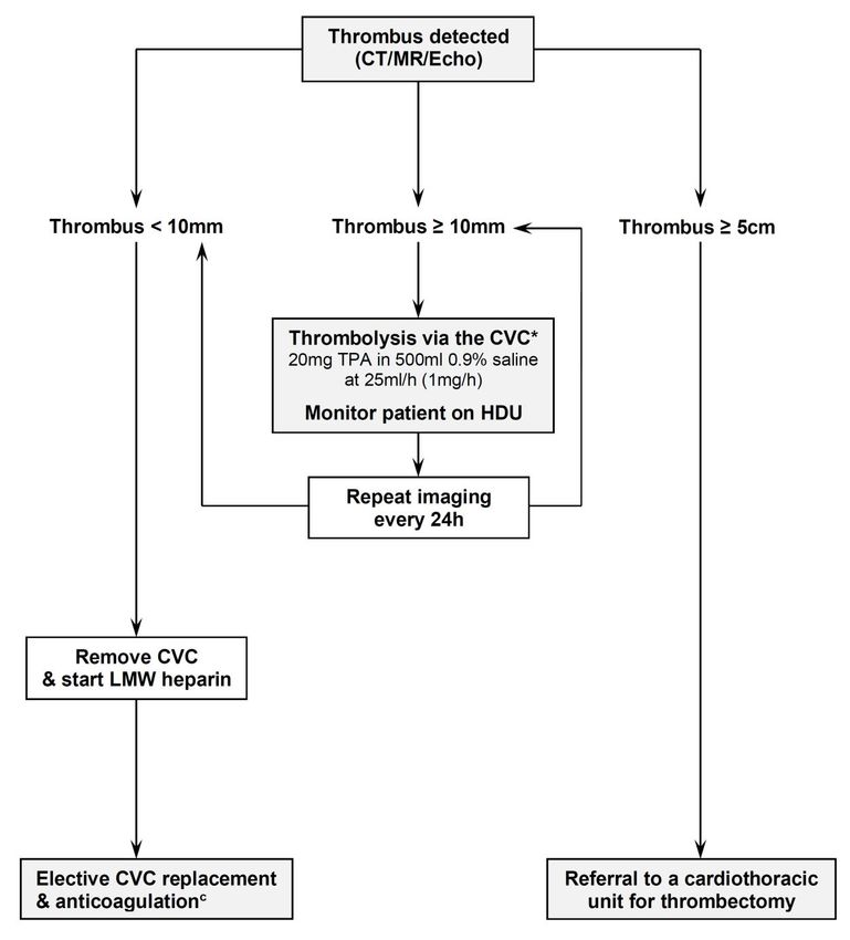
17. The management depends on the size of the thrombus. This can be divided into small,
medium or large thrombus attached to the end of the CVC. Arbitrarily small is 50mm.
18. Small thrombi: withdraw the CVC. This will not result in a clinically significant pulmonary
embolus.
19. Medium thrombi: start thrombolysis via the CVC [20mg TPA in 500ml 0.9% saline at 25ml/h
(1mg/h)] and monitor the patient on HDU. If the CVC is blocked then a thrombolysis
catheter can be placed by interventional radiology adjacent to the thrombus. The imaging
should be repeated every 24h until the thrombus isDate of Preparation September 2021
Flowchart 2: clinical practice based algorithm for thrombosis at the
end of the CVC
Referral to a cardiothoracic
unit for thrombectomy
BAPEN Office, Seven Elms, Dark Lane, Astwood Bank, Redditch, Worcestershire, B96 6HB, ENGLAND
Tel: +44 (0)1527 457 850 Email: bapen@bapen.org.uk www.bapen.org.ukDate of Preparation September 2021
Central venous stenosis
A central venous stenosis may be present in patients who have had an indwelling central venous
catheter over a long period of time. In addition, central venous stenosis can often occur treatment
for a central venous thrombosis.
Symptoms and signs
The clinical signs are similar to those for a central venous thrombosis but are much less acute. Often
there are subtle signs with dilated veins over the chest or neck, dilated collateral veins or mild signs
of swelling when an infusion is running.
Diagnosis
A diagnosis of CV stenosis can be made by CT venogram, MR venogram or contrast venogram.
Ultrasound with doppler can be used but this is only reliable to detect subclavian, jugular or femoral
stenoses. It is not reliable when the thrombosis is more central.
Management
22. Once diagnosed, the decision to treat will depend upon symptoms, the location of the
stenosis as well as local interventional or vascular expertise. The most appropriate treatment
requires close discussion with the interventional radiologists and, if necessary, the vascular
surgeons.
23. Patients with a CV stenosis who are not undergoing any interventional procedures should be
anticoagulated to prevent a subsequent thrombosis
24. Patients with a CV stenosis who undergo an interventional procedure should receive
antiplatelet therapy for 3-6 months and subsequently receive anticoagulation if no other
procedures are being considered
BAPEN Office, Seven Elms, Dark Lane, Astwood Bank, Redditch, Worcestershire, B96 6HB, ENGLAND
Tel: +44 (0)1527 457 850 Email: bapen@bapen.org.uk www.bapen.org.ukDate of Preparation September 2021
Flowchart 3: clinical practice-based algorithm for CV stenosis
BAPEN Office, Seven Elms, Dark Lane, Astwood Bank, Redditch, Worcestershire, B96 6HB, ENGLAND
Tel: +44 (0)1527 457 850 Email: bapen@bapen.org.uk www.bapen.org.ukDate of Preparation September 2021
References
1. Dibb M, Lal S. Home Parenteral Nutrition: Vascular Access and Related Complications. Nutr
Clin Pract Off Publ Am Soc Parenter Enter Nutr. United States; 2017 Dec;32(6):769–76.
2. Brandt CF, Hvistendahl M, Naimi RM, Tribler S, Staun M, Brobech P, et al. Home Parenteral
Nutrition in Adult Patients With Chronic Intestinal Failure: Catheter-Related Complications
Over 4 Decades at the Main Danish Tertiary Referral Center. J Parenter Enter Nutr [Internet].
2016;(Cvc):1–10. Available from:
http://pen.sagepub.com/cgi/doi/10.1177/0148607116655449
3. Christensen LD, Holst M, Bech LF, Drustrup L, Nygaard L, Skallerup A, et al. Comparison of
complications associated with peripherally inserted central catheters and Hickman???
catheters in patients with intestinal failure receiving home parenteral nutrition. Six-year
follow up study. Clin Nutr. 2016;
4. Bozzetti F, Mariani L, Bertinet DB, Chiavenna G, Crose N, De Cicco M, et al. Central venous
catheter complications in 447 patients on home parenteral nutrition: an analysis of over
100.000 catheter days. Clin Nutr. 2002;21:475–85.
5. Mukau L, Talamini MA, Sitzmann J V, Burns RC, McGuire ME. Long-term central venous access
vs other home therapies: complications in patients with acquired immunodeficiency
syndrome. JPEN J Parenter Enteral Nutr. United States; 1992;16(5):455–9.
6. Bakker H, Bozzetti F, Staun M, Leon-Sanz M, Hebuterne X, Pertkiewicz M, et al. Home
parenteral nutrition in adults: a european multicentre survey in 1997. ESPEN-Home Artificial
Nutrition Working Group. Clin Nutr [Internet]. 1999;18(3):135–40. Available from:
http://www.ncbi.nlm.nih.gov/pubmed/10451476
7. Lorentsen R, Munck LK, Wildt S. Parenteral therapy and complications in patients with
intestinal failure in a regional unit. Scand J Gastroenterol. England; 2017 Dec;52(12):1326–
30.
8. Durkin MJ, Dukes JL, Reeds DN, Mazuski JE, Camins BC. A Descriptive Study of the Risk Factors
Associated With Catheter-Related Bloodstream Infections in the Home Parenteral Nutrition
Population. JPEN J Parenter Enteral Nutr [Internet]. 2015/01/16. 2016 Sep;40(7):1006–13.
Available from: https://pubmed.ncbi.nlm.nih.gov/25596210
9. Szeinbach SL, Pauline J, Villa KF, Commerford SR, Collins A, Seoane-Vazquez E. Evaluating
catheter complications and outcomes in patients receiving home parenteral nutrition. J Eval
Clin Pract. England; 2015 Feb;21(1):153–9.
10. Touré A, Duchamp A, Peraldi C, Barnoud D, Lauverjat M, Gelas P, et al. A comparative study
of peripherally-inserted and Broviac catheter complications in home parenteral nutrition
patients. Clin Nutr. 2015;34(1).
11. Cotogni P, Pittiruti M, Barbero C, Monge T, Palmo A, Boggio Bertinet D. Catheter-related
complications in cancer patients on home parenteral nutrition: a prospective study of over
51,000 catheter days. JPEN J Parenter Enteral Nutr. United States; 2013;37(3):375–83.
12. Cuerda C, Joly F, Corcos O, Concejo J, Puiggrós C, Gil C, et al. Prospective study of catheter-
related central vein thrombosis in home parenteral nutrition patients with benign disease
using serial venous Doppler ultrasound. Clin Nutr. England; 2016 Feb;35(1):153–7.
13. Harrison E, Herrick AL, Dibb M, McLaughlin JT, Lal S. Long-term outcome of patients with
systemic sclerosis requiring home parenteral nutrition. Clin Nutr. 2015;34(5):991–6.
BAPEN Office, Seven Elms, Dark Lane, Astwood Bank, Redditch, Worcestershire, B96 6HB, ENGLAND
Tel: +44 (0)1527 457 850 Email: bapen@bapen.org.uk www.bapen.org.ukDate of Preparation September 2021
14. Lloyd DAJ, Vega R, Bassett P, Forbes A, Gabe SM. Survival and dependence on home
parenteral nutrition: Experience over a 25-year period in a UK referral centre. Aliment
Pharmacol Ther. 2006;24(8):1231–40.
15. Matsumoto CS, Subramanian S, Fishbein TM. Adult Intestinal Transplantation. Gastroenterol
Clin North Am. 2018 Jun;47(2):341–54.
16. Eastridge BJ, Lefor AT. Complications of indwelling venous access devices in cancer patients. J
Clin Oncol Off J Am Soc Clin Oncol. United States; 1995 Jan;13(1):233–8.
17. Spencer TR, Mahoney KJ. Reducing catheter-related thrombosis using a risk reduction tool
centered on catheter to vessel ratio. J Thromb Thrombolysis 2017; 44(4):427-434.
18. Baxi SM, Shuman EK, Scipione CA, Chen B, Sharma A, Rasanathan JJK, Chenoweth CE. Impact
of postplacement adjustment of peripherally inserted central catheters on the risk of
bloodstream infection and venous thrombus formation. Infect Control Hosp Epidemiol 2013;
34(8):785-92.
19. Gould JR, Carloss HW, Skinner WL. Groshong catheter-associated subclavian venous
thrombosis. Am J Med. United States; 1993 Oct;95(4):419–23.
20. Cadman A, Lawrance JAL, Fitzsimmons L, Spencer-Shaw A, Swindell R. To clot or not to clot?
That is the question in central venous catheters. Clin Radiol. England; 2004 Apr;59(4):349–55.
21. Crowley AL, Peterson GE, Benjamin DKJ, Rimmer SH, Todd C, Cabell CH, et al. Venous
thrombosis in patients with short- and long-term central venous catheter-associated
Staphylococcus aureus bacteremia. Crit Care Med. United States; 2008 Feb;36(2):385–90.
22. van Rooden CJ, Schippers EF, Barge RMY, Rosendaal FR, Guiot HFL, van der Meer FJM, et al.
Infectious complications of central venous catheters increase the risk of catheter-related
thrombosis in hematology patients: a prospective study. J Clin Oncol Off J Am Soc Clin Oncol.
United States; 2005 Apr;23(12):2655–60.
23. Berea-Baltierra R, Rivas-Ruiz R, Vela-Martinez E, Sevilla-Gonzalez M de la L, Talavera-Pina JO,
Valencia-Jimenez E, et al. Risk factors for subclavian vein thrombosis in cancer patients with
total parenteral nutrition. J Clin Med Res [Internet]. 2014/07/28. Elmer Press; 2014
Oct;6(5):345–53. Available from: https://pubmed.ncbi.nlm.nih.gov/25110538
24. Buchman AL, Misra S, Moukarzel A, Ament ME. Catheter thrombosis and superior/inferior
vena cava syndrome are rare complications of long term parenteral nutrition. Clin Nutr.
England; 1994 Dec;13(6):356–60.
25. van Ommen CH, Tabbers MM. Catheter-related thrombosis in children with intestinal failure
and long-term parenteral nutrition: how to treat and to prevent? Thromb Res. United States;
2010 Dec;126(6):465–70.
26. Lal S, Chadwick P, Nightingale J and the BIFA Committee. Diagnosis of catheter related blood
stream infections (CRBSIs). April 2018.
https://www.bapen.org.uk/pdfs/bifa/recommendations-for-crbsi-diagnosis.pdf
27. Lal S, Chadwick P, Nightingale J and the BIFA Committee. Management of catheter related
blood stream infections (CRBSIs). January 2019.
https://www.bapen.org.uk/pdfs/bifa/recommendations-on-management-of-crbsi.pdf
BAPEN Office, Seven Elms, Dark Lane, Astwood Bank, Redditch, Worcestershire, B96 6HB, ENGLAND
Tel: +44 (0)1527 457 850 Email: bapen@bapen.org.uk www.bapen.org.ukYou can also read