A Rare Autosomal Dominant Variant in Regulator of Calcineurin Type 1 (RCAN1) Gene Confers Enhanced Calcineurin Activity and May Cause FSGS
←
→
Page content transcription
If your browser does not render page correctly, please read the page content below
BASIC RESEARCH            www.jasn.org

A Rare Autosomal Dominant Variant in Regulator of
Calcineurin Type 1 (RCAN1) Gene Confers Enhanced
Calcineurin Activity and May Cause FSGS
Brandon M. Lane ,1 Susan Murray,2 Katherine Benson,3 Agnieszka Bierzynska,4
Megan Chryst-Stangl,1 Liming Wang ,5 Guanghong Wu,1 Gianpiero Cavalleri ,3
Brendan Doyle,6 Neil Fennelly,6 Anthony Dorman,6 Shane Conlon,2 Virginia Vega-Warner,7
Damian Fermin ,7 Poornima Vijayan ,8 Mohammad Azfar Qureshi,8 Shirlee Shril,9
Moumita Barua,8 Friedhelm Hildebrandt,9 Martin Pollak,10 David Howell,11
Matthew G. Sampson,9,12 Moin Saleem ,4 Peter J. Conlon ,2,13 Robert Spurney,5 and
Rasheed Gbadegesin 1,5
Due to the number of contributing authors, the affiliations are listed at the end of this article.

ABSTRACT
Background Podocyte dysfunction is the main pathologic mechanism driving the development of FSGS
and other morphologic types of steroid-resistant nephrotic syndrome (SRNS). Despite significant prog-
ress, the genetic causes of most cases of SRNS have yet to be identified.
Methods Whole-genome sequencing was performed on 320 individuals from 201 families with familial and
sporadic NS/FSGS with no pathogenic mutations in any known NS/FSGS genes.
Results Two variants in the gene encoding regulator of calcineurin type 1 (RCAN1) segregate with disease
in two families with autosomal dominant FSGS/SRNS. In vitro, loss of RCAN1 reduced human podocyte
viability due to increased calcineurin activity. Cells expressing mutant RCAN1 displayed increased calci-
neurin activity and NFAT activation that resulted in increased susceptibility to apoptosis compared with
wild-type RCAN1. Treatment with GSK-3 inhibitors ameliorated this elevated calcineurin activity, suggest-
ing the mutation alters the balance of RCAN1 regulation by GSK-3b, resulting in dysregulated calcineurin
activity and apoptosis.
Conclusions These data suggest mutations in RCAN1 can cause autosomal dominant FSGS. Despite the
widespread use of calcineurin inhibitors in the treatment of NS, genetic mutations in a direct regulator of
calcineurin have not been implicated in the etiology of NS/FSGS before this report. The findings highlight
the therapeutic potential of targeting RCAN1 regulatory molecules, such as GSK-3b, in the treatment
of FSGS.

JASN 32: ccc–ccc, 2021. doi: https://doi.org/10.1681/ASN.2020081234

Glomerular diseases, including diabetic nephropathy,
are the primary known cause of CKD in the United
States and the rest of the world.1,2 Most glomerular            Received August 27, 2020. Accepted February 25, 2021.
diseases are due to primary dysfunction or secondary            Published online ahead of print. Publication date available at
injury to the podocyte, the visceral epithelial cell of the     www.jasn.org.
trilayer glomerular filtration barrier. Primary podocyte
                                                                Correspondence: Dr. Rasheed Gbadegesin, Division of Ne-
dysfunction, referred to as podocytopathy, typically            phrology, Duke Molecular Physiology Institute, Carmichael
manifests as steroid-resistant nephrotic syndrome               building, RM 51-104, 300 N Duke St., Durham, NC 27701-2047.
                                                                Email: rasheed.gbadegesin@duke.edu
(SRNS) with morphologic changes of FSGS or minimal
change disease apparent on kidney biopsy specimens.3,4          Copyright © 2021 by the American Society of Nephrology

JASN 32: ccc–ccc, 2021                                                                                             ISSN : 1046-6673/3207-ccc   1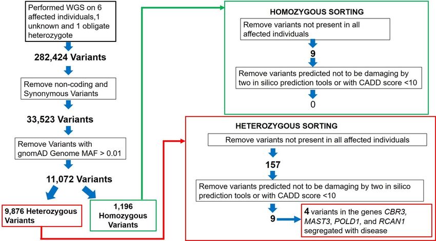
BASIC RESEARCH    www.jasn.org

   It is estimated that 5%–30% of all podocytopathies are due
                                                                    Significance Statement
to mutation in single genes, especially in children and young
adults.5–7 More than 60 genes have been identified as causes of      Whole-genome sequencing of 320 individuals with nephrotic syn-
monogenic SRNS; however, these genes are responsible for            drome (NS) of unclear genetic etiology and data from several
                                                                    independent patient cohorts provided insight into the genetic ar-
only 20% of all genetic SRNS, suggesting there are other, un-
                                                                    chitecture of the condition. The strategy identified a disease-
identified, single-gene causes of SRNS.6,8–11 Identification of       causing autosomal dominant mutation in regulator of calcineurin
these causal genes has the potential to improve our under-          type 1 (RCAN1) that increased cellular calcineurin (CN) activity,
standing of disease pathogenesis, the identification of disease      NFAT (NF of activated T cells) activation, and susceptibility to ap-
biomarkers, the identification of new therapeutic agents, and        optosis of podocytes in vitro. Inhibition of an RCAN regulator, GSK-
                                                                    3b, rescued the increased CN activation. Mutations in RCAN1 are a
the repurposing of existing agents to treat nephrotic
                                                                    novel cause of NS and reveal a potential target for developing
syndrome (NS).                                                      personalized therapy.
   To identify new, single-gene causes of SRNS, we carried out
whole-genome sequencing (WGS) on 320 individuals from
201 families with familial and sporadic NS, and reviewed           METHODS
whole-exome sequencing data from patients with NS of un-
clear genetic etiology. We identified two segregating, hetero-      WGS
zygous mutations in the regulator of calcineurin (CN) type 1       WGS was performed at GENEWIZ (South Plainfield, NJ).
(RCAN1) in two large Northern European families. There are         Briefly, genomic DNA samples were assessed for purity, quan-
three genes in the RCAN family RCAN1–3, all of which encode        tity, and quality by using the NanoDrop 2000 Spectrophotom-
proteins capable of interacting with CN and inhibiting CN-         eter (Thermo Fisher), Qubit 2.0 Fluorometer, Qubit dsDNA
dependent signaling pathways.12–22 Therefore, we screened          HS Assay Kit (Thermo Fisher), and agarose gel electrophore-
families with hereditary and sporadic NS in other independent      sis. Library construction was then performed using Illumina’s
cohorts for rare variants in RCAN1–3 genes. We identified           TruSeq DNA PCR-Free library preparation kit following the
four possible disease-causing variants: three in RCAN2, and        manufacturer’s protocol. Genomic DNA was fragmented by
one in RCAN3.                                                      acoustic shearing with a Covaris S220 instrument. Sheared
   The RCAN family of proteins form a complex with the             DNA was then end repaired and A-tailed, followed by adaptor
catalytic subunit of CN, and regulate both CN phosphatase          ligation. Final libraries were analyzed on the Agilent TapeSta-
activity and its ability to bind key substrates like NF of acti-   tion, for library sizing, and quantified using the Qubit dsDNA
vated T cells (NFAT).12–14,16–18,20,23–27 Unregulated CN acti-     HS Assay Kit along with the KAPA Library Quantification Kit
vation is central to the pathogenesis of multiple glomerular       for quantitative PCR. DNA libraries were sequenced using
disease processes, and CN inhibitors (CNIs) are often used to      Illumina platforms to generate $120 Gb of raw data per sam-
treat glomerular diseases.28–36 The rationale for treating ac-     ple, with a 23150-bp, paired-end sequencing configuration.
quired forms of NS with CNIs has historically been that the
immune system was thought to play a significant role in ac-         Variant Calling and Annotation
quired forms of NS, such as minimal change disease                 DNA-sequencing data were processed using fastp1 to trim low-
and FSGS.28,29 However, CN has nonimmunologic actions              quality bases and Illumina sequencing adapters from the 39 end
that are important in the pathogenesis of kidney diseases.         of reads.39 Reads were then aligned to the GRCh37 version of the
For example, CN causes cytoskeletal instability by dephosphor-     human genome with the BWA2 algorithm.40 PCR duplicates
ylating synaptopodin and promoting its degradation.28–31,33        were flagged using the PICARD Tools3 software suite.41 Align-
Moreover, podocyte loss plays a key role in pathogenesis           ment processing and variant calling were performed using the
of FSGS, and CN promotes a decrease in the number of               GATK4 toolkit following the Broad Institute’s Best Practices
glomerular podocytes by both genetic and nongenetic                Workflow.42,43 Functional consequences and genotype prove-
mechanisms.28–31,34,37,38                                          nances of variants were annotated using Ensembl Variant
   In this study, we discovered that a disease-causing RCAN1       Predictor.44 After annotation, variants meeting the following
variant in individuals with FSGS had a reduced ability to in-      criteria were selected for further analysis: having a “pass” status
hibit activated CN compared with wild-type (WT) RCAN1.             after GATK’s Variant Quality Score Recalibration, found to re-
The increase in CN activation induced by the RCAN1 variant         side in a coding region, and had an allele frequency of ,5% in at
was inhibited by treatment with antagonists of glycogen syn-       least one population of the Genome Aggregation Database (gno-
thase kinase 3 (GSK-3). In addition, cells expressing this         mAD).45 Second-level filtering to identify disease-causing vari-
RCAN1 variant were more sensitive to apoptotic stimuli,            ants is as shown in Supplemental Figure 1. Variants of interest
which could be rescued by CNI treatment. Collectively, our         were confirmed by Sanger sequencing.
findings suggest mutations in RCAN1 are a novel genetic cause
of NS, and use of CNIs and GSK antagonists may represent           RCAN1 Knockdown Podocytes
targeted or personalized therapy for individuals with NS/FSGS      Multiple, conditionally immortalized, human podocyte lines
due to RCAN1 mutations.                                            (courtesy of Dr. Jeffrey Kopp) with reduced RCAN1 expression

2        JASN                                                                                                      JASN 32: ccc–ccc, 2021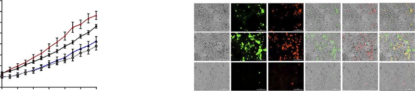
www.jasn.org    BASIC RESEARCH

were created using lentiviral transduction of short hairpin RNA   total phosphatase levels. EGTA-supplemented buffer was
(shRNA) against RCAN1 (TRCN0000019848; Millipore                  added to additional wells for each sample to measure the
Sigma). Lentiviral control lines were created using shRNA         non-CN phosphatase activity. All sample wells received water
with no known target (SHC016V; Millipore Sigma). Podocyte         and CN substrate (RII phosphopeptide), except for back-
lentiviral transduction was performed as described previ-         ground wells in which water was substituted for substrate. A
ously.46 RCAN1 KD was confirmed through immunoblotting             positive control (CN enzyme supplied by the kit) was used to
(LS-C162511; LifeSpan Biosciences).                               ensure assay effectiveness. The plate was incubated at 30°C for
                                                                  10 minutes, lysate was added to all sample wells, and then it
Immunoprecipitation Studies                                       was incubated at 30°C for 30 minutes. BioMol Green reagent
Immunoprecipitation was performed using a protocol modi-          (100 ml) was then added to all wells and incubated for 25 min-
fied from Fuentes et al.13 For the studies, human embryonic        utes at room temperature before reading the OD620nm using a
kidney cells (HEK293) cells were grown in DMEM supple-            Tecan (Männedorf, Switzerland) Infinite 200 microplate
mented with 10% FCS, penicillin (100 U/ml), and streptomy-        reader, with two reads per well. Background well readings
cin (100 mg/ml) (all from Gibco, Gaithersburg, MD), as            were subtracted from all experimental well readings, and mo-
previously described.47 For transfection, HEK293 cells were       les of phosphate were calculated using the phosphate standard
plated in six-well Costar tissue culture plates (Corning, Corn-   curve. The CN-specific phosphatase activity was calculated by
ing, NY) and grown to approximately 80% confluency. Cells          subtracting the phosphate present in EGTA-treated cells
were then cotransfected with the FLAG-tagged CN construct         (non–CN-related phosphatase activity) from calmodulin-
(GenScript, Piscataway, NJ) and the Myc-tagged RCAN1              treated cells (total phosphatase activity). All experiments
construct (WT or mutant as indicated; GenScript) using Lip-       were repeated in triplicate.
ofectamine 2000, according to the manufacturer’s recommen-
dations (ThermoFisher Scientific, Waltham, MA). Cells were         GSK-3 Inhibition
harvested 48 hours after transfection, and cell pellets were      We diluted LY2090314 (Selleck Chemicals, Houston, TX) and
lysed in ice-cold 50 mM Tris-hydrochloride (pH 7.5),              tideglusib (Selleck Chemicals) in water (1:2000) from DMSO
150 mM sodium chloride, and 1% nonidet P-40 in the pres-          stock (10 mM and 2 mM, respectively) and replaced 2 ml of
ence of protease inhibitors (Protease Inhibitor Cocktail;         water in the CN activity assay described above with an equal
Sigma-Aldrich, St. Louis, MO). For immunoprecipitation of         volume of diluted GSK inhibitors to reach a final concentra-
RCAN1 constructs, protein lysates were incubated with anti-       tions of 200 nM LY2090314 and 1 mM tideglusib. Untreated
Myc antibodies (ThermoFisher Scientific) at 4°C for 1 hour,        samples wells received 2 ml of 1:2000 diluted DMSO. To ac-
and then Protein A Plus Protein G (Millipore, Bedford, MA)        commodate the additional sample conditions for these inhi-
was added to the lysate and rocked for approximately 4 hours      bition studies, we used cell lysate(SPACE)te diluted 1:1 in lysis
at 4°C. For immunoprecipitation of CN constructs, protein         buffer. CN activity was calculated by subtracting the phos-
lysates were incubated with anti-FLAG antibodies linked to        phate activity in EGTA-treated wells from the activity in
sepharose beads (Cell Signaling Technology, Danvers, MA)          treated or untreated wells. This experiment was repeated
and rocked for approximately 4 hours at 4°C. After three          in triplicate.
washes with ice-cold lysis buffer, Laemmli sample buffer was
added to the pellet and boiled for approximately 10 minutes,      NFAT Luciferase Assay
and immunoblotting was then performed. This experiment            The NFAT luciferase assay was performed using the Dual Fire-
was repeated in triplicate.                                       fly and Renilla Luciferase Assay Kit (Biotium, Freemont, CA),
                                                                  according to the manufacturer’s protocol. Briefly, HEK293 cells
CN Activity Assay                                                 were grown in 24-well plates and transfected with Lipofecta-
CN activity was examined using the Cellular Calcineurin Ac-       mine 2000 according to the manufacturer’s protocol. Cells were
tivity assay kit according to the manufacturer’s protocol (Enzo   transfected with equal parts of an NFAT-luciferase reporter
Life Sciences, Farmingdale, NY). Conditionally immortalized       construct (Promega), loading control construct (pRL-TSK;
podocytes were grown on collagen-coated, six-well dishes un-      Promega), PPP3CA, and one of the RCAN1 constructs
til confluent. HEK293 cells were grown in six-well dishes and      (0.6 ng DNA per construct per well). Lysates were harvested
transfected with PPP3CA and RCAN1 constructs using Lip-           using the supplied lysis buffer after 48 hours and plated in
ofectamine 2000, as described above, for 48 hours. Immuno-        duplicate on 96-well plates. Measurements were taken using
blotting was performed to ensure equal levels of PPP3CA and       an Infinite Pro 200 microplate reader with automated injection
RCAN1 transfection between HEK293 cell samples. All exper-        (Tecan), which injected 100 ml of luciferase assay reagent into
imental cells were washed with Tris-buffered saline before ly-    each well, recorded the fluorescence, added 100 ml of Renilla
sates were harvested and cleared of free nucleotides using a      assay reagent, and performed another fluorescence reading.
desalting column. Phosphate standards were loaded in dupli-       The relative luminescence units for each well were then calcu-
cate on a 96-well plate. Calmodulin-supplemented buffer was       lated by dividing the luciferase reagent readings by the Renilla
added to appropriate wells for measuring the background and       reagent readings. The experiment was repeated in triplicate.

JASN 32: ccc–ccc, 2021                                                                          RCAN1 Variants Can Cause NS      3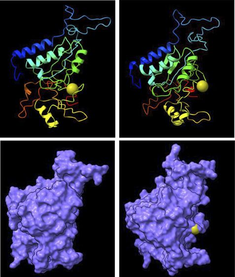
BASIC RESEARCH www.jasn.org
Automated Cell Apoptosis Imaging readings. The experiment was repeated in quadruplicate with a total
To both visualize and quantify the apoptosis and total cell death, we N of at least 16 for each cell type, and full videos of the representative
used a Lionheart FX automated microscope from BioTek along HEK293 cell images are available in Supplemental Videos 1–3.
with fluorescent apoptosis reagents. Podocytes were plated and
grown to confluency before beginning the assay. HEK293 cells Three-Dimensional In Silico Protein Modeling
were grown in 96-well plates, as described above, and transfected Molecular graphics and analyses of PDB files created in the
with PPP3CA and RCAN1 constructs using Lipofectamine LTX, I-TASSER software48,49 was performed with UCSF ChimeraX,
according to the manufacturer’s protocol, for 48 hours before be- which was developed by the Resource for Biocomputing, Vi-
ginning the assay. Cells were exposed to serum-free media contain- sualization, and Informatics at the University of California,
ing a 1:500 dilution of NucView Caspase-3 Alexa 488 (Biotium) San Francisco, with support from National Institutes of Health
and a 1:2000 dilution of propidium iodide (Sigma-Aldrich). The (NIH) (R01-GM129325) and the Office of Cyber Infrastruc-
NucView reagent consists of a substrate of caspase-3 that emits ture and Computational Biology, National Institute of Allergy
green fluorescence when cleaved, whereas propidium iodide fluo- and Infectious Diseases.50
resces in late apoptotic and necrotic cells. These media also con-
tained either 1 mM FK506 or an equal concentration of vehicle Illustrations
(ethanol). Bright-field images, along with green and red fluorescent The summary graphic was created using Biorender.com.
images, were collected every 2 hours for 48 hours. Using automated
GEN5 software from BioTek, the images were processed to remove Immunoblotting
background, and the number of fluorescent cells was quantified Immunoblotting was performed using standard methods
for each well using label-free cell counting. Wells containing full and visualized by enhanced chemiluminescence, as previously de-
serum were used as a control to test the validity of the apoptosis scribed.51 Antibodies were used at the following concentrations:
A 40030 PEDIGREE
?
* ? ? ? ?
10017 10015 10126 10559 10124 11041 10557 10016 10585 10586 28141
L I/T S L I/T S L I/T S L I/T S L I S L I/T S
C T G A T/ C C T C C C T G A T/ C C T C C C T G A T/ C C T C C C T G A T/ C C T C C C T G AT C T C C C T G A T/ C C T C C
? ? ? ? ? ? ?
10027 10568 10231 10230 10056 11067 10228 10229 10052 10486
L I S L I/T S L I/T S
C T G AT C T C C C T G A T/ C C T C C C T G A T/ C C T C C
?
11220
B C D E
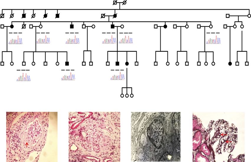
Figure 1. The RCAN1 p.I162T mutation segegrates with disesase in a family with FSGS. (A) Pedigree of European family 40030 with
FSGS and the RCAN1 I162T variant segregating with the disease in the family. Family members that are currently unnaffected but may
develop disease later in life are depicted with a question mark. Sequenced individuals are shown with a chromatogram and associated
amino acid sequence (L5Leucine, I5 Isoleucine, T5 Threonine, S5Serine). Asterisk indicates obligate carrier. (B–E) Kidney histology
from individual 10557 in family 40030. (B and C) FSGS on hematoxylin and eosin staining at (red arrow). (D) Mild foot process ef-
facement (red arrows) and thinned glomerular basement membrane. (E) Capillary loop double contour formation (red arrows) on silver
staining. Original magnification, 320 in (B), 340 in (C) and (D).
4 JASN JASN 32: ccc–ccc, 2021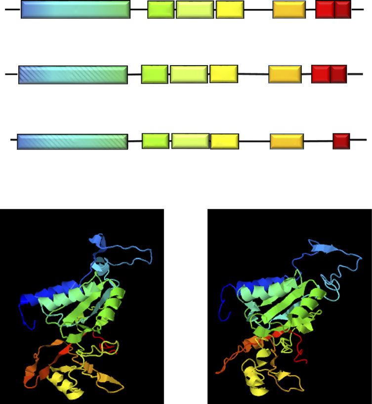
www.jasn.org          BASIC RESEARCH

Table 1. In silico prediction of disease-causing RCAN1 heterozygous variant
Variant                      gnomAD (EUR) MAF (EUR) MAF (all population) CADD                        PolyPhen                SIFT      MutationTaster
RCAN1 c. T485C, p. I162T       2 of 128,738       0.00001           0.000007             26.7   Probably damaging Damaging Disease causing
GRCH37: RCAN1-001, transcript ENST00000313806.4. EUR, European; MAF, minor allele frequency; CADD, combined annotation dependent depletion; SIFT,
sorting intolerant from tolerant.

1:500 for caspase-3 (Cell Signaling Technology), 1:1000 for MYC              harvesting, RNA extraction (RNAeasy kit; Qiagen), and gener-
tag (Cell Signaling Technology), 1:1000 for DYKDDDDK tag (Cell               ation of cDNA (Promega), as previously described.52 TaqMan
Signaling Technologies), and 1:3000 for b-actin (Sigma-Aldrich).             probes (Invitrogen) were used in to analyze gene expression for
For apoptosis experiments, HEK293 cells were grown in six-well               CD2AP (Hs00961451_m1), RCAN1 (Hs01120954_m1),
plates and transfected with PPP3CA and RCAN1 constructs using                RCAN2 (Hs00195165_m1), RCAN3 (Hs00203728_m1), and
Lipofectamine LTX, according to the manufacturer’s protocol. After           PTPRO (Hs00958177_m1). The analysis was repeated in trip-
48 hours of serum starvation, the cells were washed with PBS,                licate, with multiple wells per sample in each replicate.
harvested, and the lysates were analyzed with immunoblotting.
The experiments were repeated in triplicate and quantified using              Electron Microscopy of Renal Biopsy Specimens
ImageJ software. Unmodified Western blot images are shown in                  The harmonic mean of the glomerular basement membrane
the Supplemental Materials.                                                  thickness was calculated using multiple measurements, using
                                                                             reference ranges for men and women as reported by Das et al.53
Quantitative Real-Time PCR
Conditionally immortalized, human podocytes (generously                      Statistical Analyses
provided by Dr. Jeffrey Kopp) were grown until confluent in                   The two-tailed t test was used for the comparison of RCAN1-
T75 and differentiated at 37° for 14 days before lysate                      KD podocyte immunoblotting against RCAN1 (t59.802;

                                                                            FLISPPxSPP                    PK||Q TxxP
                         A
                               RCAN1                RRM               LxxP             ExxP      Px|x|T                252

                                                          K128E               I162T*

                                                                          FLISPPxSPP                      PK||Q TxxP
                               RCAN2               RRM               LxxP           ExxP        Px|x|T                 243

                                                                  P149T                                    R234H       N243H

                                                                        FLISPPxSPP                             TxxP
                                RCAN3               RRM              LxxP        ExxP            Px|x|T                241

                                                                                   C173G

                         B
                                       RCAN1 WT                                     RCAN1 I162T

Figure 2. Potentially pathogenic variants disrupt conserved RCAN protein domains. (A) Depictions of RCAN1, RCAN2, and RCAN3 peptides
showing the conserved protein domains within the RCAN1 family of proteins and locations (arrows) of the newly identified variants. The variants
identified in these patients are all located in or near conserved domains, including the RNA recognition motif (RRM; RNA binding–like) domain
(blue-green); the carboxy-terminal CN binding motifs (orange and red); and the SP motif (green-yellow), which contains the LxxP, FLISPPxSPP,
and ExxxP sequences. (B) Three-dimensional modeling of WT RCAN1 and the p.I162T variant revealed disruption of the amino- and carboxy-
terminal regions (blue and red, respectively), and the region around the SP motif (green), when compared with WT RCAN1.

JASN 32: ccc–ccc, 2021                                                                                         RCAN1 Variants Can Cause NS           5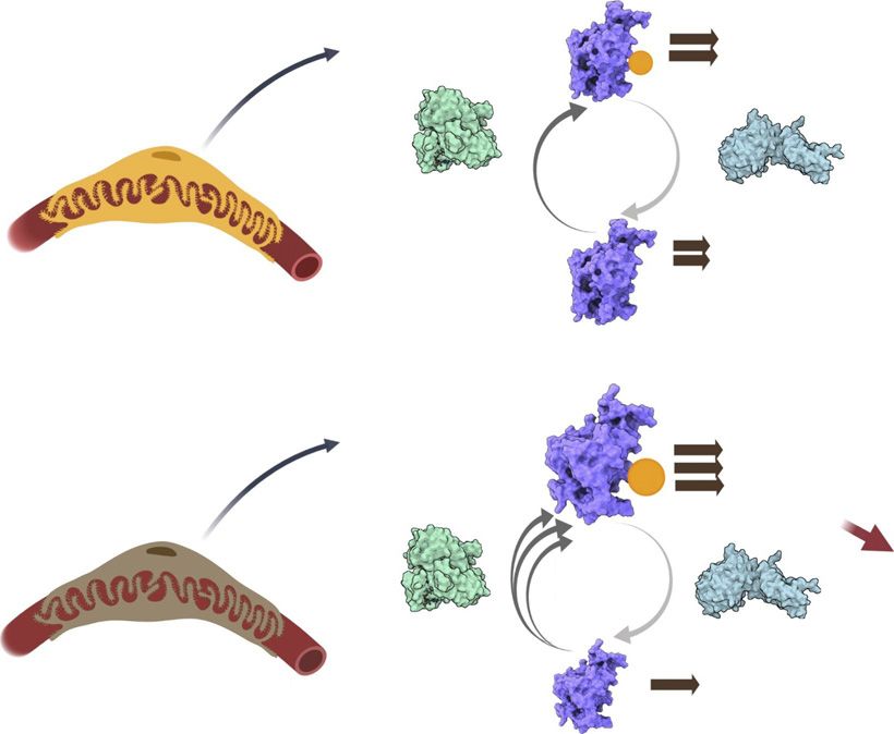
BASIC RESEARCH         www.jasn.org

Table 2. Phenotype of individuals with RCAN1 mutations
Family Number Individual Number Age at Onset (yr) Proteinuria (quantity) Biopsy (histology) CKD Stage Transplant Recurrence
40030                       10056                     29                     UNK                         N                 5               Y              N
40030a                      10126                    NA                       N                          N                 0               N             NA
40030                       10559                     48                      Y                      Y (UNK)               5               Y             UNK
40030                       10552                     35                 Y (825 mg)                  Y (FSGS)              5               N             NA
40030                       10557                     65                 Y (5000 mg)                 Y (FSGS)              5               N             NA
40030a                      10586                    NA                       N                          N                 0               N             NA
40030                       10017                    UNK                     UNK                       UNK                UNK             UNK            UNK
40030                       10229                     45                Y (.3000 mg)                     N                 1               N             NA
6559                           1                       5                 Y (1860 mg)                 Y (FSGS)              5               Y              N
6559                          101                     40                 Y (2410 mg)                   UNK                 1               N             NA
6559a                        0106                    NA                       N                          N                 0               N             NA
UNK, unknown; N, no; Y, yes; NA, not available.
a
 Asymptomatic.

degrees of freedom [df]56) and cleaved caspase-3 (t54.764;                         variants using previously published algorithms for the identi-
df56). Two-way ANOVA, followed by a Dunnett multiple                               fication of causal variants in families with Mendelian kidney
comparison analysis, was used to analyze automated live im-                        disease (Supplemental Figure 1). We identified four variants in
aging of RCAN1-KD podocytes (df52; F57.698). One-way                               four genes that are present in all affected individuals
ANOVA, followed by a Tukey multiple comparisons test, was                          (Supplemental Table 1) in a large Irish family recruited as
used to determine the differences between means for the anal-                      part of our collaboration with the Irish Kidney Gene Project
ysis of RCAN1-variant apoptosis immunoblotting results                             (Figure 1).54,55 One of these variants was in RCAN1 (c. T485C,
(df53; F556.94). A two-tailed t test analysis was used for                         p.I162T; transcript, ENST00000313806.4; GRCh37). A search
the variant NFAT luciferase assay (t511.84; df510) and the                         for pathogenic variants in these four genes in existing whole-
CN activity assay (t56.579; df522). Two-way ANOVA, fol-                            exome sequencing data from 191 families with NS of unclear
lowed by a Dunnett multiple comparisons analysis, was used                         etiology identified a second family with another segregating
to compare groups for RCAN1-variant automated live-cell                            variant in RCAN1 (Supplemental Figure 2), but did not find
apoptosis imaging (df53; F510.92) and GSK-3 inhibition                             pathogenic variants in the other three candidate genes. The
experiments (df52; F527.03).                                                       second variant, c.A382G, p.K128E, is rare, with a minor allele
                                                                                   frequency of 0.0004172 in the gnomAD database, and both
                                                                                   RCAN1 variants are conserved in evolution (Supplemental
RESULTS                                                                            Table 2).

Clinical Ascertainment and WGS                                                     In Silico Modeling
We identified 320 individuals from 201 families with familial                       In silico modeling revealed the p.I162T RCAN1 variant is
and sporadic NS/FSGS with no pathogenic mutations in any                           predicted to be damaging by at least three prediction tools,
known NS/FSGS genes. We performed WGS and filtered                                  with a Combined Annotation Dependent Deletion score

Table 3. Rare heterozygous RCAN variants in NS cohorts
Study Number        Phenotype       Variant       Allele Count gnomAD MAF (All) PolyPhen                        SIFT          MutationTaster Conservation
159                SRNS, MCD    RCAN2       0                         0.000000          Damaging           Probably           Disease causing        Zebrafish
                               c.C445A                                                                     damaging
                               p.P149T
260                SRNS, FSGS RCAN2 2 of 248,944                      0.000008          Damaging           Probably           Disease causing           Frog
                               c.A728C                                                                     damaging
                               p.N243H
3                      NS       RCAN2 3 of 249,306                     0.00001          Damaging           Probably           Disease causing        Zebrafish
                               c.C700T                                                                     damaging
                               p.R234H
459                SSNS, FR/SD RCAN3        0                         0.000000           Tolerated         Probably           Disease causing        Zebrafish
                               c.T517G                                                                     damaging
                               p.C173G
GRCH37: RCAN1-001, transcript ENST00000313806.4; RCAN2-002, transcript ENST00000371374.1; RCAN3-001, transcript ENST00000374395.4. MAF, minor
allele frequency; MCD, minimal change disease; SSNS, steroid-sensitive NS; FR/SD, frequent relapsing/steroid-dependent; SIFT, sorting intolerant from tolerant.

6         JASN                                                                                                                           JASN 32: ccc–ccc, 2021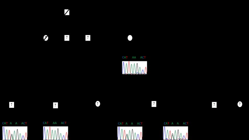
www.jasn.org         BASIC RESEARCH

      A                                                                                 B                                   Podocyte RCAN1 KD Quantification    C                                                          RCAN1 KD CN activity
                                               Control        RCAN1 KD                                            1.5                                                                                          15

                                                                                                                                                                               Relative Calcineurin Activity
                                              Podocyte         podocyte                                                                   *p
BASIC RESEARCH www.jasn.org
and RCAN3 (Table 3). The RCAN3 variant and one of the cell lysates using a cellular CN activity assay revealed the
three RCAN2 variants are novel and they are not found in p.I162T variants disrupted the regulatory function of
the gnomAD (approximately 250,000 chromosomes ana- RCAN1, resulting in increased CN activity compared with
lyzed). The other two variants in RCAN2 have minor allele WT RCAN1–expressing cells (P,0.001). An NFAT luciferase
frequency of #0.00001 in gnomAD. All of the variants are assay confirmed this increased CN activity resulted in elevated
predicted to be damaging by three in silico prediction tools, NFAT activation compared with WT RCAN1–expressing cells
and they are all conserved in evolution. Other missense vari- (P,0.001) (Figure 4). To verify the effectiveness of our con-
ants found in the three genes are listed in Supplemental structs, we also examined CN activity in cells transfected with
Table 4. either PPP3CA or RCAN1 WT alone to ensure CN activation
increased and decreased, respectively, in these assays, as com-
Loss of RCAN1 Disrupts Podocyte CN Regulation and pared with untransfected controls (Supplemental Figure 5).
Decreases Podocyte Viability
To determine the relevance of RCAN1 in the maintenance of RCAN1 p.I162T Induces Increased Apoptosis that Can
podocyte functional integrity, we first confirmed expression in Be Rescued by CN Inhibition
podocytes through quantitative real-time PCR of condition- Transfected HEK293 cell lines expressing PPP3CA and WT or
ally immortalized, human podocyte cell lines. RCAN1, mutant RCAN1 were exposed to serum starvation–induced
RCAN2, and RCAN3 are all expressed in podocytes at compa- apoptosis and evaluated using both automated live-cell imag-
rable levels with key podocyte genes, such as CD2AP and ing and Western blot quantification of caspase-3 activity.
PTPRO (GLEPP1) (Supplemental Figure 3). With the known Overexpression of RCAN1 p.I162T induced a significant in-
role of RCAN proteins in CN regulation, we examined the crease in apoptosis and total cell death relative to the WT
effects of loss-of-function RCAN1 mutations on podocyte RCAN1–expressing cells (Figure 5, Supplemental Figures 6
CN activity using shRNA-mediated RCAN1 KD in condition- and 7, and Supplemental Videos 3 and 4). Pretreatment with
ally immortalized podocytes. As expected, podocytes with re- FK506 rescued the increased apoptosis phenotype in the mu-
duced functional RCAN1 displayed increased CN activity tant cell lines, confirming the increased apoptosis in the
compared with WT controls (Figure 3). RCAN1 p.I162T–expressing cell lines is due to increased CN
Increased CN activity is known to induce podocyte apo- activity (Figure 5, Supplemental Video 5).
ptosis both in vitro and in vivo, a key phenotype associated
with FSGS.38 To examine the effects of decreased RCAN1-
mediated CN regulation on podocyte viability, we examined
the susceptibility of podocytes to serum starvation using au-
A Calcineurin Activity Assay
tomated live-cell imaging and quantification of cleaved 200 pwww.jasn.org BASIC RESEARCH
A RCAN1 Apoptosis Imaging
B Cleaved Propidium
Brightfield Caspase-3 Iodide CC3 + BF PI + BF Merge
40
35
PPP3CA +
PPP3CA + RCAN1 |162T
30 RCAN1 WT
Apoptosis cell %
PPP3CA + RCAN1 WT
25
FK506 treated
20 PPP3CA + RCAN1 |162T PPP3CA +
15 FK506 treated RCAN1 I162T
PPP3CA + RCAN1 WT
10
FK506 treated
5 PPP3CA +
0 RCAN1 I162T
0 4 8 12 16 20 24
Hours with Serum Starvation
C D RCAN1 Immunoblot Apoptosis
Cleaved 1.5 p=0.0378
Caspase 3
Relative CC3 / E-Actin
PPP3CA + RCAN1 WT
Myc PPP3CA + RCAN1 |162T
1.0
FK506 treated
E-actin
PPP3CA+ RCAN1 |162T
Flag PPP3CA + + + + 0.5 FK506 treated
Myc RCAN1 WT + - - + PPP3CA+ RCAN1 WT
Myc RCAN1 I162T - + + -
0.0
FK506 600nm - - + +
Figure 5. Mutant RCAN1 causes increased apoptosis that can be rescue by CNI FK506. (A) HEK293 cells were transfected with
constructs containing PPP3CA (CN) and either WT RCAN1 or the p.I162T variant, and the cells were exposed to serum deprivation. We
analyzed the susceptibility to apoptosis using a fluorescent reporter of caspase-3 activity over 24 hours. RCAN1 I162T–expressing cells
(red) displayed increased apoptosis compared with WT RCAN1 cells (black) (P,0.02 for all time points between 18 and 24 hours, two-
way ANOVA). This increased apoptosis in the RCAN1 mutants was rescued by treatment with 1 mM FK506 (P.0.3 for all time points),
demonstrating this aberrant apoptosis in mutant cells is due to the increased CN activity (n.16 for all samples). (B) This increased
apoptosis could be seen in still images taken 24 hours after serum starvation, which showed increased apoptosis (green, cleaved
caspase-3 [CC3]) and necrosis (red, propidium iodide [PI]) in RCAN1 I162T–expressing cells compared with WT. (C and D) The in-
creased apoptosis and rescue was confirmed through Western blot analysis of cleaved caspase-3 expression after 48 hours of serum
starvation (P50.02, n53, one-way ANOVA). BF, bright-field imaging.
RCAN1 Mutations Can Cause Disease through Altered dysregulated CN activity, which promotes apoptosis in podo-
GSK-3 Signaling cytes. Furthermore, the deficiencies in CN regulation caused
Having ruled out deficiencies in CN binding as a driving force by RCAN1 p.I162T are likely due to structural changes that
of increased CN activity in mutant RCAN1–expressing cells, we affect critical interactions with GSK-3b. These particular
examined the regulation of RCAN1 activity. A key feature of RCAN1 mutations disrupt the balance of the feedback cycle
proteins in the RCAN family is the presence of an SP motif with of CN regulation by promoting phosphorylation of RCAN1
its “signature” amino acid sequence FLISPPxSPP that begins at proteins by GSK-3, ultimately leading to increased CN activa-
amino acid 160 of RCAN1 isoform 1. This highly conserved tion and apoptosis (Figure 7).26
region of the protein is known to be phosphorylated by GSK-3
kinases, although the consequences of these modifications on
RCAN1 protein function and CN activity may be context de- DISCUSSION
pendent and have yet to be fully elucidated. The p.I162T variant
disrupts this motif directly and is predicted to alter the struc- In this study, we carried out WGS in a cohort of families with
ture of this region to make the GSK-3b site at serine 163 more hereditary FSGS/NS that is not due to pathogenic variants in
accessible for modification (Figure 6, A and B). To determine if .60 known FSGS/NS genes, and identified a loss-of-function
GSK-3 activity is a component of the pathogenic CN activation, variant in RCAN1 in a family with autosomal dominant FSGS.
we examined CN activity of HEK293 cells overexpressing CN Despite the widely known deleterious effects of uncontrolled
and RCAN1 constructs in the presence of a dual GSK-3a/b activation of CN in the glomerulus and other compartments
inhibitor, LY2090314, and GSK-3b–specific inhibitor, tideglu- of the kidney, this is the first time that mutations in genes
sib (Figure 6C). Both of these potent GSK-3 inhibitors were encoding a direct regulator of CN will be implicated in the
able to correct the aberrant CN activity of RCAN1 p.I162T, etiology of NS/FSGS.34,35,38 Although RCAN1 is the most
suggesting GSK-3b activity likely plays a pivotal role in the plausible candidate based on the genetic data and the function
pathogenesis of RCAN1-mediated kidney disease. of RCAN1, the role of the other variants that segregated with
On the basis of the combined data, we propose that genetic the FSGS phenotype observed in the leading family still re-
variants in RCAN1 can induce glomerular disease due to mains unknown.
JASN 32: ccc–ccc, 2021 RCAN1 Variants Can Cause NS 9BASIC RESEARCH www.jasn.org
A with Alzheimer disease. RCAN2 and RCAN3, located on chro-
RCAN1 WT RCAN1 I162T
mosome 6p12.3 and 1p33.11, respectively, are also highly ex-
pressed in the developing brain and the heart.13,21,22,61,62
Previous studies have reported that RCAN1, RCAN2, and
RCAN3 are expressed in the podocyte and other kidney cellu-
lar components; however, their role in human kidney disease
is unknown.63–65 In mice with doxorubicin-induced nephrop-
athy, a murine model of human FSGS, knockout of RCAN1
increased susceptibility to podocyte injury and albuminuria.63
B RCAN1 WT RCAN1 I162T The regulation of RCAN activity by molecules such as GSK-
3b and its subsequent regulation of CN activation are com-
plex, with most studies to date limited to RCAN1. Numerous
potential regulators of RCAN1 activity have been identified,
including a potential priming phosphorylation by Big MAP
kinase 1 at serine 167 in RCAN1-1 (serine 112 in isoform
RCAN1-4) that precedes phosphorylation of serine 163 by
GSK-3b (serine 108 in isoform RCAN1-4).19,57,58 RCAN1
has also been shown to be a potential facilitator of CN activity
C when phosphorylated by TGF-b–activated kinase 1 and phos-
Calcineurin Assay with GSK3 Inhibition phorylation by NF-kB–inducing kinase can increase RCAN1
100
p=0.005 stability.66,67 Whereas RCAN1 phosphorylation may activate
Relative Calcineurin Activity
50
or repress CN activity, depending on the context, phosphor-
ylated RCAN1 is also a target of CN (Figure 7).68 Furthermore,
0 RCAN1 expression can be altered by the NFAT transcriptional
network, providing an additional feedback regulatory
-50 mechanism.69,70
Regulation of CN by RCAN1 can either inhibit or activate
-100 CN, depending on the context.20,24–26 The highly conserved
SP motif with its signature amino acid sequence FLISPPxSPP
-150
is able to inhibit CN activation in vitro, although RCAN1 mu-
Control LY2090314 Tideglusib
tants truncated after the SP motif display a lower affinity than
PPP3CA + RCAN1 WT
the full-length RCAN1 protein.12,16,17,19,26 In vivo, however,
PPP3CA + RCAN1 |162T
the SP motif is not sufficient for inhibition of CN.18,19 In this
Figure 6. Inhibition of GSK-3b can rescue the aberrant CN ac- regard, inhibition of CN by RCAN1 requires: (1) the LxxP
tivation caused by mutant RCAN1. (A) Three-dimensional mod- motif within the SP domain; and (2) the PxIxIT-like domain,
eling of the RCAN1 p.I162T variant revealed alterations to the which is also used for docking of many CN substrates.18 More-
positioning of the serine 163 (S163) residue (gold sphere marker) over, stimulation of CN signaling requires both the LxxP and
that is a target of GSK-3 kinase. (B) Analysis of the surface
Exx(x)P domains within the SP motif, and the highly con-
structure of the protein shows that the S163 residue appears to
served GSK-3 phosphorylation site within the FLISPPxSPP
be more accessible for modification in RCAN1 p.I162T compared
with WT. (C) HEK293 cells were transfected with constructs
sequence.18,26 In addition to multiple RCAN family members,
containing PPP3CA and either WT RCAN1 or the p.I162T variant, each RCAN gene produces multiple splicing variants. Isoform
and the lysates were analyzed for CN activity after 48 hours using RCAN1-4, in particular, has been highly studied in cardiovas-
a CN cellular activity assay. The CN activity is increased in un- cular disease and may also contribute to CN regulation in
treated I162T samples (red) compared with WT RCAN1 (P50.005). the kidney.24,71–73
The CN activity was restored to WT levels when the lysates were We have shown that rare variants in RCAN1 can cause FSGS
treated with 0.2 mM of the dual GSK-3a/b inhibitor LY2090314 through uncontrolled activation of CN. Due to the similar
(P50.67) and 1 mM of the GSK-3b–specific inhibitor tideglusib protein functions between RCAN family members, it is rea-
(P50.09, n56 for all samples, one-way ANOVA). sonable to expect that variants in RCAN2 and RCAN3 are also
capable of disrupting CN regulation. With the known roles of
The gene encoding RCAN1 is located in chromosome CN activation and NFAT signaling in the regulation of im-
21q22.12, the region classically referred to as the minimal mune responses and podocyte cytoskeletal dynamics, CN
candidate region for the Down syndrome phenotype. regulatory molecules make attractive therapeutic targets for kid-
RCAN1 has been reported to be overexpressed in the brain ney disease. CNIs are widely used in the treatment of FSGS and
of babies with Down syndrome during development and it other morphologic forms of NS. However, CNIs are not uni-
has been associated with neurofibrillary tangles in patients formly effective, for example, only about 30%–50% of patients
10 JASN JASN 32: ccc–ccc, 2021www.jasn.org BASIC RESEARCH
Activated
P CN activity
CN
RCAN1
GSK-3E
WT podocyte Inhibited
CN activity
Activated
P CN activity
CN
Mutant Apoptosis
RCAN1
GSK-3E
Inhibited
Mutant RCAN1 podocyte
CN activity
Figure 7. RCAN1 mutants decrease cell viability by altering the GSK-3b–mediated regulation of CN activity. On the basis of the data
acquired in this study, we posit that in WT cells (top), RCAN1 regulates CN activity through a regulatory feedback loop that requires
GSK-3b kinase activity. When RCAN1 is phosphorylated (P) by GSK-3b, it either activates CN or dampens RCAN1’s ability to fully inhibit
CN, both of which promote cellular apoptosis. The increased phosphatase activity of CN also decreases the levels of phosphorylated
RCAN1, which then allows RCAN1 to resume inhibition of CN activity. In cells with mutant RCAN1 (bottom), the structural changes in
RCAN1 promote increased phosphorylation by GSK-3b, which shifts the balance of the feedback loop in favor of increased CN activity
and, ultimately, apoptosis.
with SRNS will achieve partial or full remission after CNI treat- In summary, we identified, for the first time, contributions to
ment.74 There are currently no biomarkers to predict response causality from mutations in RCAN1 in families with autosomal
to CNI therapy, despite major renal and nonrenal toxicities. dominant FSGS. We showed that the RCAN1 variant, p.I162T,
Our in vitro studies suggest that certain functional variants in disrupts the ability of RCAN1 to regulate CN activation, result-
RCAN genes may be able to identify patients with SRNS who ing in reduced cell survival that can be rescued by CNIs. In
are likely to respond to CNIs. Unfortunately, none of the pa- addition, GSK-3 inhibitors can rescue the increased CN activa-
tients in the two index RCAN1 families were treated with CNIs; tion caused by the RCAN1 mutation. Therefore, the use of CNI
therefore, we do not have human data to corroborate our cell and GSK antagonists may represent targeted or personalized
culture findings. therapy for individuals with NS due to RCAN1 mutations.
With limited treatment options available for patients with
glomerular disease, the identification of new and repurposed
pharmaceutical therapies is critical to increasing therapeutic DISCLOSURES
options for these conditions. In this study, we found that GSK-
3 inhibitors could reverse the increased CN activity induced by M. Barua reports having ownership interest in AstraZeneca; serving on the
RCAN1 mutations. The GSK-3 inhibitors used in this study, editorial board of Glomerular Diseases; and receiving research funding from
tideglusib and LY2093014, are potent and highly selective Otsuka, Regulus, and Sanofi. K. Benson reports serving as chair of the ClinGen
Kidney Cystic and Ciliopathy Disorders Variant Curation Expert Panel. R.
small-molecule inhibitors that have both been examined in
Gbadegesin reports receiving research funding from AstraZeneca, Bristol
human phase 2 clinical trials for a variety of diseases. Although Myers Squibb, and Goldfinch Biotech; and having consultancy agreements
GSK-3 activity is an attractive therapeutic target, any potential with Keryx Pharmaceutical. F. Hildebrandt reports having consultancy agree-
inhibition in patients would need to be carefully titrated due to ments with, ownership interest in, and serving as a scientific advisor for or
the importance of GSK-3 activity for maintaining podocyte member of Goldfinch Bio as cofounder; and receiving honoraria from Sanofi.
viability and kidney function.75 With numerous molecules S. Murray reports receiving research funding from Amgen. M. Pollak reports
having ownership interest in Apolo1Bio; having patents and inventions with
implicated in the regulation of RCAN1 activity, additional Athena Diagnostics; serving on the NephCure Foundation scientific advisory
therapeutic targets will likely emerge as the RCAN protein board; having consultancy agreements with, and receiving research funding
interactome becomes more defined. from, Vertex; and receiving honoraria from various academic talks. Because M.
JASN 32: ccc–ccc, 2021 RCAN1 Variants Can Cause NS 11BASIC RESEARCH www.jasn.org
Pollak is an editor of the JASN, he was not involved in the peer review process for SUPPLEMENTAL MATERIAL
this manuscript. A guest editor oversaw the peer review and decision-making
process for this manuscript. M. Saleem reports receiving research funding from This article contains the following supplemental material online at http://
Evotec, Retrophin, and UCB; having consultancy agreements with Mission Ther- jasn.asnjournals.org/lookup/suppl/doi:10.1681/ASN.2020101525/-/
apeutics, Pfizer, and Retrophin; having ownership interest in Purespring Thera- DCSupplemental.
peutics; and receiving honoraria from Purespring Therapeutics as director and Supplemental Figure 1. Filtering of rare variants in WGS data from
chief scientific officer. M. G. Sampson reports having consultancy agreements family 40030.
with Janssen Pharmaceutical, Kohlberg Kravis Roberts & Co.; and serving as a Supplemental Figure 2. Pedigree of second FSGS family with segregating
scientific advisor for, or member of, Natera. R. Spurney reports serving as scien- RCAN1 variant.
tific advisor for, or member of, the American Journal of Physiology; and having Supplemental Figure 3. RCAN gene expression in cultured human
consultancy agreements with Amgen and Tectonic. Additional funding and/or podocytes.
programmatic support for NEPTUNE has also been provided by the University Supplemental Figure 4. RCAN1 and CN binding.
of Michigan, NephCure Kidney International, and the Halpin Foundation.Ad- Supplemental Figure 5. RCAN1 single transfection calcineurin activity.
ditional funding and/or programmatic support for NEPTUNE has also been Supplemental Figure 6. Late apoptosis/necrosis quantification.
provided by the University of Michigan, NephCure Kidney International, Supplemental Figure 7. Unmodified Western blots.
and the Halpin Foundation. All remaining authors have nothing to disclose. Supplemental Table 1. Segregating heterozygous variants found in
family 40030.
Supplemental Table 2. Evolutionary conservation of RCAN1 variant
FUNDING residues.
Supplemental Table 3. Description of patient cohorts.
R. Gbadegesin is supported by National Institute of Diabetes and Digestive Supplemental Table 4. Heterozygous missense variants in RCAN1-3 genes.
and Kidney Diseases (NIDDK) grants 5R01DK098135 and 5R01DK094987, Supplemental Summary 1. Members of the Nephrotic Syndrome Study
Doris Duke Charitable Foundation Clinical Scientist Development Award (NEPTUNE).
2009033, Borden Scholars Award, and the Duke Health Scholars Award. B. Supplemental Video 1. RCAN1 WT protein modeling.
M. Lane is supported by NIDDK Duke Nephrology Award, grant T32- Supplemental Video 2. RCAN1 p.I162T protein modeling.
DK007731. A. Bierzynska is funded by Kidney Research UK (personal non- Supplemental Video 3. RCAN1 WT apoptosis.
clinical fellowship). M. Barua has received Canadian Institutes of Health Supplemental Video 4. RCAN1 p.I162T apoptosis.
Research grant 432980, McLaughlin Accelerator Award (2019), NephCure Kid- Supplemental Video 5. RCAN1 p.I162T apoptosis rescue with FK506.
ney International–NEPTUNE Ancillary Studies Grant (2016), and Physicians
Services Incorporated Health Research Grant 14-04 (2015); and support from
the Can-SOLVE CKD Network (https://www.cansolveckd.ca/) and Toronto Gen-
eral Hospital Foundation. M.G. Sampson is supported by National Institutes of REFERENCES
Health (NIH) grants R01-DK108805 and R01-DK119380. NEPTUNE is a part of
the NIH Rare Disease Clinical Research Network, supported through a collabo- 1. Bikbov B, Purcell CA, Levey AS, Smith M, Abdoli A, Abebe M, et al.;
ration between the Office of Rare Diseases Research, National Center for Advanc- GBD Chronic Kidney Disease Collaboration: Global, regional, and na-
ing Translational Sciences, and NIDDK, under grant U54‐DK‐083912. tional burden of chronic kidney disease, 1990-2017: A systematic
analysis for the Global Burden of Disease Study 2017. Lancet 395:
709–733, 2020
ACKNOWLEDGMENTS 2. USRDS annual data report: Epidemiology of kidney disease in the
United States. Available at: https://www.usrds.org/annual-data-report.
The authors acknowledge all of the participants in the study. We appreciate the Accessed May 20, 2020
technical support provided by the Duke Molecular Physiology Institute Ge- 3. Barisoni L, Kriz W, Mundel P, D’Agati V: The dysregulated podocyte
nomics Core and the personnel of Duke Center for Genomic and Computa- phenotype: A novel concept in the pathogenesis of collapsing idio-
tional Biology. The authors acknowledge Goldfinch Bio for their support with pathic focal segmental glomerulosclerosis and HIV-associated ne-
WGS of patient samples. The authors acknowledge the Irish Kidney Gene phropathy. J Am Soc Nephrol 10: 51–61, 1999
Project and the Genome England consortium for their collaboration. 4. Wiggins R-C: The spectrum of podocytopathies: A unifying view of
The full list of members of NEPTUNE are listed in Supplemental glomerular diseases. Kidney Int 71: 1205–1214, 2007
Summary 1. 5. Lovric S, Fang H, Vega-Warner V, Sadowski CE, Gee HY, Halbritter J,
B.M. Lane, R. Spurney, and R. Gbadegesin designed the experiments and et al.; Nephrotic Syndrome Study Group: Rapid detection of mono-
wrote the manuscript; B.M. Lane, R. Gbadegesin, G. Wu, L. Wang, M. Chryst- genic causes of childhood-onset steroid-resistant nephrotic syndrome.
Stangl, S. Murray, S. Conlon, K. Benson, and R. Spurney performed the ex- Clin J Am Soc Nephrol 9: 1109–1116, 2014
periments. Subject enrollment, sequencing, and analysis of sequencing data 6. Sadowski CE, Lovric S, Ashraf S, Pabst WL, Gee HY, Kohl S, et al.; SRNS
were carried out by M. Chryst-Stangl, S. Murray, K. Benson, A. Bierzynska, Study Group: A single-gene cause in 29.5% of cases of steroid-resistant
G. Cavalleri, B. Doyle, N. Fennelly, S. Conlon, V. Vega-Warner, D. Fermin, nephrotic syndrome. J Am Soc Nephrol 26: 1279–1289, 2015
P. Vijayan, M. A. Qureshi, S. Shril, M. Barua, F. Hildebrandt, M. Pollak, 7. Yao T, Udwan K, John R, Rana A, Haghighi A, Xu L, et al.: Integration of
M.G. Sampson, M. Saleem, P.J. Conlon, and R. Gbadegesin. D. Howell and genetic testing and pathology for the diagnosis of adults with FSGS.
A. Dorman evaluated kidney biopsy samples. All of the authors read, edited, Clin J Am Soc Nephrol 14: 213–223, 2019
and approved the manuscript for submission. 8. Vivante A, Hildebrandt F: Exploring the genetic basis of early-onset
chronic kidney disease. Nat Rev Nephrol 12: 133–146, 2016
9. Preston R, Stuart HM, Lennon R: Genetic testing in steroid-resistant nephrotic
syndrome: Why, who, when and how? Pediatr Nephrol 34: 195–210, 2019
DATA SHARING STATEMENT 10. Trautmann A, Bodria M, Ozaltin F, Gheisari A, Melk A, Azocar M, et al.;
PodoNet Consortium: Spectrum of steroid-resistant and congenital
Material requests and all correspondence should be sent to nephrotic syndrome in children: The PodoNet registry cohort. Clin
rasheed.gbadegesin@duke.edu. J Am Soc Nephrol 10: 592–600, 2015
12 JASN JASN 32: ccc–ccc, 2021www.jasn.org BASIC RESEARCH
11. Warejko JK, Tan W, Daga A, Schapiro D, Lawson JA, Shril S, et al.: 31. Bensman A, Niaudet P: Non-immunologic mechanisms of calcineurin
Whole exome sequencing of patients with steroid-resistant nephrotic inhibitors explain its antiproteinuric effects in genetic glomer-
syndrome. Clin J Am Soc Nephrol 13: 53–62, 2018 ulopathies. Pediatr Nephrol 25: 1197–1199, 2010
12. Rothermel B, Vega RB, Yang J, Wu H, Bassel-Duby R, Williams RS: A 32. Yoo T-H, Fornoni A: Nonimmunologic targets of immunosuppressive
protein encoded within the Down syndrome critical region is enriched agents in podocytes. Kidney Res Clin Pract 34: 69–75, 2015
in striated muscles and inhibits calcineurin signaling. J Biol Chem 275: 33. Faul C, Donnelly M, Merscher-Gomez S, Chang YH, Franz S, Delfgaauw
8719–8725, 2000 J, et al.: The actin cytoskeleton of kidney podocytes is a direct target of
13. Fuentes JJ, Genescà L, Kingsbury TJ, Cunningham KW, Pérez-Riba M, the antiproteinuric effect of cyclosporine A. Nat Med 14: 931–938,
Estivill X, et al.: DSCR1, overexpressed in Down syndrome, is an in- 2008
hibitor of calcineurin-mediated signaling pathways. Hum Mol Genet 9: 34. Wang Y, Jarad G, Tripathi P, Pan M, Cunningham J, Martin DR, et al.:
1681–1690, 2000 Activation of NFAT signaling in podocytes causes glomerulosclerosis.
14. Mulero MC, Aubareda A, Schlüter A, Pérez-Riba M: RCAN3, a novel J Am Soc Nephrol 21: 1657–1666, 2010
calcineurin inhibitor that down-regulates NFAT-dependent cytokine 35. Gooch JL, Barnes JL, Garcia S, Abboud HE: Calcineurin is activated in
gene expression. Biochim Biophys Acta 1773: 330–341, 2007 diabetes and is required for glomerular hypertrophy and ECM accu-
15. Cao X, Kambe F, Miyazaki T, Sarkar D, Ohmori S, Seo H: Novel human mulation. Am J Physiol Renal Physiol 284: F144–F154, 2003
ZAKI-4 isoforms: Hormonal and tissue-specific regulation and function 36. D’Agati VD, Kaskel FJ, Falk RJ: Focal segmental glomerulosclerosis.
as calcineurin inhibitors. Biochem J 367: 459–466, 2002 N Engl J Med 365: 2398–2411, 2011
16. Görlach J, Fox DS, Cutler NS, Cox GM, Perfect JR, Heitman J: Identi- 37. Pedigo CE, Ducasa GM, Leclercq F, Sloan A, Mitrofanova A, Hashmi T,
fication and characterization of a highly conserved calcineurin binding et al.: Local TNF causes NFATc1-dependent cholesterol-mediated
protein, CBP1/calcipressin, in Cryptococcus neoformans. EMBO J 19: podocyte injury. J Clin Invest 126: 3336–3350, 2016
3618–3629, 2000 38. Wang L, Chang J-H, Paik S-Y, Tang Y, Eisner W, Spurney RF: Calcineurin
17. Kingsbury TJ, Cunningham KW: A conserved family of calcineurin (CN) activation promotes apoptosis of glomerular podocytes both
regulators. Genes Dev 14: 1595–1604, 2000 in vitro and in vivo. Mol Endocrinol 25: 1376–1386, 2011
18. Mehta S, Li H, Hogan PG, Cunningham KW: Domain architecture of the 39. Chen S, Zhou Y, Chen Y, Gu J: fastp: an ultra-fast all-in-one FASTQ
regulators of calcineurin (RCANs) and identification of a divergent preprocessor. Bioinformatics 34: i884–i890, 2018
RCAN in yeast. Mol Cell Biol 29: 2777–2793, 2009 40. Li H, Durbin R: Fast and accurate short read alignment with Burrows-
19. Vega RB, Yang J, Rothermel BA, Bassel-Duby R, Williams RS: Multiple Wheeler transform. Bioinformatics 25: 1754–1760, 2009
domains of MCIP1 contribute to inhibition of calcineurin activity. J Biol 41. PicardToolkit 2019. Broad Institute, GitHub Repository. Available at:
Chem 277: 30401–30407, 2002 http://broadinstitute.github.io/picard. Accessed February 17, 2020
20. Sanna B, Brandt EB, Kaiser RA, Pfluger P, Witt SA, Kimball TR, et al.: 42. DePristo MA, Banks E, Poplin R, Garimella KV, Maguire JR, Hartl C,
Modulatory calcineurin-interacting proteins 1 and 2 function as calci- et al.: A framework for variation discovery and genotyping using next-
neurin facilitators in vivo. Proc Natl Acad Sci U S A 103: 7327–7332, generation DNA sequencing data. Nat Genet 43: 491–498, 2011
2006 43. Van der Auwera GA, Carneiro MO, Hartl C, Poplin R, Del Angel G, Levy-
21. Strippoli P, Lenzi L, Petrini M, Carinci P, Zannotti M: A new gene family Moonshine A, et al.: From FastQ data to high confidence variant calls:
including DSCR1 (Down Syndrome Candidate Region 1) and ZAKI-4: The genome analysis toolkit best practices pipeline. Curr Protoc Bio-
Characterization from yeast to human and identification of DSCR1-like informatics 43: 11.10.1–11.10.33, 2013
2, a novel human member (DSCR1L2). Genomics 64: 252–263, 2000 44. McLaren W, Gil L, Hunt SE, Riat HS, Ritchie GRS, Thormann A, et al.: The
22. Miyazaki T, Kanou Y, Murata Y, Ohmori S, Niwa T, Maeda K, et al.: Ensembl variant effect predictor. Genome Biol 17: 122, 2016
Molecular cloning of a novel thyroid hormone-responsive gene, ZAKI- 45. Karczewski KJ, Francioli LC, Tiao G, Cummings BB, Alföldi J, Wang Q,
4, in human skin fibroblasts. J Biol Chem 271: 14567–14571, 1996 et al.: The mutational constraint spectrum quantified from variation in
23. Chang KT, Shi Y-J, Min K-T: The Drosophila homolog of Down’s syn- 141,456 humans [published correction appears in Nature 590: E53,
drome critical region 1 gene regulates learning: Implications for mental 2021 10.1038/s41586-020-03174-8]. Nature 581: 434–443, 2020
retardation. Proc Natl Acad Sci U S A 100: 15794–15799, 2003 46. Hall G, Lane BM, Khan K, Pediaditakis I, Xiao J, Wu G, et al.: The human
24. Vega RB, Rothermel BA, Weinheimer CJ, Kovacs A, Naseem RH, FSGS-causing ANLN R431C mutation induces dysregulated PI3K/AKT/
Bassel-Duby R, et al.: Dual roles of modulatory calcineurin-interacting mTOR/Rac1 signaling in podocytes. J Am Soc Nephrol 29: 2110–2122,
protein 1 in cardiac hypertrophy. Proc Natl Acad Sci U S A 100: 2018
669–674, 2003 47. Spurney RF: Role of C-terminal serines in desensitization and phos-
25. Ryeom S, Greenwald RJ, Sharpe AH, McKeon F: The threshold pattern phorylation of the mouse thromboxane receptor. J Biol Chem 273:
of calcineurin-dependent gene expression is altered by loss of the 28496–28503, 1998
endogenous inhibitor calcipressin. Nat Immunol 4: 874–881, 2003 48. Zhang Y: I-TASSER: Fully automated protein structure prediction in
26. Hilioti Z, Gallagher DA, Low-Nam ST, Ramaswamy P, Gajer P, CASP8. Proteins 77[Suppl 9]: 100–113, 2009
Kingsbury TJ, et al.: GSK-3 kinases enhance calcineurin signaling by 49. Yang J, Zhang Y: Protein structure and function prediction using
phosphorylation of RCNs. Genes Dev 18: 35–47, 2004 I-TASSER. Curr Protoc Bioinformatics 52: 5.8.1–5.8.15, 2015
27. Martínez-Martínez S, Genescà L, Rodríguez A, Raya A, Salichs E, Were 50. Goddard TD, Huang CC, Meng EC, Pettersen EF, Couch GS, Morris JH,
F, et al.: The RCAN carboxyl end mediates calcineurin docking- et al.: UCSF ChimeraX: Meeting modern challenges in visualization and
dependent inhibition via a site that dictates binding to substrates and analysis. Protein Sci 27: 14–25, 2018
regulators. Proc Natl Acad Sci U S A 106: 6117–6122, 2009 51. Wang L, Chang J-H, Buckley AF, Spurney RF: Knockout of TRPC6
28. Spurney RF: Non-immunologic actions of calcineurin inhibitors in pro- promotes insulin resistance and exacerbates glomerular injury in Akita
teinuric kidney diseases. Front Endocrinol (Lausanne) 5: 181, 2014 mice. Kidney Int 95: 321–332, 2019
29. Schönenberger E, Ehrich JH, Haller H, Schiffer M: The podocyte as a 52. Hall G, Lane B, Chryst-Ladd M, Wu G, Lin J-J, Qin X, et al.: Dysregu-
direct target of immunosuppressive agents. Nephrol Dial Transplant lation of WTI (-KTS) is associated with the kidney-specific effects of the
26: 18–24, 2011 LMX1B R246Q mutation. Sci Rep 7: 39933, 2017
30. Hall G, Wang L, Spurney RF: TRPC channels in proteinuric kidney dis- 53. Das AK, Pickett TM, Tungekar MF: Glomerular basement membrane
eases. Cells 9: 44, 2019 thickness—a comparison of two methods of measurement in patients
JASN 32: ccc–ccc, 2021 RCAN1 Variants Can Cause NS 13You can also read