A SINGLE DOSE OF CHADOX1 MERS PROVIDES PROTECTIVE IMMUNITY IN RHESUS MACAQUES
←
→
Page content transcription
If your browser does not render page correctly, please read the page content below
Science Advances Publish Ahead of Print, published on May 1, 2020 as doi:10.1126/sciadv.aba8399

                                                                                                                                                      RESEARCH ARTICLES

                                                                                                                          Cite as: N. van Doremalen et al., Sci. Adv
                                                                                                                                    10.1126/sciadv.aba8399 (2020).

A single dose of ChAdOx1 MERS provides protective
immunity in rhesus macaques
Neeltje van Doremalen1*, Elaine Haddock1, Friederike Feldmann2, Kimberly Meade-White1, Trenton Bushmaker1, Robert J. Fischer1,
Atsushi Okumura3, Patrick W. Hanley2, Greg Saturday2, Nick J. Edwards4, Madeleine H.A. Clark4,5, Teresa Lambe4, Sarah C. Gilbert4*,
Vincent J. Munster1*
1 Laboratory of Virology, Division of Intramural Research, National Institute of Allergy and Infectious Diseases, National Institutes of Health, Rocky Mountain Laboratories,

Hamilton, MT, USA.2 Rocky Mountain Veterinary Branch, Division of Intramural Research, National Institute of Allergy and Infectious Diseases, National Institutes of Health,
Hamilton, MT, USA. 3 Center for Infection and Immunity, Mailman School of Public Health, Columbia University, New York, NY, USA. 4 The Jenner Institute, University of
Oxford, Oxford, UK. 5 Transmission Biology Group, The Pirbright Institute, Pirbright, Woking, United Kingdom.
*Corresponding author. Email: neeltje.vandoremalen@nih.gov; sarah.gilbert@ndm.ox.ac.uk; vincent.munster@nih.gov

Developing a vaccine to protect against the lethal effects of the many strains of coronavirus is critical given

                                                                                                                                                                                Downloaded from http://advances.sciencemag.org/ on December 29, 2020
the current global pandemic. For Middle East respiratory syndrome coronavirus (MERS-CoV), we show that
rhesus macaques seroconverted rapidly after a single intramuscular vaccination with ChAdOx1 MERS. The
vaccine protected against respiratory injury and pneumonia and reduced viral load in lung tissue by several
orders of magnitude. MERS-CoV replication in type I and II pneumocytes of ChAdOx1 MERS vaccinated
animals was absent. A prime-boost regimen of ChAdOx1 MERS boosted antibody titers, and viral replication
was completely absent from the respiratory tract tissue of these rhesus macaques. We also found that
antibodies elicited by ChAdOx1 MERS in rhesus macaques neutralized six different MERS-CoV strains.
Transgenic hDPP4 mice vaccinated with ChAdOx1 MERS were completely protected against disease and
lethality for all different MERS-CoV strains. The data support further clinical development of ChAdOx1
MERS.

Introduction                                                                              July, and 16,752 people were isolated with MERS-CoV-like
Coronaviruses pose a continuous emerging virus threat, as                                 symptoms. At least three superspreaders were identified in
demonstrated by the emergence of three novel coronaviruses                                the outbreak, who infected 28, 85 and 23 patients respectively
in the last 18 years. Severe acute respiratory syndrome coro-                             (5). The introduction and spread of MERS-CoV in South Ko-
navirus (SARS-CoV) was first detected in 2003, and went on                                rea underscores the potential of this virus to cause epidemics
to infect >8000 people, resulting in 774 fatalities (1). In 2012,                         outside of the Arabian Peninsula. MERS-CoV has continued
Middle East respiratory syndrome coronavirus (MERS-CoV)                                   to cause disease in humans and in 2019, 152 cases have been
was first detected. MERS-CoV continues to infect humans                                   reported in the Kingdom of Saudi Arabia (KSA), of which 33%
from the dromedary camel host and has thus far infected                                   were fatal (2). The ongoing circulation of MERS-CoV and sub-
>2500 people resulting in 866 fatalities (2). An outbreak of                              sequent outbreaks in the human population highlight the
pneumonia with an unknown cause in Wuhan, China, was                                      need for an efficient MERS-CoV vaccine.
first reported in December 2019. Although information is still                                MERS-CoV is mainly prevalent in the Arabian Peninsula,
limited, we now know this outbreak is caused by a third                                   with the majority of cases occurring in KSA (84%) (6). How-
emerging coronavirus. Currently, >3,000,000 cases are asso-                               ever, phylogenetically diverse MERS-CoV strains have been
ciated with this outbreak (3).                                                            isolated from Africa and the Middle East (7–9) and antigenic
    The clinical spectrum of MERS-CoV infection in humans                                 differences have been reported between spike (S) proteins,
varies from asymptomatic to severe respiratory disease and                                the main antigen utilized in MERS-CoV vaccines, from the
death. Patients present with influenza-like symptoms such as                              Middle East and Africa (8). Thus, it is important that a MERS-
a fever and shortness of breath. Thereafter, they may develop                             CoV vaccine is protective against a variety of diverse MERS-
pneumonia which can require mechanical ventilation and                                    CoV strains.
support in an intensive care unit (2). Human-to-human trans-                                  Thus far, only four vaccines have been tested in rhesus
mission of MERS-CoV is relatively limited and occurs mainly                               macaques. These are a DNA vaccine (10), a vaccine based on
in nosocomial settings but has been reported in local com-                                the receptor binding domain (RBD) of the MERS-CoV S adju-
munities as well (1). In 2015, a traveler from the Middle East                            vanted with alum (11), a combination of S plasmid DNA and
to South Korea caused an outbreak involving 186 people and                                S1 protein (12), and virus-like particles based on MERS-CoV
38 fatalities (4). The nosocomial outbreak lasted from May to                             S, matrix, and envelope proteins (13). Of the four vaccine

First release: 1 May 2020                                           www.advances.sciencemag.org (Page numbers not final at time of first release) 1

                              Copyright 2020 by American Association for the Advancement of Science.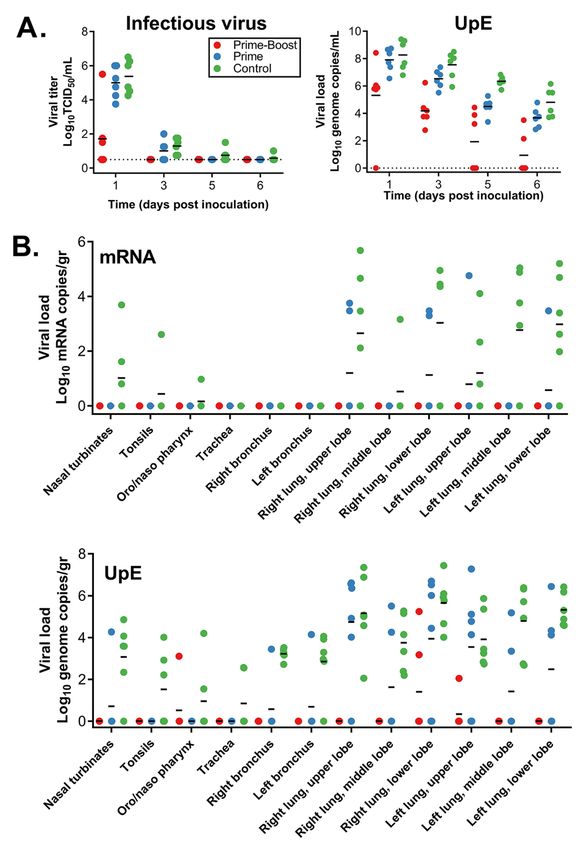
studies, only three studies included challenge studies, all of    between prime-boost and prime only animals at 0 DPI (Fig.
which used MERS-CoV isolates from 2012. To date there have        1C). A second ChAdOx1 MERS vaccination at -28 DPI resulted
been no reports of efficacy of a single-dose MERS-CoV vac-        in a statistically significant increase in S protein-specific
cine in NHPs.                                                     ELISA titer (geometric mean titer -28 DPI = 1600; 0 DPI =
    We recently demonstrated that vaccination of mice with a      10,159; P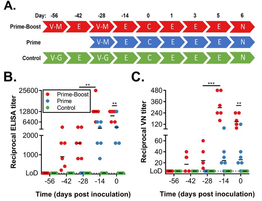
ChAdOx1 MERS, either via a prime-boost or prime-only regi-        at 1 DPI (N=5) and 3 DPI (N=1). In contrast, viral mRNA in
men, had either no lesions or limited small multifocal areas      BAL fluid from animals who received a single vaccination
of consolidation and congestion.                                  with ChAdOx1 MERS mRNA was detected on all days, but the
    An increased lung:body weight ratio is an indicator of pul-   amount was reduced compared to animals vaccinated with
monary edema. Animals in the control group had signifi-           ChAdOx1 GFP (Figure S1B). Viral RNA as measured by UpE
cantly higher lung:body weight ratios compared to ChAdOx1         qRT-PCR assay could be detected in all groups up to 6 DPI.
MERS vaccinated animals and there was minimal difference          However, the number of genome copies/mL detected was
between prime-boost and prime-only ChAdOx1 MERS vac-              lower in animals vaccinated with ChAdOx1 MERS compared
cinated animals (Fig. 3F).                                        to animals vaccinated with ChAdOx1 GFP (Fig. 4A). A signif-
    Lung tissue sections were stained with hematoxylin and        icant association was found between higher ELISA titer or
eosin or with MERS-CoV specific antibodies. All slides were       VN titer and lower levels of viral RNA, mRNA or infectious
evaluated by a board-certified veterinary pathologist blinded     virus in BAL fluid for all days, except 6 DPI for infectious vi-
to study group allocations. In animals which received a vac-      rus (Spearman’s rank correlation coefficient, Table S2).
cination with ChAdOx1 MERS, either prime-boost or prime-              All animals were euthanized on 6 DPI and tissues were

                                                                                                                                      Downloaded from http://advances.sciencemag.org/ on December 29, 2020
only, a minimal to mild bronchointerstitial pneumonia was         analyzed for the presence of viral RNA, mRNA or infectious
present characterized by mild thickening of alveolar septae       virus. Infectious virus titers were only found in nasal turbi-
by lymphocytes and macrophages. Pulmonary vessels were            nate tissue (N=1) and lung lobe tissue (N=4) from control an-
bound by moderate numbers of lymphocytes. In stark con-           imals. No viral mRNA was found in all tissues obtained from
trast, in lung tissue obtained from animals vaccinated with       animals that received a prime-boost regimen. In tissue from
ChAdOx1 GFP, moderate to marked bronchointerstitial pneu-         animals that received a prime-only regimen, limited viral
monia was present throughout the lung lobes characterized         mRNA could be found in upper and lower lung lobe tissues
by thickening of the alveolar septae by lymphocytes and mac-      (N=4). In contrast, mRNA could be found in respiratory tract
rophages, edema and fibrin. Alveoli contained abundant            tissues of all control animals, as well as in conjunctiva (Fig.
edema and fibrin and moderate to abundant numbers of al-          4B). Viral RNA was detected in tissues from all groups but
veolar macrophages, neutrophils and necrotic debris. Inflam-      was mainly found in lung lobes and bronchi. Viral load was
mation often surrounded bronchioles and pulmonary                 higher for lower respiratory tract tissue obtained from ani-
vasculature and type II pneumocyte hyperplasia was promi-         mals vaccinated with ChAdOx1 GFP (N=6) than from animals
nent. Additionally, presence of MERS-CoV antigen by im-           receiving a prime-only (N=6) or a prime-boost regimen of
munohistochemistry was found only in lungs of animals             ChAdOx1 MERS (N=2), (Fig. 4B).
vaccinated with ChAdOx1 GFP within type I & II pneumo-
cytes and was not found in lung tissue of ChAdOx1 MERS            Cytokines are upregulated in lung tissue of ChAdOx1
vaccinated animals. Severity of bronchointerstitial pneumo-       GFP vaccinated animals compared to ChAdOx1 MERS
nia, type II pneumocyte hyperplasia and hemorrhages,              vaccinated animals
edema, and fibrin deposits was scored. Statistically signifi-     The presence of 23 cytokines was evaluated in lung tissue.
cant differences between animals vaccinated with ChAdOx1          Several cytokines were upregulated in animals vaccinated
MERS or ChAdOx1 GFP were found for all three categories.          with ChAdOx1 GFP compared to animals vaccinated with
(Fig. 3E, F, H). Thus, vaccination with ChAdOx1 MERS, either      ChAdOx1 MERS, including IL2, IL6, IL8, IL18, MCP-1, MIP-
via a prime-boost regimen or prime-only regimen, signifi-         1a, and TGF-α, although only IL2 and MCP1 were significantly
cantly decreased the severity of pulmonary pathology and          different in tissue obtained from control animals compared
protected rhesus macaques against bronchointerstitial pneu-       to vaccinated animals. A significant difference was observed
monia.                                                            in IL1ra levels in lung tissue obtained from the prime only
                                                                  and prime-boost groups. It is not clear what the clinical sig-
Vaccination with ChAdOx1 MERS limits virus replica-               nificance of this difference is (Fig. S2). Overall, these results
tion in the respiratory tract                                     show local increased immune activity in animals vaccinated
BAL was performed on all animals on 1, 3, 5 and 6 DPI and         with ChAdOx1 GFP, but not in animals vaccinated with ChA-
the amount of viral RNA, mRNA and infectious virus was de-        dOx1 MERS, six days after challenge with MERS-CoV. The
termined. In the prime-boost group, infectious virus was only     likely explanation for this is that vaccinated animals have
detected at 1 DPI (N=3) after inoculation, and in the prime-      controlled the infection rapidly, whereas in the control ani-
only group at 1 DPI (N=6) and 3 DPI (N=3) but not thereafter.     mals more viral replication has taken place followed by de-
In contrast, infectious virus was detected on all days in BAL     velopment of a local immune response, and increased
fluid from the control group (Fig. 4A). Viral mRNA in BAL         cytokine levels in the lungs.
fluid of animals in the prime-boost group was only detected

First release: 1 May 2020                        www.advances.sciencemag.org (Page numbers not final at time of first release) 3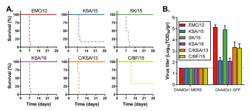
Antibodies elicited by ChAdOx1 MERS vaccination neu-                   Several studies have evaluated different types of MERS-
tralize different MERS-CoV strains                                 CoV vaccines in animal models, but few have taken these vac-
ChAdOx1 MERS is based on the spike protein from                    cines into NHPs. In the current study, we show the efficacy
Camel/Qatar/2/2014, and we currently do not have access to         of ChAdOx1 MERS in rhesus macaques. Unlike other NHP
an isolate. We thus already show cross-protection of ChAdOx1       vaccine studies (10–13), we investigate vaccine efficacy after
MERS against HCoV/EMC-2012 (99.78% spike protein amino             a single dose. Animals that received a single dose of ChAdOx1
acid identity). Here, we extend that analysis to five other        MERS showed an induction of a neutralizing antibody re-
strains. We selected six different strains of MERS-CoV (Fig.       sponse associated with mostly normal clinical parameters,
S2). Spike protein identity for all strains to the vaccine spike   showing no breathing irregularities or reduced lung function
protein was >99.3%. Amino acid identity was lowest for             by spO2 values, limited evidence of infiltration by radiograph
Camel/Burkina Faso/CIRAD-HKU785/2015 (99.33%) and                  analysis after challenge and no signs of gross pathological le-
highest for Hu/Korea/Seoul/SNU1-035/2015 (99.85%). We              sions. Vaccination reduced viral RNA load in tissues collected
tested neutralizing capability of serum obtained at 0 DPI.         at 6 DPI compared to ChAdOx1 GFP vaccinated animals by
Strains were selected based on geographical location (Saudi        several logs. Importantly, evidence of the presence of infec-

                                                                                                                                     Downloaded from http://advances.sciencemag.org/ on December 29, 2020
Arabia, South Korea, and Burkina Faso), host (dromedary            tious virus in the lungs was absent and presence of mRNA in
camel or human) and time of isolation (2012 to 2018). All six      the lungs was very limited, whereas this was abundantly pre-
strains were neutralized by antibodies elicited by ChAdOx1         sent in control animals. This was completely absent in ani-
MERS vaccination. Interestingly, although we were not able         mals vaccinated with a prime-boost regimen of ChAdOx1
to detect neutralizing ability of serum obtained from animal       MERS. Although both vaccine regimens protected rhesus ma-
12 against HCoV/EMC-2012, antibodies in the serum were             caques from clinical signs caused by MERS-CoV inoculation,
able to neutralize four out of six tested MERS-CoV strains         evidence of virus replication was reduced in animals that re-
(Table 1).                                                         ceived a prime-boost regimen compared to animals that re-
                                                                   ceived a prime only regimen, suggesting that the prime-boost
ChAdOx1 MERS protects mice against different strains               regimen is superior. It should be noted that in the current
of MERS-CoV                                                        model, animals are inoculated with a high dose of virus (7 ×
To investigate whether vaccination with ChAdOx1 MERS vac-          106 TCID50 / animal). This is likely a higher inoculum than
cination provides protection against a variety of different        most humans are exposed to. It is thus difficult to extrapolate
MERS-CoV strains, we vaccinated balb/c mice transgenic for         this observation to humans vaccinated with a single dose of
human DPP4 with ChAdOx1 MERS or ChAdOx1 GFP 28 days                ChAdOx1 MERS.
before challenge with 104 TCID50 of one of six diverse MERS-           The study was not designed to determine correlates of
CoV strains (Fig. S3) via the intranasal route. All mice vac-      protection, which must be determined separately for each
cinated with ChAdOx1 MERS survived challenge with MERS-            vaccine candidate, but it is of interest that here one animal
CoV, independent of the challenge virus used, whereas most         was protected despite not having detectable neutralizing an-
control mice were euthanized due to >20% weight loss or            tibodies against HCoV-EMC/2012 and in general the neutral-
poor body condition (Fig. 5A). Four animals per group were         izing antibody titer was not high. In clinical trials, ChAdOx1-
euthanized at 3 DPI and infectious MERS-CoV titers in lung         vectored vaccines prime strong T cell responses against the
tissue were evaluated. Whereas infectious virus could be           vaccine antigen (17-22). In a clinical study of patients recov-
found in lung tissue of control animals, we were unable to         ered from MERS infection, some had strong CD8+ T cell re-
find infectious virus in lung tissue of ChadOx1 MERS vac-          sponses without detectable antibodies (26). Further studies
cinated animals (Fig. 5B). Thus, ChAdOx1 MERS protects             addressing correlates of protection for ChAdOx1 MERS
against a variety of different MERS-CoV strains in hDPP4           should assess CD8+ T cell responses.
transgenic mice.                                                       Following the successful induction of protective immunity
                                                                   after vaccination as demonstrated here, the duration of im-
Discussion                                                         munity and ability to induce the development of memory B
Middle East respiratory syndrome coronavirus is circulating        and T cells should be assessed. The phenotype of memory
in the dromedary camel population and continuously reintro-        cells induced by vaccination may not necessarily mimic that
duced into the human population (25). MERS is associated           induced by infection with the pathogen. In the first clinical
with a high case-fatality rate (34.5%) and human-to-human          trial of ChAdOx1 MERS (ClinicalTrials.gov Identifier:
transmission is a major contributor to patient infections (1).     NCT03399578) both humoral and T cell mediated responses
Currently, no MERS-CoV vaccine is available. Ideally, such a       were assessed at multiple times and persisted up to one year
vaccine would only require a single administration and would       after vaccination. Further work will be required to assess
protect against a wide variety of different MERS-CoV strains.      memory B and T cell phenotypes. We are currently planning

First release: 1 May 2020                         www.advances.sciencemag.org (Page numbers not final at time of first release) 4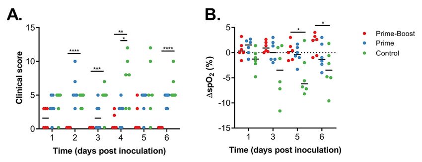
studies to look at long-term protection by ChAdOx1 MERS             Materials and Methods
vaccination in our hDPP4 mouse model.                               Ethics statement
    A variety of different MERS-CoV strains have been iso-          Animal experiment approval was provided by the Institu-
lated from dromedary camels and humans over the last eight          tional Animal Care and Use Committee (IACUC) at Rocky
years of MERS-CoV emergence (25). Dromedary camels are              Mountain Laboratories. All animal experiments were exe-
distributed throughout Africa, the Middle East, Asia and Aus-       cuted in an Association for Assessment and Accreditation of
tralia (27). Although MERS-CoV has not been detected in             Laboratory Animal Care (AALAC)-approved facility by certi-
dromedary camels in Australia (28), strains have been iso-          fied staff, following the guidelines and basic principles in the
lated from Africa (9) and the Middle East (7) and seropositive      NIH Guide for the Care and Use of Laboratory Animals, the
dromedary camels have been found in Asia (29, 30). Phyloge-         Animal Welfare Act, United States Department of Agriculture
netic analyses show a clustering of MERS-CoV by geograph-           and the United States Public Health Service Policy on Hu-
ical location (8, 9) and analysis of 219 complete MERS-CoV          mane Care and Use of Laboratory Animals. Rhesus macaques
genomes, which only included one African strain, showed the         were housed in individual primate cages allowing social in-
presence of two clades, with human isolates in both clades          teractions, in a climate-controlled room with a fixed light-

                                                                                                                                       Downloaded from http://advances.sciencemag.org/ on December 29, 2020
(31). Notably, antigenic differences have been reported be-         dark cycle (12-hours/12-hours). Rhesus macaques were mon-
tween S proteins from the Middle East and Africa (8), poten-        itored a minimum of twice daily throughout the experiment.
tially affecting the efficacy of a vaccine based on S protein. It   Commercial monkey chow, treats, and fruit were provided
is important that a MERS-CoV vaccine not only provides ho-          twice daily by trained personnel. Water was available ad libi-
mologous protection, but also protects against other MERS-          tum. Environmental enrichment consisted of a variety of hu-
CoV strains. We previously showed full protection of ChA-           man interaction, commercial toys, videos, and music. The
dOx1 MERS (based on the Camel/Qatar/2/2014 strain) vac-             Institutional Biosafety Committee (IBC) approved work with
cinated hDPP4 transgenic mice after heterologous challenge          infectious MERS-CoV virus strains under BSL3 conditions.
with HCoV-EMC/2012 (15). Here, we repeated this challenge           All sample inactivation was performed according to IBC-
and extended the experiment to include five other MERS-CoV          approved standard operating procedures for removal of spec-
strains. We utilized strains from Saudi Arabia, South Korea         imens from high containment.
and Burkina Faso, obtained from dromedary camels or hu-
mans, and isolated between 2012 and 2018 and showed full            Vaccine generation and production
protection against all strains when mice were vaccinated with       The spike protein gene from MERS-CoV strain Camel/Qa-
ChAdOx1 MERS in a prime-only regimen. Moreover, sera ob-            tar/2/2014 (GenBank accession number KJ650098.1) was co-
tained at day of challenge from rhesus macaques vaccinated          don optimized for humans and synthesized by GeneArt
with ChadOx1 MERS efficiently neutralized all six MERS-CoV          (Thermo Fisher Scientific). The synthesized S gene was
strains indicating that vaccination with ChAdOx1 MERS can           cloned into a transgene expression plasmid comprising a
protect against a variety of different MERS-CoV strains.            modified human cytomegalovirus immediate early promoter
Based on these results, future clinical development studies         (CMV promoter) with tetracycline operator (TetO) sites and
are planned with this ChAdOx1 MERS vaccine, supported by            the polyadenylation signal from bovine growth hormone
the Coalition for Epidemic Preparedness Innovations (CEPI).         (BGH). The resulting expression cassette was inserted into
    In conclusion, we show that a single vaccination with           the E1 locus of a genomic clone of ChAdOx1 using site-specific
ChAdOx1 MERS results in protection against disease progres-         recombination (32). The virus was rescued and propagated in
sion and virus replication associated with MERS-CoV chal-           T-REx-293 cells (Invitrogen). Purification was by CsCl gradi-
lenge in the rhesus macaque, and a prime-boost regimen              ent ultracentrifugation, and the virus was titered as previ-
reduced viral replication further. Furthermore, ChAdOx1             ously described (33). Doses for vaccination were based on
MERS vaccination protected against a diverse panel of con-          infectious units (IU) (34).
temporary MERS-CoV strains in hDPP4 mice. This is the first
time that broad protection after a single vaccination has been      Non-human primate study
shown for any MERS-CoV vaccine. Finally, ChAdOx1 vaccines           18 adult rhesus macaques (17M, 1F) were purchased from
can be produced rapidly, have been shown to be safe in hu-          Morgan Island and randomly divided into 3 groups of six an-
man patients and are protective against MERS-CoV in rhesus          imals each. Group 1 was vaccinated with ChAdOx1 MERS at -
macaques and hDPP4 mice. We conclude that the ChAdOx1               56 DPI and -28 DPI, group 2 was vaccinated with ChAdOx1
platform is ideal for the development of vaccines against novel     MERS at -28 DPI, and group 3 was vaccinated with ChAdOx1
emerging coronaviruses, such as HCoV-19/SARS-CoV-2.                 GFP at -56 DPI and -28 DPI. All vaccinations were done with
                                                                    3.9 × 10⁸ IU/animal/vaccination. Blood samples were ob-
                                                                    tained before vaccination and 14 days thereafter. Animals

First release: 1 May 2020                          www.advances.sciencemag.org (Page numbers not final at time of first release) 5
were challenged with MERS-CoV strain HCoV-EMC/2012 on Virus titration assay
0 DPI; with administration of 4 mL intratracheally, 1 mL in- Virus titrations were performed by end-point titration in
tranasally, 1 mL orally and 1 mL ocularly of 107 TCID50/mL VeroE6 cells, which were inoculated with tenfold serial dilu-
virus solution. Clinical exams were performed on -56, -42, - tions of virus. After 1hr incubation at 37°C and 5% CO2, tissue
28, -14, 0, 1, 3, and 5 and 6 DPI; animals were euthanized on homogenate dilutions were removed, cells were washed twice
6 DPI. All exams existed of the following: Weight and tem- with PBS and incubated in 100 μl 2% DMEM. Cytopathic ef-
perature measurements, radiographs, spO2 measurements fect was scored at 5 dpi and the TCID50 was calculated from
using pulse oximetry and blood sampling. BAL was per- 4 replicates by the Spearman-Karber method (34, 36).
formed on 1, 3, 5, and 6 DPI by insertion of an endotracheal
tube and bronchoscope into the trachea, then past the 3rd bi- Virus neutralization assay MERS-CoV
furcation, and subsequent installation of 10 mL of sterile sa- Sera were heat-inactivated (30 min, 56°C) and two-fold serial
line. Manual suction was applied to retrieve the BAL sample. dilutions were prepared in 2% DMEM. Hereafter, 100 TCID50
Necropsy was performed on 6 DPI. Radiographs were evalu- of MERS-CoV was added. After 60 min incubation at 37°C,
ated and scored by a board-certified veterinarian who was virus:serum mixture was added to VeroE6 cells and incu-
Downloaded from http://advances.sciencemag.org/ on December 29, 2020
blinded to the group assignment of the animals according to bated at 37°C and 5% CO2. At 5 dpi, cytopathic effect was
the following criteria: 0, normal examination; 1, mild inter- scored. The virus neutralization titer was expressed as the re-
stitial pulmonary infiltrates; 2, moderate interstitial infil- ciprocal value of the highest dilution of the serum which still
trates, perhaps with partial cardiac border effacement and inhibited virus replication (34).
small areas of pulmonary consolidation (alveolar patterns
and air bronchograms); and 3, pulmonary consolidation as Virus neutralization assay ChAdOx1
the primary lung pathology, seen as a progression from grade Chimpanzee adenovirus ChAdOx1 specific neutralizing anti-
2 lung pathology (35). body titers were assessed using a Secreted placental Alkaline
Phosphatase (SEAP) quantitation assay. Briefly, GripTite
Mouse study MSR 293 cells (Invitrogen cat no. R795-07) were cultured as
Groups of 10 mice were vaccinated with ChAdOx1 MERS or per manufacturer’s instructions and were seeded at 3x10⁴
ChAdOx1 GFP (1x10⁸ IU/mouse) intramuscularly 28 days cells per well in a 96 well plate the day prior to starting the
prior to intranasal inoculation with 104 TCID50 MERS-CoV. assay (24hrs +/− 2 hours). Cells were infected with the test
Mice were challenged with one of six MERS-CoV strains: sera dilutions (4 fold dilution series) at 1:18, 1:72, 1:288, 1:1152,
HCoV-EMC/12 (EMC/12, JX869059); Aseer/KSA- 1:4608 in phenol red free 0% FBS DMEM (Life Technologies
Rs924/62015 (KSA/15, KY688119); Korea/Seoul/SNU1- cat no. 31053028) and the ChAdOx1-SEAP reporter virus in a
035/2015 (SK/15, KU308549); Riyadh/KSA-18013832/2018 1:1 mixture (pre-incubated for 1 hour to allow any neutraliza-
(KSA/18, MN723544); Camel/Saudi Arabia/KFU-HKU1/2013 tion to occur) for 1 hour before replacing with phenol red free
(C/KSA/13, KJ650297); or Camel/Burkina Faso/CIRAD- 10% FBS DMEM for a further 24 hours (+/− 2 hours). Sample
HKU785/2015 (C/BF/15, MG923471). Animals were moni- dilutions were tested in duplicate lanes. SEAP concentration
tored daily for signs of disease. Four animals were euthanized was tested on 50μl media supernatants of the samples using
at 3 DPI and lung tissue was harvested. The remaining ani- CPSD (Tropix PhosphaLite Chemiluminescent Assay Kit, Life
mals were monitored for survival. Animals were euthanized Technologies cat no. T1017) using a minor variant of the man-
upon reaching >20% of body weight loss or poor body condition. ufacturer’s instructions and luminescence intensity was
measured using a Varioskan Flash luminometer (Thermo
Cells and virus Fisher). Serum dilution neutralization titers were measured
EMC/12 was provided by Erasmus Medical Center, Rotter- by linear interpolation of adjacent values (to 50% inhibition)
dam, The Netherlands; KSA/15 and KSA/18 were provided by to determine the serum dilution required to reduce SEAP
CDC, Atlanta, USA; SK/15 was provided by Seoul National concentration by 50% compared to wells with virus alone.
University, Seoul, South Korea; C/KSA/13 and C/BF/15 were
provided by Hong Kong University, Pok Fu Lam, Hong Kong. RNA extraction and quantitative reverse-transcription
Virus propagation was performed in VeroE6 cells in DMEM polymerase chain reaction
supplemented with 2% fetal bovine serum, 1 mM L-gluta- Tissues (30 mg) were homogenized in RLT buffer and RNA
mine, 50 U/ml penicillin and 50 μg/ml streptomycin. VeroE6 was extracted using the RNeasy kit (Qiagen) according to the
cells were maintained in DMEM supplemented with 10% fe- manufacturer's instructions. RNA was extracted from BAL
tal calf serum, 1 mM L-glutamine, 50 U/ml penicillin and 50 fluid using the QiaAmp Viral RNA kit (Qiagen) on the QIAx-
μg/ml streptomycin. tractor. The UpE MERS-CoV (37) or mRNA (38) detection as-
say was used for the detection of MERS-CoV viral RNA. 5 μl
First release: 1 May 2020 www.advances.sciencemag.org (Page numbers not final at time of first release) 6RNA was tested with the Rotor-GeneTM probe kit (Qiagen) immunohistochemistry using the Discovery ULTRA auto- according to instructions of the manufacturer. Dilutions of mated IHC/ISH staining instrument (Ventana Medical Sys- MERS-CoV virus stock with known genome copies were run tems) with a Discovery Red (Ventana Medical Systems) kit. in parallel. Genome copies were determined using Droplet All tissue slides were evaluated by a board-certified veteri- Digital PCR (Biorad) and the corresponding qRT-PCR. nary anatomic pathologist blinded to study group allocations. Enzyme-linked immunosorbent assay Statistical analyses A soluble, trimeric recombinant spike protein of MERS coro- Tukey’s multiple comparison test or a two-tailed unpaired navirus (isolate Ca/Jeddah/D42/2014) incorporating amino student’s t test was conducted to compare differences be- acids 1–1,273 and a carboxyl-terminal trimerization domain tween vaccine groups and the control group. A Bonferroni was produced in Chinese Hamster Ovary cells (expiCHO; correction was used to control the type I error rate for the ThermoFisher) and purified by immunoaffinity chromatog- two comparisons (group 1 vs control and group 2 vs control), raphy. Maxisorp plates (Nunc) were coated overnight at room and thus statistical significance was reached at p
Protective Immunity in Rhesus Macaques Against Middle East Respiratory                         N. Dieye, K. J. Ewer, S. Mboup, A. V. S. Hill, Safety and Immunogenicity of a
    Syndrome Coronavirus Challenge. EBioMedicine 2, 1438–1446 (2015).                              Heterologous Prime-Boost Ebola Virus Vaccine Regimen in Healthy Adults in the
    doi:10.1016/j.ebiom.2015.08.031 Medline                                                        United Kingdom and Senegal. J. Infect. Dis. 219, 1187–1197 (2019).
12. L. Wang, W. Shi, M. G. Joyce, K. Modjarrad, Y. Zhang, K. Leung, C. R. Lees, T. Zhou,           doi:10.1093/infdis/jiy639 Medline
    H. M. Yassine, M. Kanekiyo, Z. Y. Yang, X. Chen, M. M. Becker, M. Freeman, L.              23. E. de Wit, A. L. Rasmussen, D. Falzarano, T. Bushmaker, F. Feldmann, D. L. Brining,
    Vogel, J. C. Johnson, G. Olinger, J. P. Todd, U. Bagci, J. Solomon, D. J. Mollura, L.          E. R. Fischer, C. Martellaro, A. Okumura, J. Chang, D. Scott, A. G. Benecke, M. G.
    Hensley, P. Jahrling, M. R. Denison, S. S. Rao, K. Subbarao, P. D. Kwong, J. R.                Katze, H. Feldmann, V. J. Munster, Middle East respiratory syndrome coronavirus
    Mascola, W. P. Kong, B. S. Graham, Evaluation of candidate vaccine approaches                  (MERS-CoV) causes transient lower respiratory tract infection in rhesus
    for MERS-CoV. Nat. Commun. 6, 7712 (2015). doi:10.1038/ncomms8712 Medline                      macaques. Proc. Natl. Acad. Sci. U.S.A. 110, 16598–16603 (2013).
13. C. Wang, X. Zheng, W. Gai, Y. Zhao, H. Wang, H. Wang, N. Feng, H. Chi, B. Qiu, N. Li,          doi:10.1073/pnas.1310744110 Medline
    T. Wang, Y. Gao, S. Yang, X. Xia, MERS-CoV virus-like particles produced in insect         24. M. F. Yusof, K. Queen, Y. M. Eltahir, C. R. Paden, Z. M. A. H. Al Hammadi, Y. Tao, Y.
    cells induce specific humoural and cellular imminity in rhesus macaques.                       Li, A. I. Khalafalla, M. Shi, J. Zhang, M. S. A. E. Mohamed, M. H. Abd Elaal Ahmed, I.
    Oncotarget 8, 12686–12694 (2016). doi:10.18632/oncotarget.8475 Medline                         A. Azeez, O. K. Bensalah, Z. S. Eldahab, F. I. Al Hosani, S. I. Gerber, A. J. Hall, S.
14. N. K. Alharbi, E. Padron-Regalado, C. P. Thompson, A. Kupke, D. Wells, M. A. Sloan,            Tong, S. S. Al Muhairi, Diversity of Middle East respiratory syndrome
    K. Grehan, N. Temperton, T. Lambe, G. Warimwe, S. Becker, A. V. S. Hill, S. C.                 coronaviruses in 109 dromedary camels based on full-genome sequencing, Abu
    Gilbert, ChAdOx1 and MVA based vaccine candidates against MERS-CoV elicit                      Dhabi, United Arab Emirates. Emerg. Microbes Infect. 6, e101 (2017).
    neutralising antibodies and cellular immune responses in mice. Vaccine 35, 3780–               doi:10.1038/emi.2017.89 Medline

                                                                                                                                                                                             Downloaded from http://advances.sciencemag.org/ on December 29, 2020
     3788 (2017). doi:10.1016/j.vaccine.2017.05.032 Medline                                    25. G. Dudas, L. M. Carvalho, A. Rambaut, T. Bedford, MERS-CoV spillover at the
15. V. J. Munster, D. Wells, T. Lambe, D. Wright, R. J. Fischer, T. Bushmaker, G.                  camel-human interface. eLife 7, e31257 (2018).
    Saturday, N. van Doremalen, S. C. Gilbert, E. de Wit, G. M. Warimwe, Protective            26. J. Zhao, A. N. Alshukairi, S. A. Baharoon, W. A. Ahmed, A. A. Bokhari, A. M. Nehdi,
    efficacy of a novel simian adenovirus vaccine against lethal MERS-CoV challenge                L. A. Layqah, M. G. Alghamdi, M. M. Al Gethamy, A. M. Dada, I. Khalid, M. Boujelal,
    in a transgenic human DPP4 mouse model. NPJ Vaccines 2, 28 (2017).                             S. M. Al Johani, L. Vogel, K. Subbarao, A. Mangalam, C. Wu, P. Ten Eyck, S.
    doi:10.1038/s41541-017-0029-1 Medline                                                          Perlman, J. Zhao, Recovery from the Middle East respiratory syndrome is
16. N. K. Alharbi, I. Qasim, A. Almasoud, H. A. Aljami, M. W. Alenazi, A. Alhafufi, O. S.          associated with antibody and T-cell responses. Sci. Immunol. 2, eaan5393 (2017).
    Aldibasi, A. M. Hashem, S. Kasem, R. Albrahim, M. Aldubaib, A. Almansour, N. J.                doi:10.1126/sciimmunol.aan5393 Medline
    Temperton, A. Kupke, S. Becker, A. Abu-Obaidah, A. Alkarar, I. K. Yoon, E. Azhar,          27. D. R. Adney, M. Letko, I. K. Ragan, D. Scott, N. van Doremalen, R. A. Bowen, V. J.
    T. Lambe, F. Bayoumi, A. Aldowerij, O. H. Ibrahim, S. C. Gilbert, H. H. Balkhy,                Munster, Bactrian camels shed large quantities of Middle East respiratory
    Humoral Immunogenicity and Efficacy of a Single Dose of ChAdOx1 MERS Vaccine                   syndrome coronavirus (MERS-CoV) after experimental infection. Emerg.
    Candidate in Dromedary Camels. Sci. Rep. 9, 16292 (2019). doi:10.1038/s41598-                  Microbes Infect. 8, 717–723 (2019). doi:10.1080/22221751.2019.1618687 Medline
    019-52730-4 Medline                                                                        28. G. Crameri, P. A. Durr, J. Barr, M. Yu, K. Graham, O. J. Williams, G. Kayali, D. Smith,
17. S. H. Sheehy, C. J. Duncan, S. C. Elias, K. A. Collins, K. J. Ewer, A. J. Spencer, A. R.       M. Peiris, J. S. Mackenzie, L. F. Wang, Absence of MERS-CoV antibodies in feral
    Williams, F. D. Halstead, S. E. Moretz, K. Miura, C. Epp, M. D. Dicks, I. D. Poulton,          camels in Australia: Implications for the pathogen’s origin and spread. One Health
    A. M. Lawrie, E. Berrie, S. Moyle, C. A. Long, S. Colloca, R. Cortese, S. C. Gilbert, A.       1, 76–82 (2015). doi:10.1016/j.onehlt.2015.10.003 Medline
    Nicosia, A. V. Hill, S. J. Draper, Phase Ia clinical evaluation of the Plasmodium          29. A. Islam, J. H. Epstein, M. K. Rostal, S. Islam, M. Z. Rahman, M. E. Hossain, M. S.
    falciparum blood-stage antigen MSP1 in ChAd63 and MVA vaccine vectors. Mol.                    Uzzaman, V. J. Munster, M. Peiris, M. S. Flora, M. Rahman, P. Daszak, Middle East
    Ther. 19, 2269–2276 (2011). doi:10.1038/mt.2011.176 Medline                                    Respiratory Syndrome Coronavirus Antibodies in Dromedary Camels,
18. E. J. Hayton, A. Rose, U. Ibrahimsa, M. Del Sorbo, S. Capone, A. Crook, A. P. Black,           Bangladesh, 2015. Emerg. Infect. Dis. 24, 926–928 (2018).
    L. Dorrell, T. Hanke, Safety and tolerability of conserved region vaccines vectored            doi:10.3201/eid2405.171192 Medline
    by plasmid DNA, simian adenovirus and modified vaccinia virus ankara                       30. M. Saqib, A. Sieberg, M. H. Hussain, M. K. Mansoor, A. Zohaib, E. Lattwein, M. A.
    administered to human immunodeficiency virus type 1-uninfected adults in a                     Müller, C. Drosten, V. M. Corman, Serologic Evidence for MERS-CoV Infection in
    randomized, single-blind phase I trial. PLOS ONE 9, e101591 (2014).                            Dromedary Camels, Punjab, Pakistan, 2012-2015. Emerg. Infect. Dis. 23, 550–551
    doi:10.1371/journal.pone.0101591 Medline                                                       (2017). doi:10.3201/eid2303.161285 Medline
19. L. Coughlan, S. Sridhar, R. Payne, M. Edmans, A. Milicic, N. Venkatraman, B.               31. S. K. P. Lau, A. C. P. Wong, T. C. K. Lau, P. C. Y. Woo, Molecular Evolution of MERS
    Lugonja, L. Clifton, C. Qi, P. M. Folegatti, A. M. Lawrie, R. Roberts, H. de Graaf, P.         Coronavirus: Dromedaries as a Recent Intermediate Host or Long-Time Animal
    Sukhtankar, S. N. Faust, D. J. M. Lewis, T. Lambe, A. Hill, S. C. Gilbert,                     Reservoir? Int. J. Mol. Sci. 18, 2138 (2017). doi:10.3390/ijms18102138 Medline
    Heterologous Two-Dose Vaccination with Simian Adenovirus and Poxvirus                      32. M. D. Dicks, A. J. Spencer, N. J. Edwards, G. Wadell, K. Bojang, S. C. Gilbert, A. V.
    Vectors Elicits Long-Lasting Cellular Immunity to Influenza Virus A in Healthy                 Hill, M. G. Cottingham, A novel chimpanzee adenovirus vector with low human
    Adults. EBioMedicine 29, 146–154 (2018). doi:10.1016/j.ebiom.2018.02.011                       seroprevalence: Improved systems for vector derivation and comparative
    Medline                                                                                        immunogenicity.             PLOS          ONE          7,       e40385          (2012).
20. C. Kelly, L. Swadling, S. Capone, A. Brown, R. Richardson, J. Halliday, A. von Delft,          doi:10.1371/journal.pone.0040385 Medline
    Y. Oo, D. Mutimer, A. Kurioka, F. Hartnell, J. Collier, V. Ammendola, M. D. Sorbo, F.      33. M. G. Cottingham, F. Carroll, S. J. Morris, A. V. Turner, A. M. Vaughan, M. C. Kapulu,
    Grazioli, M. L. Esposito, S. D. Marco, L. Siani, C. Traboni, A. V. Hill, S. Colloca, A.        S. Colloca, L. Siani, S. C. Gilbert, A. V. Hill, Preventing spontaneous genetic
    Nicosia, R. Cortese, A. Folgori, P. Klenerman, E. Barnes, Chronic hepatitis C viral            rearrangements in the transgene cassettes of adenovirus vectors. Biotechnol.
    infection subverts vaccine-induced T-cell immunity in humans. Hepatology 63,                   Bioeng. 109, 719–728 (2011). doi:10.1002/bit.24342 Medline
    1455–1470 (2016). doi:10.1002/hep.28294 Medline                                            34. N. van Doremalen, T. Lambe, S. Sebastian, T. Bushmaker, R. Fischer, F. Feldmann,
21. M. Wilkie, I. Satti, A. Minhinnick, S. Harris, M. Riste, R. L. Ramon, S. Sheehan, Z. M.        E. Haddock, M. Letko, V. A. Avanzato, I. Rissanen, R. LaCasse, D. Scott, T. A.
     Thomas, D. Wright, L. Stockdale, A. Hamidi, M. K. O’Shea, K. Dwivedi, H. M.                   Bowden, S. Gilbert, V. Munster, A single-dose ChAdOx1-vectored vaccine provides
     Behrens, T. Davenne, J. Morton, S. Vermaak, A. Lawrie, P. Moss, H. McShane, A                 complete protection against Nipah Bangladesh and Malaysia in Syrian golden
     phase I trial evaluating the safety and immunogenicity of a candidate tuberculosis            hamsters.        PLOS       Negl.     Trop.      Dis.     13,   e0007462       (2019).
     vaccination regimen, ChAdOx1 85A prime - MVA85A boost in healthy UK adults.                   doi:10.1371/journal.pntd.0007462 Medline
    Vaccine 38, 779–789 (2020). doi:10.1016/j.vaccine.2019.10.102 Medline                      35. D. L. Brining, J. S. Mattoon, L. Kercher, R. A. LaCasse, D. Safronetz, H. Feldmann,
22. N. Venkatraman, B. P. Ndiaye, G. Bowyer, D. Wade, S. Sridhar, D. Wright, J.                    M. J. Parnell, Thoracic radiography as a refinement methodology for the study of
    Powlson, I. Ndiaye, S. Dièye, C. Thompson, M. Bakhoum, R. Morter, S. Capone, M.                H1N1 influenza in cynomologus macaques (Macaca fascicularis). Comp. Med. 60,
    Del Sorbo, S. Jamieson, T. Rampling, M. Datoo, R. Roberts, I. Poulton, O. Griffiths,           389–395 (2010). Medline
    W. R. Ballou, F. Roman, D. J. M. Lewis, A. Lawrie, E. Imoukhuede, S. C. Gilbert, T.        36. G. Karber, Article on the collective handling of pharmacological array effort.

First release: 1 May 2020                                               www.advances.sciencemag.org (Page numbers not final at time of first release) 8
Naunyn Schmiedebergs Arch Exp Pathol Pharmakol 162, 480–483 (1931).
37. V. M. Corman, I. Eckerle, T. Bleicker, A. Zaki, O. Landt, M. Eschbach-Bludau, S. van
Boheemen, R. Gopal, M. Ballhause, T. M. Bestebroer, D. Muth, M. A. Müller, J. F.
Drexler, M. Zambon, A. D. Osterhaus, R. M. Fouchier, C. Drosten, Detection of a
novel human coronavirus by real-time reverse-transcription polymerase chain
reaction. Euro Surveill. 17, 39 (2012). doi:10.2807/ese.17.39.20285-en Medline
38. C. M. Coleman, M. B. Frieman, Growth and Quantification of MERS-CoV Infection.
Curr Protoc Microbiol 37, 15E 12 11-19 (2015).
ACKNOWLEDGMENTS
We would like to thank Keith Chappell, University of Queensland, Australia, for the
clamped S protein used in ELISA assays, the animal care takers, Anita Mora and
Austin Athman for assistance with figures, Benjamin Carrasco for preparation of
animal studies, and Dana Scott, Jamie Lovaglio, Amanda Griffin, and Kathleen
Cordova for assistance during the animal studies. Funding: This work was
supported by the Intramural Research Program of the National Institute of
Allergy and Infectious Diseases (NIAID), National Institutes of Health (NIH)
Downloaded from http://advances.sciencemag.org/ on December 29, 2020
(1ZIAAI001179-01) and the Department of Health and Social Care using UK Aid
funding managed by the NIHR. SCG is a Jenner investigator. The views
expressed in this publication are those of the author(s) and not necessarily those
of the Department of Health and Social Care. Author contributions:
Conceptualization, NvD, TL, SCG and VJM; methodology, NvD, EH, FF, KMW, TB,
RJF, AO, PWH, GS, NJE, MHAC, TL; formal analysis, NvD, and GS; writing—
original draft preparation, NvD; writing—review and editing, TL, SCG, VJM.
Competing interests: SCG is a board member of Vaccitech and named as an
inventor on a patent covering use of ChAdOx1-vectored vaccines. The authors
declare no other competing interests. Data and materials availability: All data
needed to evaluate the conclusions in the paper are present in the paper and/or
the Supplementary Materials. ChAdOx1 MERS can be provided by the Jenner
Institute, University of Oxford pending scientific review and a completed material
transfer agreement. Requests for the ChAdOx1 MERS should be submitted to:
Sarah Gilbert. Additional data is available from authors upon request.
SUPPLEMENTARY MATERIALS
advances.sciencemag.org/cgi/content/full/sciadv.aba8399/DC1
Submitted 9 January 2020
Accepted 16 April 2020
Published First Release 1 May 2020
10.1126/sciadv.aba8399
First release: 1 May 2020 www.advances.sciencemag.org (Page numbers not final at time of first release) 9Downloaded from http://advances.sciencemag.org/ on December 29, 2020
Fig. 1. Vaccination of rhesus macaques with ChAdOx1 MERS elicits a humoral immune response.
Serum samples were collected from non-human primates at times of vaccination (-56 DPI and -28
DPI), 14 days later and at challenge. (A) Overview of experimental timeline. V-M = vaccination with
ChAdOx1 MERS; V-G = vaccination with ChAdOx1 GFP; E = exam; C = challenge and exam; N = exam
and necropsy (B) Two-fold serial-diluted serum samples were tested for MERS-CoV S-specific
antibodies using ELISA. (C) Two-fold serial-diluted serum samples were tested for neutralizing
antibodies against MERS-CoV in VeroE6 cells. Line = geometric mean, dotted line = limit of
detection. Statistical significance between -28 DPI and -14 DPI in the prime-boost group was
determined via one-tailed paired Student’s t test. Statistical significance between prime-boost and
prime only groups on 0 DPI was determined via two-tailed unpaired Student’s t test. ** = p-
valueFig. 2. Clinical scoring and spO2 values are improved in ChAdOx1 MERS vaccinated animals
compared to ChAdOx1 GFP vaccinated animals. (A) Animals were evaluated daily and clinical score
assessed using an established scoring sheet. (B) Changes in oxygen saturation from pre-inoculation
Downloaded from http://advances.sciencemag.org/ on December 29, 2020
values (Δ% spO2) were determined on exam days. Statistical significance between groups was
determined via two-tailed unpaired Student’s t test. Line = median; dotted line = baseline value; * =
p-valueDownloaded from http://advances.sciencemag.org/ on December 29, 2020 Fig. 3. Single-dose vaccination with ChAdOx1 MERS protects rhesus macaques against bronchointerstitial pneumonia caused by MERS- CoV challenge. Rhesus macaques were vaccinated with a prime-boost or prime-only regimen of ChAdOx1 MERS, or with ChAdOx1 GFP and challenged with MERS-CoV. (A) Ventrodorsal thoracic radiographs collected on 6 DPI. A marker (R) indicates right side of animal. No pathologic changes were observed in animals vaccinated with ChAdOx1 MERS via a prime-boost or prime-only regimen. Animal vaccinated with ChAdOx1 GFP shows focally extensive area of increased pulmonary opacity and deviation of the cardiac silhouette, highlighted in the circle located in the middle and caudal lung lobes. (B) Thoracic radiographs of each animal were scored per lung lobe resulting in a maximum score of 18. Values were averaged per group per day, shown is mean with standard deviation. See Table S2 for more details. (C) Gross pathology of lungs shows no pathologic changes in ChAdOx1 MERS vaccinated animals, and focally extensive areas of consolidation in left cranial, middle and caudal lung lobes in control animals (asterisks). (D) Gross lung lesions were scored for each lung lobe, ventral and dorsal. Values were averaged per group, shown is mean with standard deviation. (E) Lung tissue sections were stained with hematoxylin and eosin. Moderate numbers of lymphocyte accumulation around pulmonary arterioles (asterisks), and mild thickening of alveolar septae by lymphocytes and macrophages (arrow) in lung tissue of animals vaccinated with ChAdOx1 MERS. Marked bronchointerstitial pneumonia with abundant pulmonary edema and fibrin (asterisks), type II pneumocyte hyperplasia (arrow), and increased numbers of alveolar macrophages (arrowhead) in lung tissue of control animals. Magnification = 200x. (F) Lung to body weight ratio was determined for all animals at necropsy. Shown is mean with standard deviation. (G) Lung tissue sections were stained with antibody against MERS-CoV antigen, which is visible as a red-brown staining. No immunoreactivity was found in ChAdOx1 MERS vaccinated animals, whereas multifocal immunoreactivity of type I and II pneumocytes could be found in lung tissue of ChAdOx1 GFP vaccinated animals. (H) Lung tissue sections were scored on severity of lesions (0=no lesions, 1=1-10%, 2=11-25%, 3=26-50%, 4=51- 75%, 5=76-100%) and averaged per group. Shown is mean with standard deviation. A = bronchointerstitial pneumonia; B = type II pneumocyte hyperplasia; C = Hemorrhages, edema, fibrin deposits. Statistical significance between groups was determined via two-tailed unpaired Student’s t test. * = p-value
Downloaded from http://advances.sciencemag.org/ on December 29, 2020

          Fig. 4. Vaccination with ChAdOx1 MERS results in reduced virus replication in the respiratory tract.
          (A) Infectious virus titers and viral load were determined in BAL fluid. Individual values are depicted.
          (B) UpE and mRNA copies were determined in respiratory tract tissues collected at 6 DPI. Individual
          values are depicted. Line = geometric mean, dotted line = limit of detection.

First release: 1 May 2020                      www.advances.sciencemag.org (Page numbers not final at time of first release) 13
Fig. 5. ChAdOx1 MERS provides cross-protection against different MERS-CoV strains in the mouse
Downloaded from http://advances.sciencemag.org/ on December 29, 2020
model. (A) Survival curves of ChAdOx1 MERS vaccinated (solid line) and ChAdOx1 GFP vaccinated
(dashed line) hDPP4 mice challenged with MERS-CoV. (B) Infectious virus titers in lung tissue
collected on 3 DPI from hDPP4 mice challenged with MERS-CoV. Shown is mean titer with standard
deviation.
First release: 1 May 2020 www.advances.sciencemag.org (Page numbers not final at time of first release) 14Table 1. Neutralizing titer of serum obtained from animals vaccinated with a prime-boost regimen of ChAdOx1 MERS against different
MERS-CoV strains. U = unassigned.
GenBank Lineage
Abbrevia- Animal number
Full virus name accession (9, 24)
tion
number
1 2 3 4 5 6 7 8 9 10 11 12
Hu/HCoV/EMC/
EMC/12 JX869059 A 120 480 120 240 240 120 120 40 40 40 80A single dose of ChAdOx1 MERS provides protective immunity in rhesus macaques
Neeltje van Doremalen, Elaine Haddock, Friederike Feldmann, Kimberly Meade-White, Trenton Bushmaker, Robert J. Fischer,
Atsushi Okumura, Patrick W. Hanley, Greg Saturday, Nick J. Edwards, Madeleine H.A. Clark, Teresa Lambe, Sarah C. Gilbert and
Vincent J. Munster
published online May 1, 2020
Downloaded from http://advances.sciencemag.org/ on December 29, 2020
ARTICLE TOOLS http://advances.sciencemag.org/content/early/2020/04/30/sciadv.aba8399
SUPPLEMENTARY http://advances.sciencemag.org/content/suppl/2020/04/30/sciadv.aba8399.DC1
MATERIALS
REFERENCES This article cites 34 articles, 4 of which you can access for free
http://advances.sciencemag.org/content/early/2020/04/30/sciadv.aba8399#BIBL
PERMISSIONS http://www.sciencemag.org/help/reprints-and-permissions
Use of this article is subject to the Terms of Service
Science Advances (ISSN 2375-2548) is published by the American Association for the Advancement of Science, 1200 New York
Avenue NW, Washington, DC 20005. The title Science Advances is a registered trademark of AAAS.
Copyright © 2020 The Authors, some rights reserved; exclusive licensee American Association for the Advancement of Science.
No claim to original U.S. Government Works. Distributed under a Creative Commons Attribution NonCommercial License 4.0 (CC
BY-NC).You can also read