Breakthrough instruments and products PhotoSonus M+ laser for photoacoustic imaging
←
→
Page content transcription
If your browser does not render page correctly, please read the page content below
Breakthrough instruments and products
PhotoSonus M+ laser for photoacoustic
imaging
Cite as: Rev. Sci. Instrum. 92, 059502 (2021); https://doi.org/10.1063/5.0053559
Submitted: 08 April 2021 . Accepted: 13 April 2021 . Published Online: 11 May 2021

  Aldas Juronis, and     Mantvydas Jašinskas

ARTICLES YOU MAY BE INTERESTED IN

Dual feedback based bipolar current source with high stability for driving voice coil motors
in wide temperature ranges
Review of Scientific Instruments 92, 054708 (2021); https://doi.org/10.1063/5.0039680

A quaternion-based indirect Gaussian particle filter for nonlinear attitude estimation
Review of Scientific Instruments 92, 055007 (2021); https://doi.org/10.1063/5.0039279

Long duration x-ray source development for x-ray diffraction at the National Ignition
Facility
Review of Scientific Instruments 92, 053904 (2021); https://doi.org/10.1063/5.0043677

Rev. Sci. Instrum. 92, 059502 (2021); https://doi.org/10.1063/5.0053559              92, 059502

© 2021 Author(s).
Review of                                                                             NEW PRODUCTS               scitation.org/journal/rsi
 Scientific Instruments

     Breakthrough instruments and products
     PhotoSonus M+ laser for photoacoustic imaging
     Cite as: Rev. Sci. Instrum. 92, 059502 (2021); doi: 10.1063/5.0053559
     Submitted: 8 April 2021 • Accepted: 13 April 2021 •
     Published Online: 11 May 2021

     Aldas Juronisa)            and Mantvydas Jašinskasb)

     AFFILIATIONS
     EKSPLA, UAB, Savanoriu Ave. 237, Vilnius 02300, Lithuania

     a)
          Author to whom correspondence should be addressed: a.juronis@ekspla.com
     b)
          m.jasinskas@ekspla.com

     ABSTRACT
     This report highlights PhotoSonus M+, a tunable wavelength laser system recently introduced and developed specifically for photoacoustic
     imaging applications, where the highest imaging depth and resolution are required.
     © 2021 Author(s). All article content, except where otherwise noted, is licensed under a Creative Commons Attribution (CC BY) license
     (http://creativecommons.org/licenses/by/4.0/). https://doi.org/10.1063/5.0053559

     I. INTRODUCTION                                                         cooling unit, all integrated into a single robust cart-type housing
           Photoacoustic imaging is one of the fastest growing research      with customizable fiber coupled output to provide mobility, ease of
     areas of non-invasive, high-resolution, and high-contrast visualiza-    use, and low maintenance cost. This allows the use of PhotoSonus
     tion of both superficial and deep tissues. This method has a number     M+ as a mobile stand-alone laser system that can be easily moved
     of advantages over widely used conventional research and diagnos-       to another location, be it another laboratory or another bed-side
     tic methods as it does not use hazardous irradiation such as x-rays     location. Expert knowledge, proprietary technological solutions, and
     and has a significantly higher imaging resolution than conventional     many years of experience in the development and manufacturing of
     ultrasound. Photoacoustic imaging has proven to be a very effi-         various tunable wavelength laser systems contributed to designing
     cacious diagnostic modality of breast tumors, skin cancer, thyroid      of a mobile, rigid, and highly reliable laser system (Fig. 1), which
     nodules, osteoarthritis and rheumatoid arthritis, early diagnosis of    does not require reinstallation or any additional adjustment after its
     blood vessel disorders, and much more. In addition, photoacous-         reallocation.
     tic imaging can be used for visualization of non-living objects, such         The wide gap-less wavelength tuning range 660–2300 nm
     as nondestructive inspection of the internal structure and property     makes PhotoSonus M+ an essential laser source for any photo-
     changes of composite materials and food inspection.                     acoustic imaging system. The tuning range can also be extended
           One of the most important components of any photoacoustic         down to 330 nm. For real-time photoacoustic imaging applications,
     imaging system is a proper light source. EKSPLA—a developer and         a proprietary Fast Wavelength Switching (FWS) feature is very use-
     manufacturer of reliable tunable wavelength laser systems—recently      ful. This unique feature enables us to pre-set a wavelength scanning
     introduced the PhotoSonus M+ laser system developed specifically        cycle, which contains almost any number of wavelengths that are
     for photoacoustic imaging applications requiring a high imaging         distributed in any order and at any step. Each laser shot can be
     depth and resolution.                                                   emitted with a different wavelength. However, there is also a pos-
                                                                             sibility to set the number of shots per wavelength up to 100. The
                                                                             maximum wavelength range that can be switched between two con-
     II. LASER PARAMETERS AND FUNCTIONALITY                                  secutive pulses is the entire extended signal range: 660–1300 nm.
          The PhotoSonus M+ tunable wavelength laser system deliv-           Wavelength switching accuracy is better than ±0.5 nm. For better
     ers more than 250 mJ at 680 nm, which is the highest pulse energy       synchronization with data acquisition equipment and easier wave-
     available in the market from an integrated optical parametric oscil-    length real-time tracking, two synchronization signals are used by
     lator (OPO) system. The system contains a high-energy 10 Hz             the FWS: One indicates the start of each scanning cycle, while the
     Q-switched laser, a parametric oscillator, power supply, and a          other indicates that the laser has fired a pre-set wavelength.

Rev. Sci. Instrum. 92, 059502 (2021); doi: 10.1063/5.0053559                                                                              92, 059502-1
© Author(s) 2021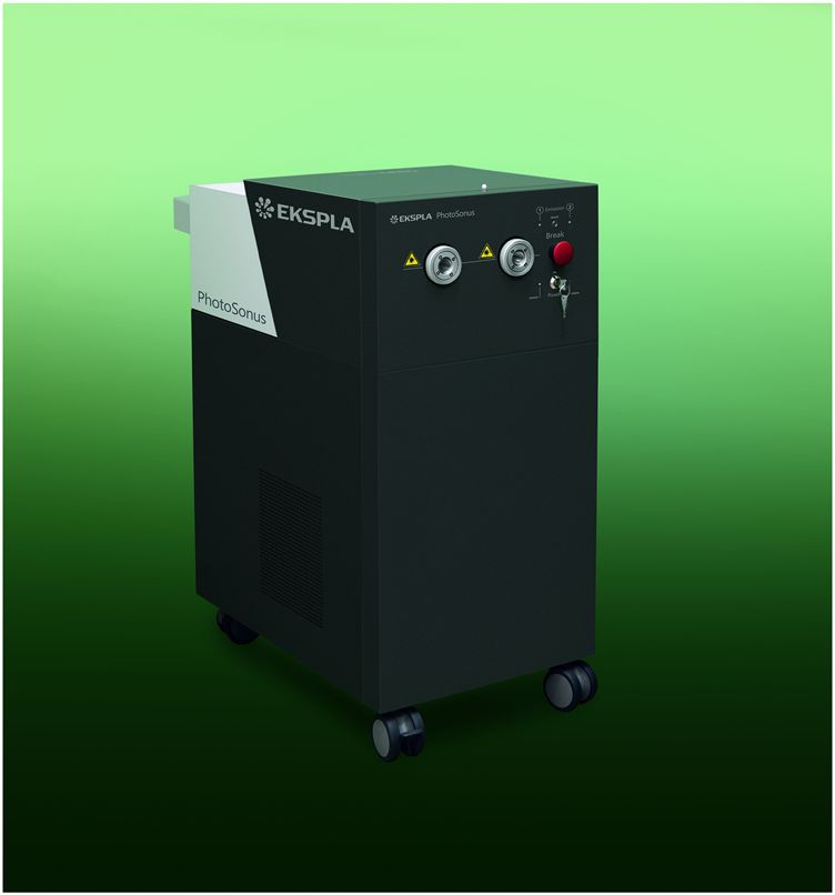
Review of NEW PRODUCTS scitation.org/journal/rsi
Scientific Instruments
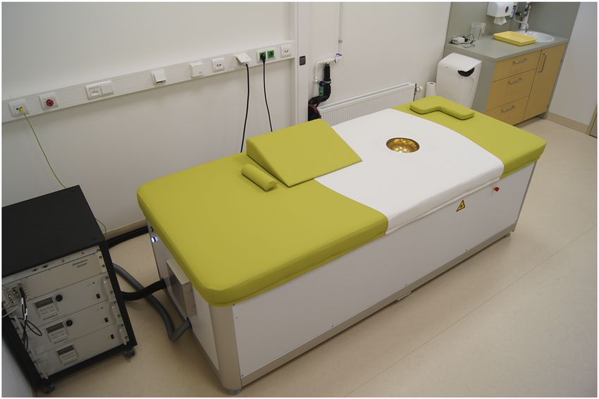
FIG. 3. Clinical setup of the latest generation PAMMOTH imager equipped with the
modified twin PhotoSonus M+ laser system. Courtesy of PAMMOTH (Courtesy of
PA Imaging R & D B.V.).
FIG. 1. PhotoSonus M+ laser system containing a pump laser, an OPO, a power
supply unit, and a water-cooling system, all integrated into a single, compact, and III. APPLICATIONS IN EARLY BREAST CANCER
mobile cart. IMAGING
PhotoSonus M+ lasers are used in the EU funded PAMMOTH
project (acronym of Photoacoustic Mammoscopy for evaluating
The highly flexible PhotoSonus M+ platform makes it easy to
screening-detected lesions in the breast),1 where EKSPLA is one
integrate and be used with any photoacoustic imaging system. It
of the project partners. PAMMOTH’s objective is to develop, val-
is fully motorized and computer controlled, with user trigger out-
idate, and begin exploitation of a dedicated breast imaging device
puts and inputs and special options such as motorized switching
for a significant impact in breast cancer early diagnosis. The pro-
between the OPO signal and idler through the same output, motor-
posed device combines non-invasive 3D photoacoustic imaging and
ized attenuator, internal pulse energy meter, and electromechanical
ultrasound imaging. From the ultrasound mode, the radiologist can
output shutter. For user convenience, the output of the PhotoSonus
visualize anatomical features and extent of tumors, and from the
M+ laser has a fiber connector that could be customized for being
multi-wavelength photoacoustic mode, he/she can assess tumor
coupled with almost any type of fiber bundle. A version of the Pho-
vascularity.2 Quantitative spectroscopic photoacoustic images are
toSonus M laser system with slightly lower pulse energy and lower
extracted off-line, providing the radiologist with information relat-
price (topped with 180 mJ pulse energy) is currently successfully
ing to tumor physiology and function such as angiogenesis and
used in preclinical photoacoustic tomography systems for imaging
hypoxia.
of small animal vasculature, organs, skeletal system, and skin (Fig. 2).
The latest clinical prototype contains a modified twin mutu-
ally synchronized PhotoSonus M+ laser system, which can pro-
vide a max combined pulse energy of 500 mJ while operating at
10 Hz, or 250 mJ at 20 Hz. The output of such a system is cou-
pled into a fiber bundle with one input at the laser end and 40
separate fiber outputs that are evenly distributed across the imag-
ing bowl where a patient’s breast is immobilized during the photo-
acoustic mammoscopy examination. A clinical demonstration trial
of the latest generation of the PAMMOTH imager equipped with
the modified twin PhotoSonus M+ laser systems has just started at
the Medisch Spectrum Twente Hospital (The Netherlands) (Fig. 3).
Despite the significant instrumentation progress, the entire system
is not yet optimized completely; however, the first acquired pho-
toacoustic images are promising for the optimal expected imaging
depth to exceed 50 mm and image resolution to exceed 0.35 mm.
Examination of one breast takes less than 3 min.
IV. SUMMARY
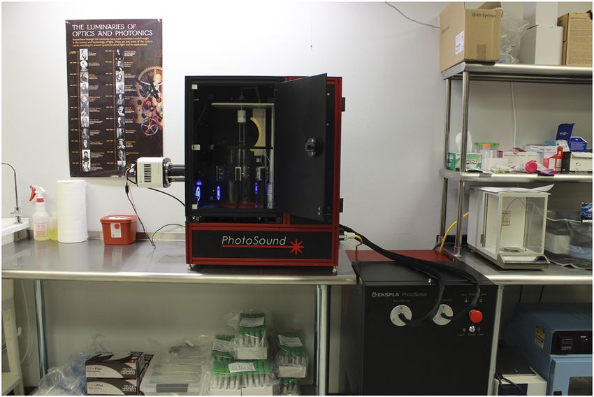
FIG. 2. A small animal whole body in vivo imaging platform TriTom based on The PhotoSonus M+ laser system is proving to be an excellent
photoacoustic fluorescence tomography with an integrated PhotoSonus M laser tunable wavelength irradiation source suitable for high depth and
system. Courtesy of PhotoSound Technologies, Inc.
high-resolution photoacoustic imaging in a large area of live tissues.
Rev. Sci. Instrum. 92, 059502 (2021); doi: 10.1063/5.0053559 92, 059502-2
© Author(s) 2021
Review of                                                                                   NEW PRODUCTS                  scitation.org/journal/rsi
 Scientific Instruments

                                                                                  2
     REFERENCES                                                                    S. M. Schoustra, D. Piras, R. Huijink, T. J. P. M. Op’t Root, L. Alink, W. M.
     1
                                                                                  Kobold, W. Steenbergen, and S. Manohar, “Twente photoacoustic mammoscope
      See https://www.pammoth-2020.eu for Photoacoustic Mammoscopy for evaluat-   2: System overview and three-dimensional vascular network images in healthy
     ing screening-detected lesions in the breast.                                breasts,” J. Biomed. Opt. 24(12), 121909 (2019).

Rev. Sci. Instrum. 92, 059502 (2021); doi: 10.1063/5.0053559                                                                                         92, 059502-3
© Author(s) 2021
You can also read