Bronchoscopic Features and Correlative Factor Analysis of Severe Mycoplasma pneumoniae Pneumonia in Children.

Page created by Alexander Pierce
 
CONTINUE READING
Bronchoscopic Features and Correlative Factor Analysis of Severe Mycoplasma pneumoniae Pneumonia in Children.
Posted on Authorea 20 May 2021 — The copyright holder is the author/funder. All rights reserved. No reuse without permission. — https://doi.org/10.22541/au.162152127.79623314/v1 — This a preprint and has not been peer reviewed. Data may be preliminary.

                                                                                                                                                                                                                                                               Bronchoscopic Features and Correlative Factor Analysis of Severe
                                                                                                                                                                                                                                                               Mycoplasma pneumoniae Pneumonia in Children.
                                                                                                                                                                                                                                                               haiming yang1 , Gan Li1 , and Xicheng Liu2
                                                                                                                                                                                                                                                               1
                                                                                                                                                                                                                                                                   Beijing Children’s Hospital
                                                                                                                                                                                                                                                               2
                                                                                                                                                                                                                                                                   Beijing Children’s Hospital Capital Medical University

                                                                                                                                                                                                                                                               May 20, 2021

                                                                                                                                                                                                                                                               Abstract
                                                                                                                                                                                                                                                               Objectives: We aimed to determine the bronchoscopic features of children with severe Mycoplasma pneumoniae pneumonia
                                                                                                                                                                                                                                                               (SMPP), and correlation with obliterative bronchitis (OB), so as to help early clinical evaluation and treatment of pediatric
                                                                                                                                                                                                                                                               SMPP. Methods: 213 pediatric SMPP cases admitted to Beijing Children’s Hospital were included. Medical records and
                                                                                                                                                                                                                                                               bronchoscopic manifestations at different SMPP stages were retrospectively analyzed . Results: Of 213 acute-stage pediatric
                                                                                                                                                                                                                                                               SMPP patients, bronchoscopy revealed 22 cases (10.3%) with subacute-stage or recovery-stage OB, for an OB incidence rate
                                                                                                                                                                                                                                                               in cases with mucous embolus of 48.8% (22/47), a significantly higher rate than the rate without mucous embolus (0/166, 0%)
                                                                                                                                                                                                                                                               (P < 0.001). Notably, the OB incidence rate for children undergoing bronchoscopy within 10 days (9/142, 6.3%) of SMPP
                                                                                                                                                                                                                                                               onset was significantly lower than in children undergoing bronchoscopy 10 days post-disease onset (13/71, 76.5%) (P < 0.007).
                                                                                                                                                                                                                                                               Conclusions: In pediatric SMPP patients, airway mucus and debris from epithelial necrosis and exfoliation can block airway
                                                                                                                                                                                                                                                               subbranches, readily leading to OB.

                                                                                                                                                                                                                                                               INTRODUCTION
                                                                                                                                                                                                                                                               Mycoplasma pneumoniaepneumonia (MPP) is one of the most common types of community-acquired pneu-
                                                                                                                                                                                                                                                               monia (CAP) in children.1 Macrolide antibiotics are effective treatments for most patients, but in some
                                                                                                                                                                                                                                                               cases MPP may progress to severe MPP (SMPP). SMPP patients exhibit poor responses to single macrolide
                                                                                                                                                                                                                                                               antibiotics and progression of disease that is accompanied by persistent high fever, aggravation of pulmonary
                                                                                                                                                                                                                                                               lesions, and additional complications, such as necrotizing pneumonia, atelectasis, bronchiectasis, obliterative
                                                                                                                                                                                                                                                               bronchiolitis (OB), and other disorders. SMPP complications can lead to repeated infections and decreased
                                                                                                                                                                                                                                                               quality of life for afflicted children.2,3
                                                                                                                                                                                                                                                               The pathogenesis of SMPP likely involves many processes, such as excessive immune inflammatory reactions,
                                                                                                                                                                                                                                                               mixed infections, and so on. Therefore, the abovementioned factors should be considered during SMPP treat-
                                                                                                                                                                                                                                                               ment to reduce the development of pulmonary sequelae. Notably, using bronchoscopy we have found that
                                                                                                                                                                                                                                                               MPP is associated with specific manifestations such as airway mucus blockage and mucosal necrosis as main
                                                                                                                                                                                                                                                               findings that cannot be completely eliminated by conservative treatments such as antibiotics and glucocor-
                                                                                                                                                                                                                                                               ticoids. Indeed, airway blockage is of particular concern in that it can occur within a short period of time
                                                                                                                                                                                                                                                               and progress to permanent atelectasis. Therefore, avoidance of OB should be an early treatment priority for
                                                                                                                                                                                                                                                               practitioners caring for pediatric MPP patients in China, especially in the northern region.4 Consequently,
                                                                                                                                                                                                                                                               a better understanding of the effects of early abnormal dynamic inflamed airway changes on later incidence
                                                                                                                                                                                                                                                               of airway occlusions and their implications for MMP treatment have not yet been reported. Such infor-
                                                                                                                                                                                                                                                               mation may be acquired via bronchoscopy, which can facilitate SMPP diagnosis during acute-phase MMP
                                                                                                                                                                                                                                                               by providing bronchial lavage fluid for pathogen detection and information pertaining to microscopic dis-
                                                                                                                                                                                                                                                               ease manifestations. Also, bronchoscopy can reveal complete membranous obstructions within subsegmental
                                                                                                                                                                                                                                                               bronchi of OB cases.

                                                                                                                                                                                                                                                                                                                            1
Bronchoscopic Features and Correlative Factor Analysis of Severe Mycoplasma pneumoniae Pneumonia in Children.
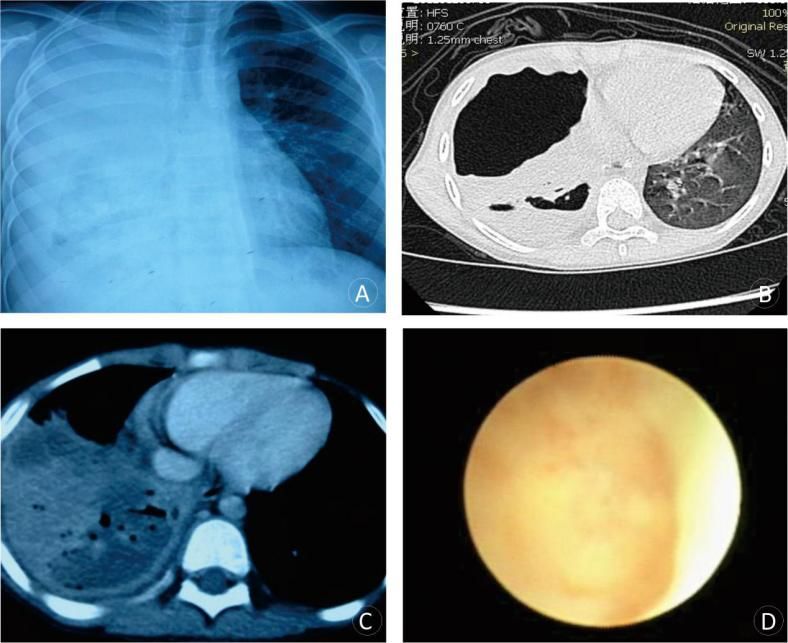
In this retrospective study, we analyzed bronchoscopic findings of pediatric SMPP patients admitted to
Posted on Authorea 20 May 2021 — The copyright holder is the author/funder. All rights reserved. No reuse without permission. — https://doi.org/10.22541/au.162152127.79623314/v1 — This a preprint and has not been peer reviewed. Data may be preliminary.

                                                                                                                                                                                                                                                               Beijing Children’s Hospital affiliated with Capital Medical University from October 2017 to January 2019
                                                                                                                                                                                                                                                               to reveal correlations between bronchoscopically determined disease manifestations and SMPP prognosis.
                                                                                                                                                                                                                                                               METHODS
                                                                                                                                                                                                                                                               Subjects
                                                                                                                                                                                                                                                               This study was a retrospective analysis of pediatric patients afflicted with SMPP and admitted to the
                                                                                                                                                                                                                                                               hospital from June 2017 to January 2019. Diagnostic criteria of MPP were as follows: (1) fever, cough,
                                                                                                                                                                                                                                                               and other clinical manifestations; (2) chest image findings indicative of pneumonia; (3) MPP-specific IgM
                                                                                                                                                                                                                                                               antibody titer persisting at a level [?] 1:320, or an antibody level that increased 4-fold during convalescent
                                                                                                                                                                                                                                                               and acute SMPP phases (passive agglutination method, Fujirebio Inc., Japan). Based on MPP diagnosis
                                                                                                                                                                                                                                                               combined with the latest diagnostic criteria for severe CAP in China, any one of the following criteria was
                                                                                                                                                                                                                                                               used as a reference for the diagnosis of SMPP:5,6 obvious shortness of breath or tachycardia (criteria: 1-
                                                                                                                                                                                                                                                               5 years old, RR [?] 40 times/min; >5 years old, RR [?] 30 times/min, respectively); hypoxemia during
                                                                                                                                                                                                                                                               inhalation and pulse oximetry-based oxygen saturation (SaO2 ) less than 0.93; chest images demonstrating
                                                                                                                                                                                                                                                               multi-lobe or segment involvement or pulmonary area involvement of greater than 2/3 of the total lung area;
                                                                                                                                                                                                                                                               pleural effusion, atelectasis, pulmonary necrosis, pulmonary abscess, and other pulmonary complications;
                                                                                                                                                                                                                                                               complications associated with severe damage of other systems, such as central nervous system infection,
                                                                                                                                                                                                                                                               heart failure, myocarditis, gastrointestinal hemorrhage, obvious electrolyte/acid-base balance disorder, etc.
                                                                                                                                                                                                                                                               Exclusion criteria included previous congenital or secondary immunosuppression or defect, chronic pulmonary
                                                                                                                                                                                                                                                               disease, kidney or cardiovascular disease, and connective tissue disorders, etc. Patients were also excluded
                                                                                                                                                                                                                                                               if they were infected with other pathogens within 7 days post-admission or did not receive bronchoscopy at
                                                                                                                                                                                                                                                               admission. All recruited children had received treatment with macrolide antibiotics for one week without
                                                                                                                                                                                                                                                               noticeable improvement.
                                                                                                                                                                                                                                                               Bronchoscopy
                                                                                                                                                                                                                                                               Bronchoscopy was performed under the following conditions: decreased respiratory sounds corresponding
                                                                                                                                                                                                                                                               to the lesion site during auscultation; ineffective anti-infection treatment; chest image showing large area
                                                                                                                                                                                                                                                               of pulmonary consolidation. Bronchoscopic reexamination was conducted when chest imaging showed poor
                                                                                                                                                                                                                                                               absorption of lung lesions and the airway still harbored a mucous embolus one week after bronchoscopy had
                                                                                                                                                                                                                                                               been conducted in spite of systemic hormone and anti-MP treatment.
                                                                                                                                                                                                                                                               Bronchoscopic procedures were as follows: for each patient, the nasal cavity and pharynx were locally
                                                                                                                                                                                                                                                               anesthetized with 1% lidocaine prior to the procedure. After sedation via intravenous injection of 0.1-0.3
                                                                                                                                                                                                                                                               ml/kg midazolam, bronchoscopy was performed using either an Olympus BFXP-260F model (outer diameter
                                                                                                                                                                                                                                                               2.8 mm) or Olympus BF P-260F model (outer diameter 4.0 mm) bronchoscope via entry through the nostril.
                                                                                                                                                                                                                                                               Changes in mucosa of the nasal cavity, pharynx, trachea, bronchus, and subsegmental mucosa were observed
                                                                                                                                                                                                                                                               in turn. If the mesh foreign body basket was blocked by mucous plugs or the cell brush removed mucus plugs
                                                                                                                                                                                                                                                               from the airway, deep subsegmental bronchi lumens were infused with not more than 5 ml/kg 0.9% sodium
                                                                                                                                                                                                                                                               chloride according to patient body weight. Routine examination of lavage fluid was conducted to determine
                                                                                                                                                                                                                                                               etiology.
                                                                                                                                                                                                                                                               The study was approved by the ethics committee of Beijing Children’s Hospital. All parents or legal guardians
                                                                                                                                                                                                                                                               provided written informed consent before conducting the study and any study-related procedures.
                                                                                                                                                                                                                                                               Data Collection
                                                                                                                                                                                                                                                               Bronchoscopic manifestations, time between disease onset and bronchoscopy, lung images, serum C-reactive
                                                                                                                                                                                                                                                               protein (CRP), and body temperature changes were analyzed retrospectively. The following data were as-
                                                                                                                                                                                                                                                               sessed: bronchoscopic manifestations of pediatric SMPP patients with different imaging findings; lavage fluid
                                                                                                                                                                                                                                                               pathogen detection; time of body temperature recovery to normal, and total fever course after bronchoscopy;
                                                                                                                                                                                                                                                               OB incidence, time to OB onset, and disease characteristics; pulmonary imaging results, including lesion

                                                                                                                                                                                                                                                                                                                     2
Bronchoscopic Features and Correlative Factor Analysis of Severe Mycoplasma pneumoniae Pneumonia in Children.
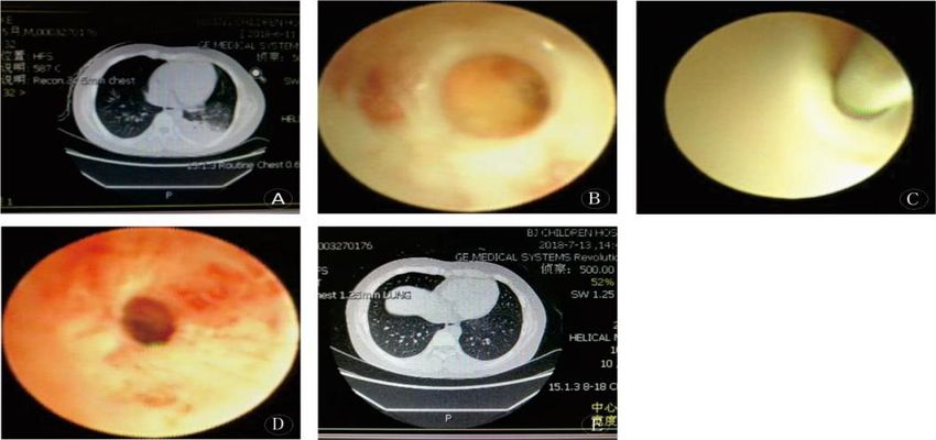
site, type, imaging findings, and therapeutic outcome; duration of fever during disease course and after
Posted on Authorea 20 May 2021 — The copyright holder is the author/funder. All rights reserved. No reuse without permission. — https://doi.org/10.22541/au.162152127.79623314/v1 — This a preprint and has not been peer reviewed. Data may be preliminary.

                                                                                                                                                                                                                                                               bronchoscopy; inflammatory indexes, such as routine blood and CRP test results.
                                                                                                                                                                                                                                                               Statistical Analysis
                                                                                                                                                                                                                                                               SPSS statistics Version 22.0 (International Business Machines (IBM) Corporation) was employed for data
                                                                                                                                                                                                                                                               analysis. Enumeration data were expressed as percentages and the χ2 test was used for comparisons between
                                                                                                                                                                                                                                                               groups. Measurement data conforming to the normal distribution were expressed as the mean ± standard
                                                                                                                                                                                                                                                               deviation (SD) and the t-test was used to make comparisons between two groups. The median [interquartile
                                                                                                                                                                                                                                                               range (IQR) M(P25 -P75 )] was used for measurements of data not conforming to the normal distribution and
                                                                                                                                                                                                                                                               the rank-sum test was used to make comparisons between two groups. Count data were expressed by rate
                                                                                                                                                                                                                                                               or constituent ratio and chi-squared testing was performed. A value of P < 0.05 was considered statistically
                                                                                                                                                                                                                                                               significant.
                                                                                                                                                                                                                                                               RESULTS
                                                                                                                                                                                                                                                               Study Population
                                                                                                                                                                                                                                                               A total of 248 cases were diagnosed with SMPP, of which 213 cases met the enrollment criteria. The average
                                                                                                                                                                                                                                                               time period that elapsed from disease onset to first bronchoscopy was 8.9 ± 2.4 days, with the average
                                                                                                                                                                                                                                                               number of prior bronchoscopies found to be 2.2 ± 1.8. Of the enrolled cases, 142 cases (142/213, 66.7%)
                                                                                                                                                                                                                                                               had their first bronchoscopy within 10 days post-disease onset, while 71 cases (71/213, 33.3%) had their first
                                                                                                                                                                                                                                                               bronchoscopy 10 days post-onset of disease. 183 cases (183/213, 85.9%) underwent bronchoscopy no more
                                                                                                                                                                                                                                                               than twice, while 30 cases (30/213, 14.0%) underwent bronchoscopy at least three times (Table 1).
                                                                                                                                                                                                                                                               Clinical and Bronchoscopic Manifestations
                                                                                                                                                                                                                                                               In this study, disease course was assessed based on clinical manifestations, lung imaging, and bronchoscopic
                                                                                                                                                                                                                                                               findings then was classified into three disease course categories: acute-stage, subacute-stage, and recovery-
                                                                                                                                                                                                                                                               stage.
                                                                                                                                                                                                                                                               Symptoms during acute-stage SMPP included fever, elevated CRP, and large areas of consolidation detected
                                                                                                                                                                                                                                                               via lung imaging. Generally, detection of bronchial mucosal congestion with or without airway mucosal
                                                                                                                                                                                                                                                               follicular hyperplasia and bronchial secretions increased with use of bronchoscopy. Symptoms of subacute-
                                                                                                                                                                                                                                                               stage SMPP were characterized by decreased or normal fever peak, improved signs as assessed via lung
                                                                                                                                                                                                                                                               imaging, increased blood CRP, and decreased bronchial secretions and airway blockages accompanied by
                                                                                                                                                                                                                                                               mucous membrane necrosis and exfoliation found via bronchoscopy. Symptoms of recovery-stage SMPP
                                                                                                                                                                                                                                                               included normal body temperature, normal blood CRP, and either improved findings via lung imaging or
                                                                                                                                                                                                                                                               atelectasis.
                                                                                                                                                                                                                                                               Bronchoscopic manifestations of acute-stage SMPP: all cases exhibited different degrees of congestion and
                                                                                                                                                                                                                                                               roughness of tracheal and bronchial mucosa, mucosal longitudinal fold changes, and mucinous secretion.
                                                                                                                                                                                                                                                               During the first bronchoscopy, follicular hyperplasia of airway mucosa was found in 162 cases (162/213,
                                                                                                                                                                                                                                                               76%), with lung lesions located in grade I and grade II trachea. Erosion of airway mucosa was found in 105
                                                                                                                                                                                                                                                               cases (49.2%), with lesions located in grade III or lower grade bronchi. Mucous plug blockages involving
                                                                                                                                                                                                                                                               grade III or lower bronchi were found in 47 cases (47/213, 22.0%). The mean disappearance time of mucous
                                                                                                                                                                                                                                                               plugs was 15 ± 1.7 days. The mean number of previous bronchoscopies was 2.6 ± 3.5. Of cases with mucous
                                                                                                                                                                                                                                                               plugs, 8 cases (8/213, 3.8%) had severe mucous plug obstruction (with tree-like bronchial-shaped changes).
                                                                                                                                                                                                                                                               Locations of lesions were as follows: lesions of 11 cases (11/213, 10.5%) were in the left upper lung, of 15
                                                                                                                                                                                                                                                               cases were in the left lingual lobe (15/213, 14.3%), of 48 cases were in the left lower basal segment (48/213,
                                                                                                                                                                                                                                                               45.7%), of 13 cases (13/213, 12.4%) were in the right upper lobe, of 10 cases (10/213, 9.5%) were in the right
                                                                                                                                                                                                                                                               middle lobe, of 64 cases (64/213, 60.9%) were in the right lower basal segment, of 63 cases (63/213, 60%)
                                                                                                                                                                                                                                                               were in one lobe, of 32 cases (32/213, 30.5%) were in two lobes, of 7 cases (7/213, 6.7%) were in three lobes,
                                                                                                                                                                                                                                                               of 3 cases (3/213, 2.8%) were in four lobes; no cases involved five lobes.
                                                                                                                                                                                                                                                               Bronchoscopic manifestations in subacute and recovery stages: 22 cases (22/213, 10.3%) had OB, including

                                                                                                                                                                                                                                                                                                                     3
13 males (13/22, 59%) and 9 females (9/22, 41%). The median time to diagnosis of Sub-bronchial stenosis
Posted on Authorea 20 May 2021 — The copyright holder is the author/funder. All rights reserved. No reuse without permission. — https://doi.org/10.22541/au.162152127.79623314/v1 — This a preprint and has not been peer reviewed. Data may be preliminary.

                                                                                                                                                                                                                                                               was 15 days [IQR 12-18]. The initial obstruction was a thin film that was easily broken with biopsy forceps
                                                                                                                                                                                                                                                               in order to reopen the airway. With time, the untreated occluded airway section gradually thickened and the
                                                                                                                                                                                                                                                               center became concave and became associated with a peripheral radial hyperplasia-like obstruction. Proximal
                                                                                                                                                                                                                                                               tracheal examination revealed the presence of fishbone-like bronchiectases that were difficult to reopen using
                                                                                                                                                                                                                                                               biopsy forceps (Figures 1 and 2).
                                                                                                                                                                                                                                                               Analysis of Factors Associated with SMPP Progression to OB
                                                                                                                                                                                                                                                               Of 47 (46.8%) pediatric SMPP patients with mucous emboli, 22 patients developed OB and no OB was found
                                                                                                                                                                                                                                                               in children without mucous emboli; thus, OB incidence in children with mucous emboli was significantly
                                                                                                                                                                                                                                                               higher than in children without mucous emboli (P < 0.001). All 8 cases (100%) with plastic bronchitis
                                                                                                                                                                                                                                                               determined via bronchoscopy exhibited different degrees of OB. During the convalescent stage, OB occurred
                                                                                                                                                                                                                                                               in 4 of 183 cases (2.2%) reporting two or fewer prior bronchoscopies and 18 of 30 cases (60%) with at least
                                                                                                                                                                                                                                                               3 prior bronchoscopies. The incidence of OB in children with two or fewer bronchoscopies was significantly
                                                                                                                                                                                                                                                               lower than OB incidence in children with at least three prior bronchoscopies (P < 0.001). Importantly,
                                                                                                                                                                                                                                                               of 142 children (6.3%) who had the first bronchoscopy within 10 days of disease onset, 9 developed OB
                                                                                                                                                                                                                                                               (6.3%); of 71 children who had their first bronchoscopy on or after 10 days of disease onset, 13 (76.5%)
                                                                                                                                                                                                                                                               developed OB. Therefore, OB incidence of children undergoing bronchoscopy within 10 days of disease onset
                                                                                                                                                                                                                                                               was significantly lower than that of children undergoing bronchoscopy at or after 10 days post-disease onset
                                                                                                                                                                                                                                                               (P < 0.007). Moreover, the average chest CT number for children with acute stage obstructive bronchitis
                                                                                                                                                                                                                                                               was 26 u, while the average CT number for children without OB was 19 u. Thus, chest CT values of children
                                                                                                                                                                                                                                                               with OB were significantly higher than those of children without OB, with the difference between groups
                                                                                                                                                                                                                                                               reaching statistical significance (P < 0.05). Notably, OB was found in all 6 cases afflicted with necrotizing
                                                                                                                                                                                                                                                               pneumonia (100%), as detected via pulmonary imaging (Table 2, Figure 3).
                                                                                                                                                                                                                                                               Adverse Events
                                                                                                                                                                                                                                                               No massive hemorrhage occurred. However, during acute-stage SMPP some bronchoalveolar lavage or cell
                                                                                                                                                                                                                                                               brush interventions caused a small amount of bleeding. In such cases, a local application of 1:10000-diluted
                                                                                                                                                                                                                                                               adrenaline may help. No case of pneumothorax was detected.
                                                                                                                                                                                                                                                               Discussion
                                                                                                                                                                                                                                                               Children afflicted with SMPP have severe clinical symptoms and more complications than those with MPP,
                                                                                                                                                                                                                                                               including necrotizing pneumonia, atelectasis, bronchiectasis, OB, and permanent airway structural damage.
                                                                                                                                                                                                                                                               As a result, lung function declines and repeated infections can impact quality of life for these patients.
                                                                                                                                                                                                                                                               According to previous studies, main clinical manifestations of pediatric SMPP include continuous high fever
                                                                                                                                                                                                                                                               and the presence of large areas of lung consolidation detected via pulmonary imaging. These findings reflect
                                                                                                                                                                                                                                                               an overactive immune response that often requires hormone therapy for suppression of harmful inflammatory
                                                                                                                                                                                                                                                               processes.7-9
                                                                                                                                                                                                                                                               The clinicalClinical manifestations of MPP in China differ from those reported in other countries, with sym-
                                                                                                                                                                                                                                                               ptoms more serious in China, especially in northern areas.4 In recent years, the widespread application of
                                                                                                                                                                                                                                                               pediatric bronchoscopy for SMPP diagnosis and treatment has led to great progress in treating this disorder
                                                                                                                                                                                                                                                               in China. Although some practitioners doubt the value of bronchoscopic findings for MPP prognosis, the
                                                                                                                                                                                                                                                               value of bronchoscopy has been demonstrated for pediatric SMPP prognosis in China. In this study, conges-
                                                                                                                                                                                                                                                               tion and swelling of airway mucosa were obvious in acute-stage cases examined via bronchoscopy, with some
                                                                                                                                                                                                                                                               of these findings accompanied by follicular hyperplasia, a relatively specific MPP-associated manifestation.
                                                                                                                                                                                                                                                               Meanwhile, during bronchoscopy bronchoalveolar lavage fluid is readily collected for use in MPP pathogen
                                                                                                                                                                                                                                                               detection as a direct and effective method for achieving differential MPP diagnosis and treatment. Import-
                                                                                                                                                                                                                                                               antly, acute-stage SMMP mucous plug obstructions should be cleared bronchoscopically in a timely manner
                                                                                                                                                                                                                                                               in order to maintain unobstructed airway subbranches and subsequently control infection. In subacute-stage
                                                                                                                                                                                                                                                               SMMP, bronchoscopy can be used to monitor recovery from airway inflammation and detect OB sequelae
                                                                                                                                                                                                                                                               for use in guiding treatment and respiratory rehabilitation planning. Therefore, bronchoscopic examination

                                                                                                                                                                                                                                                                                                                     4
and intervention can benefit children with SMMP, with no significant adverse effects of bronchoscopy found
Posted on Authorea 20 May 2021 — The copyright holder is the author/funder. All rights reserved. No reuse without permission. — https://doi.org/10.22541/au.162152127.79623314/v1 — This a preprint and has not been peer reviewed. Data may be preliminary.

                                                                                                                                                                                                                                                               in this study.
                                                                                                                                                                                                                                                               OB, a type of chronic airflow obstruction syndrome, mainly involves small and medium bronchi and is
                                                                                                                                                                                                                                                               associated with proliferation of fibrous tissue. The main manifestations of OB are atelectasis and lobar
                                                                                                                                                                                                                                                               collapse. Under bronchoscopic examination, the distal obliterans of some subsegmental and subsubsegmental
                                                                                                                                                                                                                                                               bronchi of OB patients is accompanied by atrophy of proximal mucosa, appearance of obvious cartilage rings,
                                                                                                                                                                                                                                                               and lumen expansion. OB often occurs secondary to severe pneumonia and has gradually gained recognition
                                                                                                                                                                                                                                                               as a common SMPP-associated sequela,10 although few studies in China or abroad have described OB caused
                                                                                                                                                                                                                                                               by M. pneumoniae infection. With increasing research attention paid to SMPP airway damage, OB detection
                                                                                                                                                                                                                                                               rates have significantly increased and highlight OB as an urgent clinical problem stemming from processes
                                                                                                                                                                                                                                                               underlying SMPP development. Meanwhile, studies have shown that MP infection can result in cellular and
                                                                                                                                                                                                                                                               humoral immune dysfunction and disturbances of cytokine networks. Moreover, an excessive inflammatory
                                                                                                                                                                                                                                                               reaction causes airway mucosal epithelial damage followed by airway subbranch injury that leads to scar
                                                                                                                                                                                                                                                               contracture and occlusion. In this study, it was observed that OB caused by SMPP mainly involved grade III
                                                                                                                                                                                                                                                               bronchi or below. The OB developmental process could be outlined as follows: in acute-stage disease, diseased
                                                                                                                                                                                                                                                               sub-bronchi begin to erode then mucous plugs develop and blockages form. Next, necrotic epithelium detaches
                                                                                                                                                                                                                                                               and blocks the airway in the subacute stage. Afterwards, subsegmental bronchi gradually became narrower,
                                                                                                                                                                                                                                                               leading to poor ventilation, insufficient local blood supply surrounding lesions, then gradual formation of
                                                                                                                                                                                                                                                               obliterans. The obliterans formation process supports the conclusion that obliterans are caused by airway
                                                                                                                                                                                                                                                               injury. Meanwhile, initial obliterans lesions first appear as a thin film that is easily reopened using biopsy
                                                                                                                                                                                                                                                               forceps. Next, the occluded part gradually thickens to form a barrier that biopsy forceps cannot reopen
                                                                                                                                                                                                                                                               in most cases. It has been suggested that it may be possible to reopen airway branches after choosing
                                                                                                                                                                                                                                                               the appropriate timing for bronchoscopic intervention, with further study needed to optimize interventional
                                                                                                                                                                                                                                                               timing for improved treatment outcomes.
                                                                                                                                                                                                                                                               In this study, the timing of bronchoalveolar lavage was also investigated to detect correlations with OB
                                                                                                                                                                                                                                                               incidence during late-stage SMPP. Specifically, the incidence of OB in pediatric patients who received first
                                                                                                                                                                                                                                                               bronchoalveolar lavage at 10 days post-SMPP onset was significantly higher than that of children recei-
                                                                                                                                                                                                                                                               ving their first bronchoscopies within 10 days post-disease onset. It has been suggested that for pediatric
                                                                                                                                                                                                                                                               patients with persistent fever and large lung consolidations, atelectasis, and other pulmonary pathological
                                                                                                                                                                                                                                                               changes detected via imaging within 10 days of onset, timely bronchoalveolar lavage is recommended to avoid
                                                                                                                                                                                                                                                               complications such as OB.
                                                                                                                                                                                                                                                               The main components of bronchial mucous plugs include a mixture of mucin and cellular debris combined
                                                                                                                                                                                                                                                               with a large number of infiltrating inflammatory cells that make this mixture extremely viscous. At present,
                                                                                                                                                                                                                                                               it is widely recognized that hyperimmune inflammation is one of the main pathogenic mechanisms underly-
                                                                                                                                                                                                                                                               ing SMPP.3,11 MP infection tends to lead to mucosal necrosis and mucus hypersecretion via a mechanism
                                                                                                                                                                                                                                                               that may involve direct damage and release of infection-triggered toxic metabolites that ultimately damage
                                                                                                                                                                                                                                                               bronchial epithelial cells and cilia. Such pathological damage may further impair mucociliary system function
                                                                                                                                                                                                                                                               and weaken ciliary discharge capacity and clearance of secretions. Afterwards, tethered secretions that form
                                                                                                                                                                                                                                                               within the local airway can trigger airway obstruction, with local obstruction of the proximal bronchus and
                                                                                                                                                                                                                                                               various sizes of mucous plugs occurring within deep subsegmental bronchi in some serious cases. This study
                                                                                                                                                                                                                                                               demonstrated that pediatric SMMP patients who also developed OB during late-stage disease exhibited dif-
                                                                                                                                                                                                                                                               ferent degrees of mucous plug development during acute-stage SMPP that were detected via bronchoscopy.
                                                                                                                                                                                                                                                               Therefore, for patients with mixed lesions that are associated mainly with mucous plugs, mucus necrosis,
                                                                                                                                                                                                                                                               and particularly with successive mucus plug development detected upon bronchoscopic reexamination, loca-
                                                                                                                                                                                                                                                               lization of mucous plugs should be done as early as possible in conjunction with administration of systemic
                                                                                                                                                                                                                                                               treatment. Moreover, regular bronchoalveolar lavage treatment would facilitate the timely removal of mu-
                                                                                                                                                                                                                                                               cous plugs in order to improve ventilation and secretion clearance at lesion sites and promote inflammatory
                                                                                                                                                                                                                                                               absorption.
                                                                                                                                                                                                                                                               This study demonstrated that the density of large consolidation areas was significantly increased during

                                                                                                                                                                                                                                                                                                                     5
acute-stage SMPP, suggesting that subbranches can be severely blocked by mucous embolisms. Thus, bron-
Posted on Authorea 20 May 2021 — The copyright holder is the author/funder. All rights reserved. No reuse without permission. — https://doi.org/10.22541/au.162152127.79623314/v1 — This a preprint and has not been peer reviewed. Data may be preliminary.

                                                                                                                                                                                                                                                               choscopy should be performed as soon as possible to remove airway secretions that in such cases consist
                                                                                                                                                                                                                                                               of thick viscous mucus that is difficult to clear away. For conducting a first bronchoscopic examination of
                                                                                                                                                                                                                                                               patients with this type of lesion, a bronchoscope with a larger diameter (such as 4.0 mm) is recommended;
                                                                                                                                                                                                                                                               because the diameter of the suction duct is large, it can loosen and brush mucous plugs to the depth of
                                                                                                                                                                                                                                                               subsegmental bronchi and can effectively remove mucous plugs from deeper airways with the help of a cell
                                                                                                                                                                                                                                                               brush. Due to acute-stage mucosal damage and congestion, adrenaline can be applied locally to shrink blood
                                                                                                                                                                                                                                                               vessels while plugs are slowly and gently brushed to minimize bleeding during plug removal. During late-stage
                                                                                                                                                                                                                                                               SMPP recovery characterized by improved lung imaging findings, the 2.8-mm diameter bronchoscope could
                                                                                                                                                                                                                                                               be selected for subsequent inspection and clearance of deep subsegmental bronchial mucous plugs.
                                                                                                                                                                                                                                                               Due to the risk of pneumothorax that may occur during bronchoscopic examination and treatment of necrotic
                                                                                                                                                                                                                                                               pediatric SMPP, the use of bronchoscopy for such cases is still controversial at present. However, it is worth
                                                                                                                                                                                                                                                               noting that of 6 patients with late-stage necrotic pneumonia receiving treatment in this study, all of these
                                                                                                                                                                                                                                                               patients developed OB even though they had different degrees of bronchoscopically detected airway blockage
                                                                                                                                                                                                                                                               due to mucous plugs and mucinous necrosis. Notably, no pneumothorax occurred as a result of bronchoscopic
                                                                                                                                                                                                                                                               treatment. Nevertheless, owing to the small sample size of this study, it is impossible to judge the advantages
                                                                                                                                                                                                                                                               and disadvantages of bronchoscopy for necrotic SMPP based solely on results of this study.
                                                                                                                                                                                                                                                               In summary, pediatric SMPP patients exhibit certain characteristics during different disease stages that can
                                                                                                                                                                                                                                                               facilitate diagnosis. However, airway mucus and mucosal epithelial necrosis and detachment could lead to
                                                                                                                                                                                                                                                               clogging of subbranches and subsequent OB. Thus, it is recommended that pediatric SMPP patients with
                                                                                                                                                                                                                                                               mucous plugs undergo timely bronchoscopy in addition to customary antibiotic treatment. Because this stu-
                                                                                                                                                                                                                                                               dy did not comprehensively summarize and analyze factors associated with OB in pediatric SMPP patients,
                                                                                                                                                                                                                                                               predictive factors predisposing patients to OB development were not definitively identified. Therefore, col-
                                                                                                                                                                                                                                                               lection of additional clinical data is needed to further evaluate possible predictors of later OB occurrence
                                                                                                                                                                                                                                                               that would be used to inform future clinical practice.
                                                                                                                                                                                                                                                               Acknowledgements: We thank the children and families who graciously consented to participate in the
                                                                                                                                                                                                                                                               bronchoscopy of SMPP children in this study.
                                                                                                                                                                                                                                                               References
                                                                                                                                                                                                                                                                 1. Jain S, Williams DJ, Arnold SR, et al. Community-acquired pneumonia requiring hospitalization
                                                                                                                                                                                                                                                                    among U.S. children. N Engl J Med,2015 ; 372(9): 835-845.
                                                                                                                                                                                                                                                                 2. Komatsu H, Tsunoda T, Inui A, Sogo T, Fujisawa T. Characteristics of hospitalized children infected
                                                                                                                                                                                                                                                                    with macrolide-resistant Mycoplasma pneumonia. Braz J Infect Dis, 2014 ; 18(3): 294-299.
                                                                                                                                                                                                                                                                 3. Izumikawa K. Clinical Features of Severe or Fatal Mycoplasma pneumoniae Pneumonia.Front Micro-
                                                                                                                                                                                                                                                                    biol, 2016 ; 7: 800
                                                                                                                                                                                                                                                                 4. Yan C, Sun HM, Zhao HQ. Latest surveillance data on Mycoplasma pneumoniae infections in children,
                                                                                                                                                                                                                                                                    suggesting a new epidemic occurring in Beijing. J Clin Microbiol, 2016 ; 54: 1400-1401.
                                                                                                                                                                                                                                                                 5. Lee KY, Lee HS, Hong JH, et al . Role of prednisolone treatment in severe Mycoplasma pneumoniae
                                                                                                                                                                                                                                                                    pneumonia in children. Pediatr Pulmonol, 2006 ; 41: 263-268.
                                                                                                                                                                                                                                                                 6. Li CC, Shang YX, Shen XZ, Chen ZM, Zhao XY. Guidelines for the management of community
                                                                                                                                                                                                                                                                    acquired pneumonia in children (revised in 2013).Chinese Journal of Pediatrics, 2013 ; (10): 745-752.
                                                                                                                                                                                                                                                                 7. Bajantri B, Venkatram S, Diaz-Fuentes G. Mycoplasma pneumoniae: a potentially severe infection. J
                                                                                                                                                                                                                                                                    Clin Med Res, 2018 ; 10: 535-544.
                                                                                                                                                                                                                                                                 8. Yan C, Xue GH, Zhao HQ, et al . Molecular and clinical characteristics of severe Mycoplasma pneu-
                                                                                                                                                                                                                                                                    moniae pneumonia in children.Pediatr Pulmonol, 2019 ; 54(7): 1012-1021.
                                                                                                                                                                                                                                                                 9. Chan ED, Welsh CH. Fulminant Mycoplasma pneumoniae pneumonia.West J Med, 1995 ; 162(2):
                                                                                                                                                                                                                                                                    133-142.
                                                                                                                                                                                                                                                                10. Colom AJ, Teper AM. Post-infectious bronchiolitis obliterans.Pediatr Pulmonol, 2019 ; 54(2): 212-219
                                                                                                                                                                                                                                                                11. Moonnumakal SP, Fan LL. Bronchiolitis obliterans in children.Curr Opin Pediatr, 2008 ; 20(3): 272-
                                                                                                                                                                                                                                                                    278.Table 1: General informations

                                                                                                                                                                                                                                                                                                                     6
Posted on Authorea 20 May 2021 — The copyright holder is the author/funder. All rights reserved. No reuse without permission. — https://doi.org/10.22541/au.162152127.79623314/v1 — This a preprint and has not been peer reviewed. Data may be preliminary.

                                                                                                                                                                                                                                                               Age(years   Male/Total Female          Pulmonary      Atelectasis Pulmonary Extrapulmonary  Average      Averag
                                                                                                                                                                                                                                                               old)        cases(%)   /Total          consolida-     cases/Total necro-       complica-    fever time   postop
                                                                                                                                                                                                                                                                                      cases(%)        tion           cases(%)    sis(cases)/Total
                                                                                                                                                                                                                                                                                                                                              tions(cases)/Total
                                                                                                                                                                                                                                                                                                                                                           (days)       ative
                                                                                                                                                                                                                                                                                                      cases/Total                cases(%)     cases(%)                  fever t
                                                                                                                                                                                                                                                                                                      cases(%)                                                          (days)
                                                                                                                                                                                                                                                               4.2 ±       114/213(53.5) 99/213(46.4) 213/213(100)   189/213(88.7) 6/213(2.8)   192/213(90.1) 8.8±1.2   1.8±4.4
                                                                                                                                                                                                                                                               2.6

                                                                                                                                                                                                                                                               Table 2: Main factors of influencing obliterative bronchiolitis: comparison between children
                                                                                                                                                                                                                                                               with and without obliterans

                                                                                                                                                                                                                                                                                  Time of first bronchoscopy since the onset of the disease      Time of first bronchoscopy sin
                                                                                                                                                                                                                                                                                  Within 10 days                                                 After 10 days
                                                                                                                                                                                                                                                               Cases              142                                                            71
                                                                                                                                                                                                                                                               Obliterans cases   9                                                              13
                                                                                                                                                                                                                                                               P value            P¡0.007                                                        P¡0.007

                                                                                                                                                                                                                                                               Figures and Legends
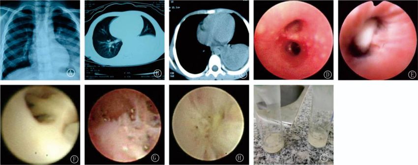
                                                                                                                                                                                                                                                               Figure 1: Chest X-ray and high-resolution CT showed large consolidation shadow in the left
                                                                                                                                                                                                                                                               lower lung(A,B,C).Obvious hyperemia of bronchial mucosa and nodular protrusion on the
                                                                                                                                                                                                                                                               surface of mucosa (D). Longitudinal fold change of subbronchiolar mucosa and mucus plug
                                                                                                                                                                                                                                                               obstruction in the lesion site of bronchial subbranch (E).The subbronchiolar mucosa looked
                                                                                                                                                                                                                                                               pale, and necrotic detachment of bronchial epithelial mucosa blocked lumen opening(F).The
                                                                                                                                                                                                                                                               subbronchiolar mucosa were pale in color and occluded with concave center and peripheral
                                                                                                                                                                                                                                                               radial hyperplasia in the occluded part(G,H). Plastic mucus plug taken by cell brush under
                                                                                                                                                                                                                                                               bronchoscope(I).

                                                                                                                                                                                                                                                               Figure 2: Chest high-resolution CT showed atelectasis in the left lower lung three weeks after
                                                                                                                                                                                                                                                               the onset of the disease(A). The color of subbronchiolar mucosa was pale and the lumen was
                                                                                                                                                                                                                                                               occluded(B). Biopsy forceps were used to recanalize the subbranch occlusion under broncho-
                                                                                                                                                                                                                                                               scope(C) and the recanalized airway subbranch(D).Chest high-resolution CT scan showed that
                                                                                                                                                                                                                                                               atelectasis in the left lower lung got better one month after bronchoscopy(E).

                                                                                                                                                                                                                                                                                                              7
Posted on Authorea 20 May 2021 — The copyright holder is the author/funder. All rights reserved. No reuse without permission. — https://doi.org/10.22541/au.162152127.79623314/v1 — This a preprint and has not been peer reviewed. Data may be preliminary.

                                                                                                                                                                            monary bronchus under bronchoscope(D).

8
                                                                                                                                                                            liquefactive necrosis and cavity in right lung (A,B,C). Extensive occlusion in left lower pul-
                                                                                                                                                                            Figure 3: Chest X-ray and high-resolution CT discovered large consolidation shadow with
You can also read