Case Report A Guttate Psoriasis That Tends to Spare Three Tattoos: A Macrophage Liaison
←
→
Page content transcription
If your browser does not render page correctly, please read the page content below
Hindawi
Case Reports in Dermatological Medicine
Volume 2021, Article ID 9448636, 5 pages
https://doi.org/10.1155/2021/9448636

Case Report
A Guttate Psoriasis That Tends to Spare Three Tattoos: A
Macrophage Liaison

          Panagiota Spyridonos,1 Vasiliki Zampeli ,2 Sophia-Nefeli Rapti,3
          and Ioannis D. Bassukas 2,3
          1
            Department of Medical Physics/Informatics, Faculty of Medicine, School of Health Sciences, University of Ioannina,
            Ioannina, Greece
          2
            Department of Skin and Venereal Diseases, Faculty of Medicine, School of Health Sciences, University of Ioannina,
            Ioannina, Greece
          3
            Department of Dermatology, University Hospital of Ioannina, Ioannina, Greece

          Correspondence should be addressed to Ioannis D. Bassukas; ibassuka@uoi.gr

          Received 23 May 2021; Accepted 6 September 2021; Published 13 September 2021

          Academic Editor: Alireza Firooz

          Copyright © 2021 Panagiota Spyridonos et al. This is an open access article distributed under the Creative Commons Attribution
          License, which permits unrestricted use, distribution, and reproduction in any medium, provided the original work is
          properly cited.
          Induction of new psoriasis sites was reported in only a small amount of psoriasis patients undergoing tattooing, despite the
          intuitive belief that tattoo trauma might awaken the disease due to the isomorphic phenomenon of Koebner. In this case report, we
          discuss a patient who presented with a remarkable sparing of his three tattoo sites during a guttate psoriasis flare-up that was
          unrelated to tattooing. The spatial concordance of tattoo and psoriasis lesions was analyzed on clinical pictures of tattoo sites taken
          during the psoriasis episode. For the quantification of the spatial distribution of the psoriasis lesions, Voronoi diagrams were
          generated, and coefficients of variation and the two-sample t-test were employed to compare the distributions of Voronoi patch
          sizes in different settings. Compared to skin areas without tattoos, a tattoo introduced a higher variation in the sizes of the Voronoi
          patches centered around psoriasis lesions. Based on our findings, we would like to discuss the possible role of macrophages as the
          key cellular link in the complex pathophysiologic relationship between tattooing/tattoo and psoriasis. Taking into account the
          relationship of autophagy and psoriasis lesions, we propose the hypothesis that tattoos represent a “psoriasis-hostile” tissue
          environment pertained by a population of LAP active M2-polarized macrophages. Further clinical studies of the relationship of
          psoriasis lesions to the tattooed skin are needed and may provide important insights into the role of macrophages in the
          pathogenesis of psoriasis.

1. Introduction                                                           unrelated to tattooing, we would like to comment on the
                                                                          putative role of macrophages as the key cellular link in the
The nosologic relationship of psoriasis and tattoo remains a              complex pathophysiologic relationship between tattooing/
matter of debate. Despite the intuitive concern that tattoo               tattoo and psoriasis.
trauma might induce the disease (Koebner phenomenon,
KP), a KP was reported in only 3.2% of psoriasis patients                 2. Case Presentation
undergoing tattooing [1], an overall rather moderate rate [2].
Moreover, Grodner and colleagues [3] observed that much                   An afebrile, 27-year-old male patient, in otherwise good
more patients with psoriasis did not recall lesions that had              general condition, presented with a rash of scattered, up to
affected their tattoo sites during episodes of disease flare-ups            1.0 cm large psoriasiform lesions that progressively affected
(47.6%) than recalling them (19.8%). Herein, on the occasion              his torso (shown in Figure 1) and the extremities during the
of a patient who presented with a distinct sparing of three               past two weeks. The rash was accompanied by moderate
tattoo sites during a guttate psoriasis episode that was                  itching and an apparent tendency to coalesce in typical for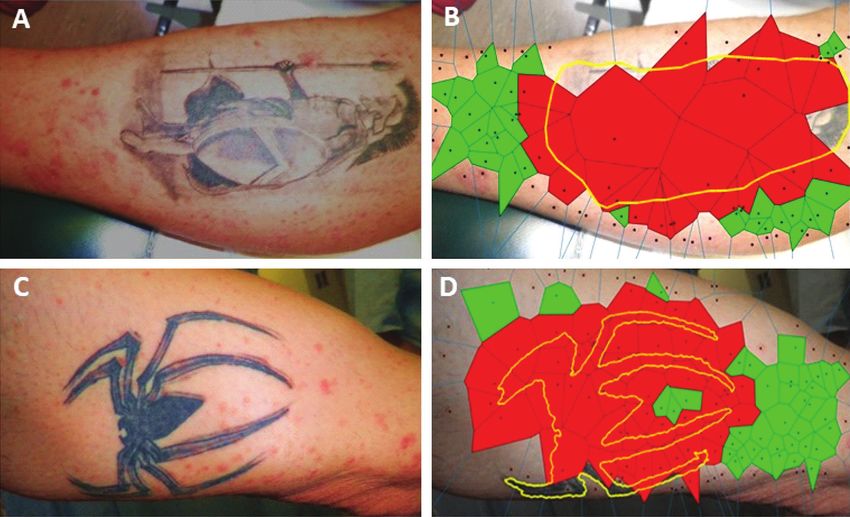
2                                                                                          Case Reports in Dermatological Medicine

Figure 1: (a) Skin area affected with psoriasis. (b) Enhanced lesion representation by a∗ color channel of lab space. (c) Segmented lesions
(yellow spots) superimposed on the initial image to visualize the result of morphological processing. (d) Lesions’ centroids generating
Voronoi tessellation.

plaque psoriasis skin localizations, i.e., the knees. The patient      of the Voronoi polygons, with the patches that intersect
was diagnosed with plaque psoriasis at the age of 15 years,            tattoo lines, i.e., those patches that are localized within the
which was treated successfully with topical regimens. The              tattooed skin area or are located in the vicinity of the tattoo
laboratory evaluation including chest X-ray, ASO, syphilis,            contours, being significantly larger compared to the rest,
and hepatitis A, B, and C serology returned no pathologic              more remote polygons (p < 0.001; Table 1).
findings. Based on patient’s history, the macromorphology
of the rash, and a lesional skin biopsy, guttate psoriasis was         3. Discussion/Conclusion
diagnosed. Except for psychosocial stress, no other trig-
gering events of the dermatosis could be elucidated. Re-               Sparing of skin lesions by an evolving psoriasis has been
markably, the rash spared almost completely the sites of his           observed, so psoriasis spared a polio-affected limb [6–8] or a
three tattoos (lower leg, forearm, and shoulder; larger di-            guttate psoriasis spared Becker’s melanosis [9]. Also, a pur-
ameters 15–25 cm, shown in Figures 2 and 3). The tattoos               puric rash and a leukocytoclastic vasculitis that spared tattoos
(two of them monochrome black and the third black con-                 have been described [10, 11]. However, to the best of authors’
tours on whitish background; Figures 2 and 3) were per-                knowledge, this is the first report of guttate psoriasis sparing
formed on different occasions over the last 3 years (the last           tattoos. Remarkable is also the case of a patient with a reverse
one about 9 months before the flare-up) in the same tattoo              Koebner phenomenon, i.e., clearing of psoriasis lesions after
studio. The patient reported no KP on previous tattooing               tattooing [12]. All these observations, together with evidence
with either local or generalized exacerbations of his psoriasis.       for lower than anticipated KP risk in association with tat-
    The spatial concordance of tattoo and psoriasis lesions            tooing, indicate to a distinct pathophysiologic interaction of
was analyzed on clinical pictures of tattoo sites taken during         tattoo and psoriasis. We suggest that the macrophage, the key
the flare-up. For the detection of psoriasis lesions, RGB               cell species involved in tattoo pathophysiology, is the cellular
clinical images (shown in Figure 1(a)) were converted into             link of tattoo and the psoriasis sparing in this patient.
the L∗ a∗ b∗ color space. The high-peak intensity areas                     Dermal macrophages, both resident and blood-borne
(“spots”) in the a∗ color channel (shown in Figure 1(b)),              monocyte-derived, comprise a slowly renewing and in loco
which correspond to psoriasis lesions, were extracted by               relatively long-lived cell species [13]. Macrophages are the
serially applying the h-maxima transform and the regional              key effector cells in the pathophysiology of tattoos, as they
maxima morphological operations [4] (shown in                          phagocytose and carry locally most of the tattoo pigment
Figure 1(c)). To quantify the spatial distribution of the              [14]. Moreover, they enable the stability of the tattoo via a
psoriasis lesions, Voronoi diagrams [5] were generated with            process of capture-release-recapture of the pigment in situ
generator points the centroids of above intensity spots                during successive cell death and renewal cycles [15]. Zaba
(shown in Figure 1(d)). Figure 2 exemplifies this procedure             et al. [16] have previously shown that macrophages in
in the case of a tattoo (arbitrary designated tattoo #1).              normal skin that have ingested tattoo pigment was not able
Coefficients of variation (CV) and two-sample t-test were                to stimulate T cell activation. This is probably the result of a
employed to compare the distributions of Voronoi patch                 LC3-associated phagocytosis (LAP) process [17], which
sizes in different settings. Skin areas without a tattoo, like the      ensures the shift of the local immune state towards sustained
front of the torso (Figure 1), are characterized by a rather           anti-inflammation by dampening of proinflammatory sig-
homogeneous, unimodal size distribution of the Voronoi                 nals and preventing the presentation of autoantigens to
patches centered on psoriasis lesions (CV � 0.46, Table 1).            other immune cells [18, 19]. Accordingly, a tattoo site can be
However, the presence of a tattoo introduces a higher                  conceptualized as a noninflammatory skin area with pig-
variation in the size distributions of the corresponding               ment-laden macrophages in a predominantly “deactivated,”
Voronoi patches in its neighborhood (CV � 0.60–1.23;                   immunologically inert, or “anti-inflammatory,” M2-polar-
shown in Figures 2 and 3; Table 1). Moreover, it seems that            ized state [20], resembling a tissue milieu that corresponds to
the presence of a tattoo induces a bimodal size distribution           a sustained resolution phase of an inflammatory process.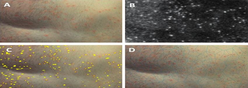
Case Reports in Dermatological Medicine 3
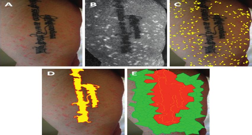
Figure 2: Psoriasis and tattoo #1. (a) Clinical picture. (b) Retained a∗ color channel of L∗ a∗ b∗ color space: high-peak intensity areas mark
psoriasis lesions and low-level intensity areas correspond to tattooed skin segments. (c) Applying h-maxima transform followed by regional
maxima, the lesional skin spots (patches) were extracted and are displayed in yellow pseudocolor. (d) Equivalently, the tattoo area was
extracted by applying h-minima transform, followed by regional minima and is shown in yellow pseudocolor to enhance visualization.
(e) The extracted psoriasis skin lesions’ centroids (panel (C) applied to generate Voronoi diagrams (tessellation) of the tattoo area. Voronoi
polygons with and without overlapping with the tattoo area (yellow border) are displayed in red and green pseudocolors, respectively.
Figure 3: Tattoo #2 (panels (a) and (b)) and tattoo #3 (panels (c) and (d)). (a)–(c) Clinical pictures. (b)–(d) Voronoi patches intersecting
(red) or not intersecting (green) tattooed skin areas (yellow contoured).
On the other hand, macrophages emerge also as a key macrophage polarization are reduced in psoriasis [22], and
cell species in the pathophysiology of psoriasis. In human the improvement of psoriasis with TNF-α inhibitors cor-
psoriasis, a subpopulation of classically activated proin- relates with the inhibition of the M1 macrophage polari-
flammatory macrophages plays a crucial role in the path- zation pathway [23]. Stimulated macrophages are the main
ogenesis of skin lesions, and a preponderance of M1 immune-effector cell species in animal psoriasis models, like
macrophage activation state is also associated with increased the inducible human TNF transgenic mouse line [24].
PASI scores [21]. In accordance with above, markers of M2 Furthermore, in the K14-VEGF-A-Tg mice psoriasis model,4 Case Reports in Dermatological Medicine
Table 1: Effect of tattoo on psoriasis lesions centered Voronoi patches.
Mean size (SD)2 t-test
Skin site CV1 4 Figure 3
Vout Vin5 P value6
Tattoo-free 0.46 13416 (6244) N/A7 N/A 1
Tattoo #1 0.60 12878 (4791) 28972 (12205)Case Reports in Dermatological Medicine 5
populations of CD11c+BDCA-1+ dendritic cells and
CD163+FXIIIA+ macrophages,” Journal of Clinical Investi-
gation, vol. 117, no. 9, pp. 2517–2525, 2007.
[17] B. L. Heckmann, E. Boada-Romero, L. D. Cunha, J. Magne,
and D. R. Green, “LC3-associated phagocytosis and inflam-
mation,” Journal of Molecular Biology, vol. 429, no. 23,
pp. 3561–3576, 2017.
[18] A. J. Clarke and A. K. Simon, “Autophagy in the renewal,
differentiation and homeostasis of immune cells,” Nature
Reviews Immunology, vol. 19, no. 3, pp. 170–183, 2019.
[19] M.-Y. Wu and J.-H. Lu, “Autophagy and macrophage
functions: inflammatory response and phagocytosis,” Cells,
vol. 9, no. 1, p. 70, 2019.
[20] T. Lawrence and G. Natoli, “Transcriptional regulation of
macrophage polarization: enabling diversity with identity,”
Nature Reviews Immunology, vol. 11, no. 11, pp. 750–761,
2011.
[21] J. Fuentes-Duculan, M. Suárez-Fariñas, L. C. Zaba et al., “A
subpopulation of CD163-positive macrophages is classically
activated in psoriasis,” Journal of Investigative Dermatology,
vol. 130, no. 10, pp. 2412–2422, 2010.
[22] H. J. Kim, J. Jang, E.-H. Lee, S. Jung, J. Y. Roh, and Y. Jung,
“Decreased expression of response gene to complement 32 in
psoriasis and its association with reduced M2 macrophage
polarization,” The Journal of Dermatology, vol. 46, no. 2,
pp. 166–168, 2019.
[23] S.-H. Lin, H.-Y. Chuang, J.-C. Ho, C.-H. Lee, and C.-C. Hsiao,
“Treatment with TNF-α inhibitor rectifies M1 macrophage
polarization from blood CD14+ monocytes in patients with
psoriasis independent of STAT1 and IRF-1 activation,”
Journal of Dermatological Science, vol. 91, no. 3, pp. 276–284,
2018.
[24] R. Leite Dantas, D. Masemann, T. Schied et al., “Macrophage-
mediated psoriasis can be suppressed by regulatory
T lymphocytes,” The Journal of Pathology, vol. 240, no. 3,
pp. 366–377, 2016.
[25] J. Zhang, Y. Lin, C. Li et al., “IL-35 decelerates the inflam-
matory process by regulating inflammatory cytokine secretion
and M1/M2 macrophage ratio in psoriasis,” The Journal of
Immunology, vol. 197, no. 6, pp. 2131–2144, 2016.
[26] C. H. Lu, C. Y. Lai, D. W. Yeh et al., “Involvement of M1
macrophage polarization in endosomal Toll-like receptors
activated psoriatic inflammation,” Mediators of Inflammation,
vol. 2018, Article ID 3523642, 14 pages, 2018.
[27] S. Morimura, T. Oka, M. Sugaya, and S. Sato, “CX3CR1
deficiency attenuates imiquimod-induced psoriasis-like skin
inflammation with decreased M1 macrophages,” Journal of
Dermatological Science, vol. 82, no. 3, pp. 175–188, 2016.You can also read